


Ultrafast Beamforming in Medical Ultrasound Imaging
“`html
Medical Ultrasound Imaging: Ultrafast Beamforming Algorithms for Real-Time and High-Quality Imaging
Sreejeesh SG, Sakthivel R, Jayaraj U Kidav
1 VLSI Group, National Institute of Electronics and Information Technology, Calicut (NIELIT) NIT Campus Post, Calicut, India
2 Vellore Institute of Technology, Vellore, Katpadi, Tamilnadu, India
3 National Institute of Electronics and Information Technology, Aurangabad, (NIELIT) Maharashtra, India
OPEN ACCESS
PUBLISHED 31 December 2024
CITATION Sreejeesh, SG., Sakthivel, R., et al., 2024. Medical Ultrasound Imaging: Ultrafast Beamforming Algorithms for Real-Time and High-Quality Imaging. Medical Research Archives, [online] 12(12). https://doi.org/10.18103/mra.v12i12.6177
COPYRIGHT © 2024 European Society of Medicine. This is an open-access article distributed under the terms of the Creative Commons Attribution License, which permits unrestricted use, distribution, and reproduction in any medium, provided the original author and source are credited.
DOI https://doi.org/10.18103/mra.v12i12.6177
ISSN 2375-1924
Abstract
Medical ultrasound imaging is a prevalent diagnostic instrument in the field of medicine. Beamforming is a signal processing methodology employed to improve the efficacy of imaging systems, especially in medical ultrasound imaging. Ultrafast (UF) beamforming algorithms (BAs) are designed to improve the speed and efficiency of beamforming operations. Ultrasound imaging algorithms have been devised to enhance the quality and efficiency of ultrasound imaging. This article will provide an overview of the research on UF medical ultrasound algorithms. We will also explore some recent developments in the field of UF beamforming algorithms. This article discusses the development and implementation of UF-BAs for medical ultrasound imaging. Traditional beamforming techniques are computationally intensive and limit the real-time imaging capability of ultrasound systems. The algorithms discussed in the article leverage modern parallel computing architectures to reduce processing time while maintaining image quality significantly. The article presents a detailed analysis of the algorithm’s performance in processing time and image quality using simulated and accurate data. The article discusses how various UF-BAs can provide real-time high-quality images facilitating novel uses in medical ultrasound imaging. The prospective advantages of these algorithms for clinical use and future research trajectories are investigated as well.
Keywords: Ultra-fast Imaging, Ultrafast Beamformer, Parallel beamformer, Real-Time Imaging, Signal Processing, High frame rate, High-Performance Computing, Ultrasound imaging, Parallel Computing, Computational Efficiency, Fast Imaging, Super-resolution imaging.
1. Introduction
UF beamforming algorithms are novel signal processing techniques that can be applied in medical ultrasound imaging to produce real-time images of interior human organs and structures. Several algorithms can speed up beamforming using high-performance computing architectures (parallel processing or GPU acceleration) using Field Programmable Gate Array FPGA. UF-BAs strive to enhance the rate and performance of conventional beamformer device setup techniques without compromising image quality. In a set of transducer elements receives conventional beamforming and ultrasound signals, which are merged to create a beam focused in a particular direction. This is done in multiple directions before the entire image is formed. However, this technique is resource-intensive and can take seconds to generate a single image.
Instead, UF-BAs rely on advanced processing techniques to speed up beamforming. These algorithms can perform real-time beamforming, showing rapid and high-quality image generation. UF-BAs can also adaptively change the step size of the beamforming process, like the focus or the steering angle, according to the specific properties of the tissue to be imaged. In medical applications, ultrasound has been used for a long period, while beginning in the 1930s, ultrasound has been used in medical imaging, and it does not require ionizing radiation. Real-time ultrasound imaging is crucial for many medical applications, enabling clinicians to see inside the body as it happens. This is especially true in diagnostic or therapeutic procedures where timely and accurate information ensures the best possible patient outcomes.
Ultrasound imaging in real-time, with high speed, is a rich source of information for clinicians to get several, such as the location, size, and nature of internal structures and the movement of blood or other fluids. A medical provider uses this information to make many diagnoses (including tumours, cysts, and other abnormalities). This technology is also helpful in tracking treatment advancements and assisting with positioning medical devices or needles. UF BAs aim to advance medical ultrasound imaging by optimizing the quality of real-time images, ultimately facilitating improved clinical decision-making and enhancing patient outcomes. Advanced ultrasound imaging methods like ultrasound localization microscopy (ULM) and functional ultrasound (fUS) rest on UF ultrasound imaging.
Contributions: In this paper, we discuss various schemes of ultra-fast imaging techniques, their merits, and demerits. We also discuss various parallel beamformer architectures and summarize a scheme that can produce a high frame rate for medical ultrasound imaging applications.
The paper is structured as follows: Section II addresses the background and pertinent literature. Section III delineates the Ultrafast Beamforming Algorithms (UF-BAs). Section IV primarily examines the trade-offs between processing time and image quality, aiming to identify areas necessitating further research to enhance the performance of UF-BAs. Section V ultimately concludes the paper by outlining potential avenues for future research.
2. Background
Ultrasound beamforming is the process by which ultrasound signals transmitted and received by a transducer array are combined to form an image of the tissue being imaged. A transducer array is made up of several separate transducer elements that can both send and receive ultrasonic signals. Each transducer element receives electrical signals during the transmit phase, which causes it to transmit ultrasonic waves. The subsequent waves transmit through the tissue and are reflected to the transducer array.
The echoes of each transducer element are detected, and the electrical signals associated are detected and arranged to produce the image. A sequence of steps, such as delay and sum, aperture synthesis, and beamforming, are performed. This eliminates additional time spent traveling to and from the target tissue/organ, the echoes captured by each transducer element are delayed and aggregated. The resultant delayed signals are consolidated into a singular beamformed signal. This method used is an aperture synthesis that combines the signals received by an element of transducer. Usually, this is done by pointing the beam in different directions and adding the signals received at each angle. Beamforming modifies the ultrasound beam’s shape to enhance image quality and resolution. In order to implement this, one can use many techniques, like focusing, steering, and apodization. Focusing is achieved by controlling the arrival time and strength of the signals transmitted from each transducer cell to construct a focused beam. Steering is achieved by adjusting the timing and strength of each signal sent from the transducer elements to alter the direction of the beam.
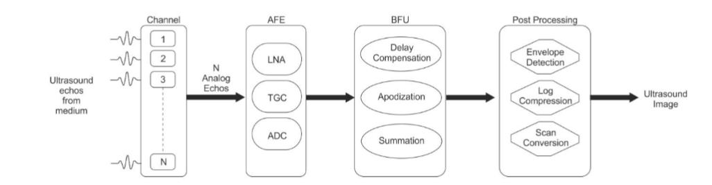
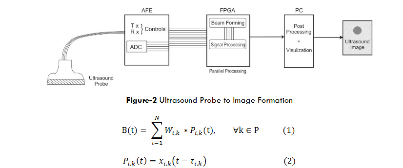
It is also referred to as the amplitude of the signals transmitted by each transducer element modified to decrease artifacts and enhance image quality. More generally, ultrasound beamforming is a key aspect of medical ultrasound imaging, as it allows clinicians to see into a patient’s internal body parts and diagnose a host of medical conditions. For example, recent advancements in ultra-fast BAs have improved imaging speed, efficiency, and image quality, resulting in better patient outcomes and improved clinical decision-making. Meanwhile, this approach signifies the importance of high-throughput imaging in analogously facilitating new approaches to disease treatment. A US imaging system must execute signal processing operations to produce an image for diagnostic purposes from the scattered back and reflecting US waves. A generalized ultrasound receive signal processing block diagram, which is evolved from, is presented in Figure-1 for better understanding.
The received ultrasonic echoes from the tissue are detected by N elements/channels. A Low Noise Amplifier (LNA) amplifies N analog radio frequency echoes in the Analog Front End (AFE), which are then further amplified or reduced by depth proportional gain through a procedure known as Time Gain Compensation (TGC). An analog-to-digital converter (ADC) is then used to transform the signals into digital form. The beamformer unit delays apodizes, and sums the digital RF signals. The beamformed signal from N channel RF echoes undergoes envelope detection, logarithmic compression, and scan conversion to generate a diagnostic ultrasound image. This article will discuss the different beamforming architectures used for imaging in medical ultrasound and the hardware implementations on different platforms.
A.1A Beamforming for Medical Imaging- Traditional Approaches
Beamforming is a commonly used signal-processing method in biomedical imaging applications, including ultrasound and photoacoustic imaging. Traditional approaches to beamforming for biomedical imaging are Delay and Sum beamforming, Minimum Variance beamforming, Synthetic Aperture Beamforming, and Adaptive Beamforming.
A.1B Delay-and-Sum Beamforming
Most ultrasound systems use the DAS approach as the foundation for their beamforming unit. As the name suggests, signal recombination is based on the geometrical propagation of reception delays. The delays will be modified for each receive range cell in dynamic receive beamforming. The delay line compensates for the ultrasonic RF echoes, multiplies them by window or apodization factors, and then adds them to create the beam output.
B(t) = ∑Wi,kN i=1 * Pi,k(t), ∀k ∈ P (1)
Pi,k(t) = xi,k(t − τi,k) (2)
Where k is the beamformed scan line in the Field of View (FOV), or scan area S, N is the number of sensor elements or channels, xi,k is the ultrasonic sensor output for the ith channel and Pi,k is the corresponding delay (τi,k) signal which is compensated. The beamformed output B is created by multiplying the delay-corrected (Pi,k) signals by the apodization coefficients (Wi,k) to form the beamformed output B(t). The side lobes are suppressed by multiplying by the apodization coefficients, which improves lateral resolution. The DAS beamformers computational complexity is O (N), and the architecture’s implementation is simple.
A.1C Minimum Variance Beamformer
Compared to non-adaptive beamformers like delay and sum (DAS), MVB is an adaptive BF that produces better images with improved resolution and contrast. MVB strives to calculate a signal’s DOA at a minimal variance. MVB can enhance the final image signal-to-noise ratio (SNR) and resolution by reducing variance. Flexible beamformers detect the weight vector with respect to the signal. As a result, each point has its own weight vector. One adaptive beamformer, the MVB, computes the weight vector by minimizing output power and preserving the target signal. MVB employs spatial smoothing and spatial averaging techniques.
W(MVB) = R−1a / aHR−1a . (3)
MVB implementation for article use diagonal loading by adding a factor to the main diagonal of estimated array covariance matrix to add robustness and stability.
R̅dl = R−1 + ΔI (4)
I is the diagonal matrix, and a is the steering vector toward the desired signal. The estimated array covariance matrix is typically used to calculate the Δ I factor in the manner described below.
Δ = δ · trace(R) (5)
In article, Eigen-space-based MVB and generalized coherence factor methods are investigated and implemented to improve results compared to DAS—the image formation from the ultrasound probe as blocks in Figure 2.
A.1D Synthetic Aperture Beamformer
Synthetic Aperture Beamforming (SAB) is an advanced signal-processing algorithm successfully applied in multiple fields, such as ultrasound imaging, radar systems, and sonar systems. It is achieved by integrating the data of several receiver or antenna array elements to generate a synthetically larger aperture than the actual physical aperture. This virtual aperture compensates for the limited resolution and has better imaging capabilities.
SAB combines the signals received at the respective positions using phase coherence. Using the signals’ relative delays and phase shifts, the system can synthesize a coherent beam focused in your desired direction. Place the physical aperture within a physical limit and use this beamforming technique in a communication system.
In a recent paper, they introduced a method that reduces the number of channels required for acquisition using a sub-aperture beam steering and compounding process. This reduction is accomplished without losing image quality, specifically FOV and resolution. The images shown in the article with a 1-D linear array simulation show the power of this method, which can also be extended to 2-D matrix arrays.
A.1E Nonlinear contrast-enhanced ultrasound
The utilization of microbubbles in nonlinear contrast-enhanced ultrasound has emerged as a valuable method for enhancing ultrasound image contrast. By introducing microbubbles into the bloodstream and employing ultrasound imaging, this technique enables the visualization of microbubble flow throughout the body. Researchers have successfully applied this approach to obtain high-resolution images of the liver and heart, demonstrating notable improvements in contrast and achieving high frame rates.
Microbubbles possess notable characteristics of high scattering and resonance when exposed to ultrasound, substantially enhancing the echo signals. Article proposes a contrast-dependent nonlinear imaging method, subharmonic imaging (SHI), to assess the vascularity of breast lesions. This paper considers the visualization and quantification of vascularity in breast lesions using contrast-enhanced nonlinear ultrasound imaging, particularly second harmonic imaging (SHI) to aid in their characterization. The authors conducted preliminary assessments, indicating that 3D SHI performs better than 3D harmonic imaging (HI) in detecting flow with ultrasound contrast agents (UCA) in vascular lesions. Furthermore, they highlight significant distinctions in the vascularity distribution within the lesions between malignant and benign cases.
3. ultra-fast beamforming algorithms
Beamforming, as the core technique, heavily relies on medical imaging and ultrasound imaging as non-invasive imaging techniques, enabling clinicians to visualize visualization and diagnosis within the human body. However, these methods have struggled to deliver real-time imaging, essential in situations demanding rapid clinical decisions and actions. To construct an image, traditional beamforming methods (like delay-and-sum (DAS)) involve processing multiple receive signals one at a time. Although an efficient model, DAS beamforming has high computational requirements and cannot be used for real-time imaging, giving rise to inherent processing delays. Inherent limitations persist in traditional beamforming methods in both sensitivity and imaging quality. The processing time of sequential signals limits the attainable frame rate, which is not practical for observing real-time dynamic physiological phenomenon.
However, a perennial dilemma exists between processing latency and resolution, which ultimately hinders diagnostic quality and remits a crucial infrastructural compromise —the more elaborate the diagnostic images, the higher the processing latency.
Ultra-fast BAs are a suitable choice for real-time medical imaging needs. Such algorithms exploit advanced signal processing techniques, parallel computing, and algorithmic innovations to accelerate image formation. Ultra-fast algorithms that accelerate reconstruction by harnessing the sheer computational power of the most modern packages are being developed to overcome these costly pipelines toward real-time imaging without compromising diagnostic data.
A.1F Advantages of ultra-fast beamforming
The previous UF BAs make medical imaging possibilities never before possible. Some reasons these innovative approaches could transform clinical practice and patient care. These ultra-fast beamforming techniques provide a considerable enhancement in frame rates over their conventional counterparts. This increased frame rate enables the real-time representation of dynamic functional processes. Ultra-fast imaging can record transient cardiac activity from complex flow dynamics inside blood vessels to the swift movement of a beating heart. The swift image acquisition facilitated by ultra-fast beamforming directly translates to improved temporal resolution. Retrospective techniques in data acquisitions are used to capture high frame rate images in real-time using ultra-fast imaging algorithms to achieve high temporal resolution. Ultra-fast BAs synergize with other technological advancements, such as artificial intelligence and machine learning. The swift data acquisition aligns well with AI-driven analytics, enabling real-time image interpretation and decision support.
A.1G Ultra-fast beamforming algorithms for medical ultrasound
Medical ultrasound and other imaging techniques image generation processes can be sped up by using ultra-fast beamforming techniques. These algorithms make use of cutting-edge computational methods to produce images of excellent quality in situations that are real-time or almost real-time. Several distinct ultra-fast BAs have been created, each with a unique strategy and set of benefits. Some prominent ultra-fast BAs are listed below:
i. Plane-Wave Imaging
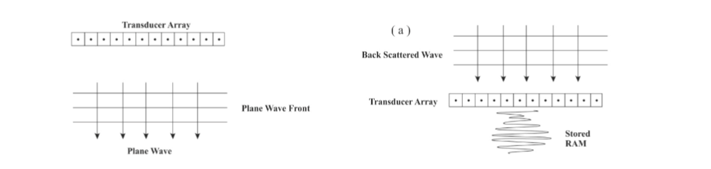
Plane-wave imaging (PWI) is a technique for creating ultrasound images using unfocused plane waves. Unlike traditional focused-beam techniques, where individual beams are sequentially transmitted and received, PWI simultaneously transmits ultrasound waves from various elements at various angles. This innovation significantly accelerates image acquisition, enabling high frame rates and real-time visualization of dynamic physiological processes. A single plane wave is transmitted from each transducer element in plane-wave imaging. These waves propagate in parallel and generate a planar wave front that intersects the imaged region. The resulting echoes are received by all elements in the array, capturing the reflections from various angles. When the received data is processed, a single image frame is rapidly formed, eliminating the need for time-consuming sequential scanning.
The working principle of Plane wave imaging depends on various factors. To achieve ultra-fast imaging, focus in transmit mode must be avoided. A single emission illuminates the entire medium, which has a quasi-plane wave. The back-scattered (BS) signals or RF signals are stored in the individual memory of every array element. The transducer can store more BS signals and generate a fresh signal when the arrival is complete. The quantity of data storage that can be used will determine how many frames can be stored. The frame rate in transmit mode is constrained by the ultrasonic travel time because there is no focus. The travel time of ultrasonic waves in a medium with a 7.5 cm deep tissue equivalent is 100 seconds, thereby limiting the frame rate to 10,000 frames per second.
Following that, parallel beam formation (PBF) is done numerically. PBF is executed in a subsequent phase utilizing the delay and sum method or alternative conventional beamforming techniques, with DAS being the most straightforward option. A factor of N can increase the data acquisition rate by using a parallel processing approach that simultaneously acquires N B-mode picture lines from each expanded transmit pulse. The greater data rate can be utilized to provide Conventional images at regular video frame rates can be used to lower patient exposure, or they can be used to raise the image frame rate to create independent images that can be averaged in the image frame to reduce noise. Without affecting the frame rate, the field of view (FOV) can be expanded over a typical scan. The advantage of plane wave imaging is enhanced frame rates, architected by transmitting all beams simultaneously, and improved temporal resolution, achieved by rapid image acquisition. It has a wide field of view and reduced motion artifacts, and it can also provide real-time guidance if we use it for interventional procedures in real-time for medical sonography. Developing advanced signal processing algorithms to improve lateral resolution, implementing adaptive focusing strategies, and integrating PWI with other imaging modalities for comprehensive assessments are the future areas that need to be addressed.
ii. Synthetic Aperture Beamforming –An Ultra-fast Ultrasound Imaging Technique
Synthetic Aperture Techniques (SAFT) represent a significant advancement in medical ultrasound imaging aimed at overcoming the limitations of traditional beamforming methods. SAFT leverages computational processing to create high-resolution images by simulating a larger aperture, leading to improved lateral resolution, penetration depth, and image quality. This technique has revolutionized the field by enabling real-time or near-real-time imaging while maintaining diagnostic accuracy.
Synthetic Aperture Beamforming (SAB) works by combining the signals from multiple transducer elements creates a virtual aperture much larger than the physical aperture of the transducer. This can improve the image quality by reducing the effects of noise and artifacts. Synthetic Aperture beamforming can be realized in both frequency domain and time domain. This algorithm is computationally very complex, so implementing the same requires suitable hardware like Field Programmable Gate Arrays –FPGAs. Frequency domain implementation is less complex but cannot be done in real-time. Using time or frequency domain SAB depends on the specific application and the desired trade-offs between image quality, frame rate, and computational complexity.
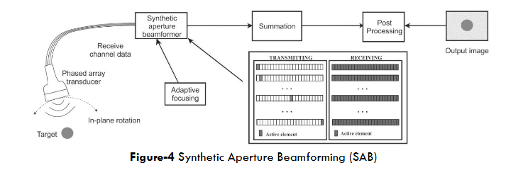
Synthetic Aperture Sequential Beamforming (SASB) is an improvised version of phased array ultrasound imaging, which shows that the lateral resolution can be improved compared to conventional dynamic receive beamforming. Figure-4 illustrates the Synthetic Aperture technique, which simultaneously records data from several sources in all directions. The entire image may then be rebuilt using these data. Every element in the array receives the echo signals after one emits a pulse. This approach’s advantage is that the best image may be produced by applying complete dynamic focusing for transmission and reception. The STA approach focuses on determining the geometric distance between the transmitting element and the imaging point and then back to the receiving element.
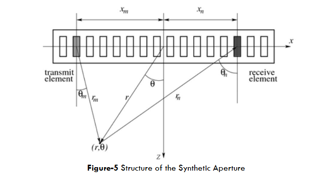
As illustrated in Figure-5, when element m transmits a brief pulse and element n receives the echo signal, a round-trip delay is τmn = τm + τn. In which combination of (m, n) represents a transmit and receive element, where the values of m and n are constrained such that 0 ≤ m, n ≤ N–1. The delays for mth element and nth element are:
τm = (1/2) * (r1 – r2) / c
τn = (1/2) * (r1 – r2) / c
where r is the distance between the center of the synthetic aperture and the point (r, θ), and mx and nx are the locations of the mth and nth elements, respectively. Equation (8) can be used to define the A-scan signal for an N-element array for every point in a picture.
A(r, θ) = (1/2) * (1/N) * Σm=0N-1 Σn=0N-1 ym,n(t – τmn) (8)
The delay associated with beamforming for both the receiving and transmitting elements are shown in equation (5) as m respectively and ym,n(t) as the echo signal. The initial and subsequent summations denote transmit and receive beamforming processes. Although SAFT has several advantages, its implementation demands sophisticated signal processing and computing capabilities. Optimization techniques may be needed to combine computing demands with imaging speed in real-time SAFT.
iii. Multi-Line Transmission based Beamforming
Multi-line transmission (MLT)- based beamforming techniques have emerged as a powerful approach to enhancing image quality and diagnostic capabilities in medical ultrasound imaging. MLT beamforming involves transmitting ultrasound pulses from multiple focal points along different transmission lines. Unlike traditional single-line transmission, where a single focused beam is used to interrogate the tissue, MLT utilizes multiple transmission lines to illuminate the tissue from various angles. The received echoes from these multiple lines are then coherently combined to form a high-quality image with improved spatial resolution, reduced artifacts, and enhanced penetration.
As explained in 2D by Tong et al., the increase in frame rate is accomplished by transmitting multiple guided focused beams simultaneously. In order to produce one of the MLT beams during each transmit event, the aperture can be divided into several sub-apertures. Alternatively, you can employ apodization on the transmit aperture to create several beams in a single transmission. Finally, it is possible to linearly superimpose the pulses usually produced on individual phased array elements to progressively form focused beams at different angles and simultaneously generate all the beams.
A phased array transducer’s separate element can be exposed to nonidentical electric pulses to transmit beams simultaneously in several directions. The number of pulses that generate conventional focused SLT beams along the various MLT lines can define the pulse applied to a given element. As an illustration, Figure 6 depicts the pulses given to certain transducer elements to transmit in two directions simultaneously.
MLT’s advantages include improved lateral resolution, reduced artifacts, enhanced depth penetration, and enhanced contrast. It also enables volumetric imaging for 3D imaging applications in ultrasonography. Volumetric imaging enables comprehensive visualization of complex structures and facilitates more accurate measurements by acquiring data from multiple directions. Contrast enhancement is achieved by providing additional information from different transmission angles. Enhanced Depth Penetration is achieved by exploiting multiple transmission angles to improve signal-to-noise Ratio (SNR), which maintains the image quality at greater depths in the tissue being observed. MLT reduces the impact of side and grating lobes, leading to sharper images with better distinction between adjacent structures, which will give you improved lateral resolution. Challenges and limitations of the MLT include increased computational complexity, complex transducer array design, and more. MLT beamforming requires processing data from multiple lines, which increases the computational load. The transducer array plays a crucial role in MLT beamforming. Issues of related crosstalk and interference must be considered when designing the transducer.
Multi-Line Transmission (MLT) based beamforming has emerged as a promising approach to overcoming the limitations of traditional beamforming methods in medical ultrasound imaging. By leveraging data from multiple transmission lines, MLT offers improved resolution, reduced artifacts, enhanced depth penetration, and better contrast. Although challenges related to computational complexity and transducer array design exist, the potential benefits of MLT make it an exciting area of research with the potential to significantly impact diagnostic accuracy and clinical outcomes in medical ultrasound imaging.
iv. Deep Learning Based Beamforming
End-to-end learning allows deep learning models to learn the beamforming procedure directly from the raw ultrasound data. Beamforming, image reconstruction, and signal processing are all aspects of traditional ultrasound imaging. Deep learning avoids these intermediary techniques and learns a straightforward mapping from raw ultrasound data to high-quality images. With this groundbreaking method, the creation of images might be accelerated while maintaining or even improving image quality. Traditional beamforming methods involve complex signal processing and computation, and deep learning-based approaches leverage neural networks to directly learn the mapping from raw ultrasound data to high-quality images. This allows for rapid image formation and aligns with ultra-fast imaging.
Data-driven beamforming: This method uses a deep neural network to learn how to combine the received echoes from the transducer elements to create an image. The deep neural network is trained on a large dataset of ultrasound images and their corresponding radio frequency (RF) data.
Model-based beamforming: This technique forecasts the parameters of a conventional beamforming algorithm using a deep neural network. An extensive set of ultrasound images and associated RF data train the deep neural network. Data-driven and model-based beamforming are combined in the hybrid beamforming technique. The deep neural network is trained to blend the outputs of the deep neural network and the conventional beamforming algorithm.
It has been demonstrated that deep learning-based beamforming can enhance the image quality of medical ultrasound imaging in several different ways, such as improving the imaging of deep targets, suppressing speckle noise, enhancing the image edges, improving the image contrast, reducing noise and other artifacts, and increasing the resolution of the images.
The advantages of deep learning-based ultra-fast beamforming are rapid image formation due to reduced computational time, improved image quality due to CNNs-based algorithms, enhanced image clarity, increasing diagnostic accuracy. The deep learning algorithms also simplify the complex signal processing chain in the image formation stage. These algorithms also provide personalization for independent patients and give adaptability to different scenarios. The challenges of these algorithms are resource-intensive training and deployment of deep learning models. Training requires many data in the learning process of the algorithms.
4. Discussion
A critical challenge in ultra-fast beamforming algorithms (BAs) for medical ultrasound imaging is managing the trade-offs between processing speed and image quality. Achieving real-time or near-real-time imaging necessitates minimizing the time required to acquire and process raw data into diagnostically reliable images. Advanced techniques, such as Plane-Wave Imaging and Multi-Line Transmission, have demonstrated the ability to achieve high frame rates. However, rapid acquisition and processing methods may compromise image quality.
Processing optimizations, such as reducing spatial or temporal averaging, are often employed to expedite frame generation. While these approaches enhance processing speed, they may introduce noise and reduce the clarity of anatomical features, potentially affecting the diagnostic utility of the images. Striking an optimal balance between processing speed and image fidelity is essential to ensure timely visualizations without sacrificing diagnostic accuracy.
This trade-off highlights the ongoing imperative to refine ultra-fast beamforming techniques by integrating advancements in both hardware and software. Enhanced processing architectures, innovative algorithms, and machine learning-driven optimizations have the potential to bridge this gap, enabling a seamless harmony between speed and image quality. Achieving this balance is crucial for supporting real-time clinical decision-making and enhancing the overall efficacy of medical ultrasound imaging.
Striking a harmonious equilibrium requires iterative refinement of algorithms to minimize the degradation of image quality while achieving optimal processing speed. There is always room for innovation in the dynamic field of ultra-fast beamforming. Targeted research is required to address the trade-offs between processing speed and image quality. We need to improve artifact mitigation, deep learning integration, hardware advancements, and hybrid approaches with clinical validations.
5. Conclusion
The science of ultra-fast beamforming in medical ultrasound imaging is evolving rapidly, and there are many new directions of research and development. Complying with the suggestions given here can go a long way toward enhancing the efficacy, adaptability and clinical usefulness of these techniques.
The combination of ultra-fast beamforming with adaptive and deep learning techniques offers a tremendous opportunity to balance the trade-off between performance and quality. Hybrid strategies that meld the strengths of these two techniques might allow for extremely accurate, real-time imaging without losing diagnostic accuracy. Additionally, studying the combined effects of ultrasound imaging with complementary technologies such as elastography or photoacoustic imaging may allow for a broader picture of tissue properties that surpasses the scope of each imaging method and yield more robust diagnostic information.
One exciting direction is re-engineering ultra-fast beamforming to address specific patient anatomical and pathological parameters. Making algorithms that change in response to every patient’s own characteristics would open up the potential for accurate medical imaging and improve diagnosis and care.
By exploring these new avenues, ultra-fast beamforming can change the way we think about medical imaging in real-time, providing improved diagnostic quality and clinical value for a wide variety of applications.
References
- Tanter, M., & Fink, M. (2014). Ultrafast imaging in biomedical ultrasound. IEEE Transactions on Ultrasonics Ferroelectrics and Frequency Control, 61(1), 102–119. https://doi.org/10.1109/tuffc.2014.2882
- 2020-2021 Index IEEE Transactions on Biomedical Engineering Vol. 68. (2021). IEEE Transactions on Biomedical Engineering, 68(12), 3753–3840. https://doi.org/10.1109/tbme.2021.3136764
- Bercoff, J., Montaldo, G., Loupas, T., Savery, D., Mézière, F., Fink, M., & Tanter, M. (2011b). Ultrafast compound doppler imaging: providing full blood flow characterization. IEEE Transactions on Ultrasonics Ferroelectrics and Frequency Control, 58(1), 134–147. https://doi.org/10.1109/tuffc.2011.1780
- Demi, L. (2018). Practical Guide to Ultrasound Beam Forming: Beam Pattern and Image Reconstruction Analysis. Applied Sciences, 8(9), 1544. https://doi.org/10.3390/app8091544
- Sg, S., Sakthivel, R., & Kidav, J. U. (2019). Beamforming Algorithm Architectures for Medical Ultrasound. International Journal of Innovative Technology and Exploring Engineering, 8(12), 2452–2459. https://doi.org/10.35940/ijitee.l2556.1081219
- Kidav, J. U., & G, S. S. (2021). An FPGA-Accelerated Parallel Digital Beamforming Core for Medical Ultrasound Sector Imaging. IEEE Transactions on Ultrasonics Ferroelectrics and Frequency Control, 69(2), 553–564. https://doi.org/10.1109/tuffc.2021.3126578
- Ibrahim, A., Hager, P. A., Bartolini, A., Angiolini, F., Arditi, M., Thiran, J., Benini, L., & De Micheli, G. (2017). Efficient Sample Delay Calculation for 2-D and 3-D Ultrasound Imaging. IEEE Transactions on Biomedical Circuits and Systems, 11(4), 815–831. https://doi.org/10.1109/tbcas.2017.2673547
- Schneider, F., Agarwal, A., Yoo, N. Y. M., Fukuoka, T., & Kim, N. Y. (2009). A Fully Programmable Computing Architecture for Medical Ultrasound Machines. IEEE Transactions on Information Technology in Biomedicine, 14(2), 538–540. https://doi.org/10.1109/titb.2009.2025653
- Kidav, J. U., Sivamangai, N. M., Pillai, M. P., & M, S. R. (2018). Architecture and FPGA prototype of cycle stealing DMA array signal processor for ultrasound sector imaging systems. Microprocessors and Microsystems, 64, 53–72. https://doi.org/10.1016/j.micpro.2018.10.005
- Karaman, M., Kolagasioglu, E., & Atalar, A. (1992). A VLSI receive beamformer for digital ultrasound imaging. IEEE International Conference on Acoustics, Speech, and Signal Processing, 657–660 vol.5. https://doi.org/10.1109/icassp.1992.226510
- Thomenius, K. (2002). Evolution of ultrasound beamformers. Proc. IEEE Ultrason. Symp., 1996, Pp. 1615–1622., 2, 1615–1622. https://doi.org/10.1109/ultsym.1996.584398
- Deylami, A. M., Jensen, J. A., & Asl, B. M. (2016). An improved minimum variance beamforming applied to plane-wave imaging in medical ultrasound. 2017 IEEE International Ultrasonics Symposium (IUS), 57, 1–4. https://doi.org/10.1109/ultsym.2016.7728895
- Capon, J. (1969). High-resolution frequency-wavenumber spectrum analysis. Proceedings of the IEEE, 57(8), 1408–1418. https://doi.org/10.1109/proc.1969.7278
- Jensen, J. A., Kortbek, J., Nikolov, S., Hemmsen, M. C., & Tomov, B. G. (2010). Implementing Synthetic Aperture Imaging in Medical Ultrasound: The Dual Stage Beamformer Approach. Synthetic Aperture Radar (EUSAR), 2010 8th European Conference On, 1–4. https://backend.orbit.dtu.dk/ws/files/4629826/paper%5B1%5D.pdf
- Jorgen Arendt Jensen, Svetoslav Ivanov Nikolov, Kim Lokke Gammelmark, Morten Hogholm Pedersen, Synthetic aperture ultrasound imaging, Ultrasonics, Volume 44, Supplement, 2006, Pages e5-e15, ISSN 0041 624X, https://doi.org/10.1016/j.ultras.2006.07.017
- Nili, V. A., Ezati, M., Yan, Y., Kavehvash, Z., & Mehrmohammadi, M. (2022). Field of View and Resolution Improvement in Coprime Sparse Synthetic Aperture Ultrasound Imaging. 2022 IEEE International Ultrasonics Symposium (IUS), 34, 1–4. https://doi.org/10.1109/ius54386.2022.9957167
- Cheng, S., Yu, J., Guo, X., Ta, D., & Xu, K. (2022). Nonlinear Simulation of Modulation Pulse Sequencing for Contrast-Enhanced Ultrasound (CEUS) Imaging. 2022 IEEE International Ultrasonics Symposium (IUS), 1–4. https://doi.org/10.1109/ius54386.2022.9957327
- Stanziola, A., Toulemonde, M., Yildiz, Y. O., Eckersley, R. J., & Tang, M. (2016). Ultrasound Imaging with Microbubbles [Life Sciences]. IEEE Signal Processing Magazine, 33(2), 111–117. https://doi.org/10.1109/msp.2015.2496914
- Sridharan, A., Eisenbrey, J. R., Stanczak, M., Daecher, A., Machado, P., Wilkes, A., Sevrukov, A., Ojeda-Fournier, H., Mattrey, R. F., Wallace, K., & Forsberg, F. (2016). Contrast-enhanced nonlinear 3D ultrasound imaging of breast lesions in a clinical population. 2017 IEEE International Ultrasonics Symposium (IUS), 1–4. https://doi.org/10.1109/ultsym.2016.7728798
- Moubark, A. M., Nie, L., Cowell, D. M. J., Ali, S. H. M., & Freear, S. (2019). A New Nonlinear Compounding Technique for Ultrasound B-mode Medical Imaging. 2017 IEEE International Ultrasonics Symposium (IUS). https://doi.org/10.1109/ultsym.2019.8926026
- Cruza, J., Perez, M., Moreno, J., & Fritsch, C. (2015). Real Time Fast Ultrasound Imaging Technology and Possible Applications. Physics Procedia, 63, 79–84. https://doi.org/10.1016/j.phpro.2015.03.013
- Chen, Y., Huang, Y., Li, C., Hou, Y. T., & Lou, W. (2020). Turbo-HB: A Novel Design and Implementation to Achieve Ultra-Fast Hybrid Beamforming. IEEE INFOCOM 2022 – IEEE Conference on Computer Communications. https://doi.org/10.1109/infocom41043.2020.9155
- Campbell, N. A., Samson, C. A., & Brown, J. A. (2019). An Ultrafast High-Frequency Hardware Beamformer for a Phased Array Endoscope. 2017 IEEE International Ultrasonics Symposium (IUS), 34, 1505–1508. https://doi.org/10.1109/ultsym.2019.8925578
“`