


Future Monitoring Strategies with Microelectrode Arrays
“`html
Translating Past Medicinal Research Advances into Future Monitoring strategies using Microelectrode Arrays: A comprehensive Review and Forward-Looking Perspective
Shivani Shukla
Ida Tiwari
1. Introduction
Ever since the pioneering research in early 1970s, there has been lot of interests in, microelectrodes which are electrodes having at least one dimension smaller than the thickness of diffusion layer of the electroactive species. Microelectrodes have several advantages over the traditional equivalents, including a smaller double layer capacitance, a quicker development of steady state signal, and a lower ohmic drop of potential. These features have opened up new opportunities for the use of microelectrodes as electrochemical sensors in environmental and biological applications. Since microelectrodes are small, respond quickly, and have good signal to noise ratios, they can be easily used to explore chemical events at the surface of tissues or individual living cells with high spatial and temporal precision. Instead of using transient approaches to measure kinetics, steady-state methods are made possible by the rapid pace of mass movement at microelectrodes. The enhanced diffusion properties of the microelectrodes enable measurements in static solutions with negligible systematic disturbance. Furthermore, because of their lower ohmic drop and capacitive effects, microelectrodes can also be used in solutions with extremely low levels in a supportive electrolyte. A high accuracy potentiostat inside a well-designed Faraday cage is required to reduce the electromagnetic interference from neighboring electronic equipment because the electrical signal at a single microelectrode is frequently in the picoampere to nanoampere range. Microelectrode arrays (MEAs), which usually comprises tens to millions of microelectrodes, facilitates getting through the experimental challenges. Compared to a single microelectrode, the output current for MEAs is significantly greater in magnitude. MEAs are electrochemical signal measurement devices made up of several individual electrodes. As a platform for simultaneous multianalyte detection, arrays of individually programmable electrodes have been used in biological sensing, environmental monitoring, pharmaceuticals, and the quantitative identification of toxins in food samples. In this article we have focused, on the number of ways to fabricate MEAs, employing lithographic techniques like photolithography, thin film deposition, lift off, and reactive ion etching, screen printing, and complementary metal oxide semiconductor. Even with major advancements in MEAs fabrication techniques, its wider applicability is still hampered by the absence of reliable and affordable techniques. Microelectrode arrays have long been used in biosensing and electroanalytical chemistry. One of the earliest studies to integrate electrochemistry with biomolecules was a glucose biosensor based on electrodes, described by Clark and Lyons in 1960s. MEAs are employed not only as sensors but also for cell-based impedance research and the investigation of cell electrical activity. Recently, reports of MEAs being used to research 3D cell cultures have also surfaced. Microelectrode fabrication is a crucial stage in the development of MEAs since these electrodes are essential to biosensing. These microelectrodes are difficult to fabricate because of their small size, which results in a high impedance value and unfavourable low signal-to-noise ratios. In order to produce microelectrodes with high signal-to-noise ratio, greater sensitivity, and lower impedance, advancements in fabrication techniques such as the utilization of conducting nanomaterials and the creation of 3D nanostructures are still being made. Microelectrode arrays has become a ground breaking techniques that allows several cells or tissues to collect electrophysiological signals simultaneously. Unparalleled insight into how cell react to pharmacological sample is made possible by their ability to provide spatial and temporal resolution of electrical activity at microscale. In order to improve drug development processes and clinical management, MEAs serve as an essential link between conventional medical information and future monitoring techniques.
2. Microelectrode arrays and its method of preparation
Multiple electrodes (10 100 µm) arranged symmetrically on a substrate to detect a bioelectric signal comprise a MEAs. The fabrication of a single or small number of microelectrodes marked the beginning of the use of MEAs for cellular electrophysiological research, yet, a single electrode is insufficient to adequately investigate a cell’s electrical activity. In 2007, Kelly et al. used a microelectrode array and a single electrode to compare recorded signals from the visual cortex. Their research led them to the conclusion that, in comparison to a single electrode, an array of microelectrodes produced data of high quality. Microelectrode arrays have been produced through three popular methods: flexible arrays, microwires, and micromachined. The first implantable electrodes that could continuously record from the brain and concentrate on a single neuron were microwires. Because the wire bends during implantation, one disadvantage of these electrodes is that it is impossible to precisely control where the electrode tips are in relation to one another. This problem might be resolved by micromachined electrode arrays that use silicon’s hard structure. There are two distinct versions of silicon-based electrodes. The first variant has electrode locations patterned on the silicon substrate shank, whereas the second model has electrically isolated sharpened silicon needles. In the flexible multi-electrode array approach, two layers of silicon-free polyimide, parylene, or benzocycobutene cover the metal-based electrode. Furthermore, MEAs can either be two-dimensional (2D) or three-dimensional (3D) depending on how the microelectrodes are distributed spatially on these substrates. Thomas et al. released one of the first few articles explaining how a 2D MEAs operates in 1972. In order to measure the electrical activity of cultivated chick heart cells, they created gold microelectrodes on a glass coverslip. 2D rigid MEAs have been widely employed in both in vitro and in vivo research on myocytes, cardiac cells, and brain cells. Rigid MEAs utilized in in vivo research, however, have the potential to harm host tissues both during and after implantation. The MEAs used to lower this risk should be as thin as possible to put the least amount of stress on the host tissue and should be mechanically compliant to fit the anatomy of the tissue. For these kinds of research, flexible MEAs are preferable over rigid MEAs since they are built on polymeric substrates and are relatively thin.
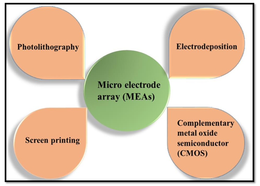
2.1 METHOD OF PREPARATION
The preparation methods of MEAs depicted in Fig. 1.
2.1.1 Photolithography method
Electrochemical sensors are made at the microscale using photolithography, a process that is extensively employed in the semiconductor sector. It depends on photoresists (chemicals that are sensitive to lights) and exposure instruments illuminated by mercury arc lamp (which produce UV wavelength of 365 or 436 nm). The highly accurate photolithographic method enables the production of microscale electrode arrays.
- Metal based Microelectrode Arrays
Among the most common lithographic techniques for developing metal based MEAs, photolithography is the most popular and in use today. An adhesive layer made up of Ti, Cr, Al, Ti-W, and other materials (10-100 nm in thickness) must be deposited in order to improve adhesion between the substrate and the electrode materials (100-1000 nm in thickness, Au, Pt, Ir, etc.). The methods use sputter or thermal vapour deposition technique. Using a desired mask (micro-disk/micro-band, linear array, etc.) and conventional photoresist (positive / negative), the layers are patterned. Lift off is another way to eliminate photoresist layer. Following removal, plasma-enhanced chemical vapour deposition (CVD) evaporates an insulating layer across the substrate s whole surface. After that, the insulating layer is etched using oxygen plasma or reactive ion etching after being designed using a different preferred mask.
- Carbon based Microelectrode Arrays
Photolithography has also been used to develop carbon based MEAs. The preferred electrode material for a long time has been carbon because of its rich surface chemistry, broad potential range, low fouling, and affordability. The carbon film s low conductivity and adherence to the substrate are the primary issues in this instance. The former produces layers that are easily removed during microfabrication process, latter results in decline in iR during microfabrication. Through a low temperature sputtering deposition method, carbon thin films exhibit exceptional substrate adherence. A lift-off process or oxygen plasma has been used because traditional wet-etching techniques on sputter deposited carbon films have not been effectively developed. Using photolithography, our research group synthesized carbon nanotube (CNTs)/Au MEAs and CNTs/Pd MEAs. - Fabrication of Carbon nanotube/Gold and Carbon nanotube/Palladium Microelectrode Arrays
The fabrication of MEAs on a glass substrate and silicon wafer involved the arrangement of numerous microelectrodes in a particular 8×8 pattern, resulting in a matrix of 64 microelectrodes. Each microelectrode is around 10 µm by 10 µm in size, and one electrode in each corner is about 100 µm by 100 µm. The glass substrate and silicon wafer were prepared for the microfabrication process by cleaning them with acetone, isopropanol, and sonication with methanol. Additionally, it was exposed to oxygen plasma for 5 minutes at a power of 100 W rf. Following cleaning, the substrate was photolithographically shaped to form contact pads, connecting lines, and microelectrodes using the first mask. Au (95 nm) on glass substrate and Pd (70 nm) on silicon wafer was used to evaporate the patterned sample after it has been DC sputtered with Ti (4 nm). Following that, the sample was immersed in NMP-1165 solvent for lift-off. After SiO2 (230 nm) evaporated onto sample, photolithographic patterning was done using the second mask. To access electrical connections and contact pads for electrode-analyte contact, the regions identified in the second patterning stage are prepared for reactive ion etching using a gas mixture of CF4 (30 sccm) and Ar (10 sccm) at 60 mT with 100 W power. Following etching, the photoresist was eliminated using NMP-1165 solvent, and Al (8 nm for glass substrate and 10 nm for silicon wafer) and Fe (2 nm for glass substrate and 3 nm for silicon wafer) were DC sputtered as a catalyst for CNT development. Lastly, photothermal chemical vapour deposition (PTCVD) is utilized to grow CNTs on MEAs at a low substrate temperature (< 400 °C) and 2 Torr pressure, using combination of C2H2 (10 sccm) and H2 (100 sccm).
2.1.2 Screen printing method
In contrast to photolithography, screen printing operates simply and doesn t require a complex flow procedure. Conductor and insulator patterns are often screen-printed using a specialized printer to prepare electrodes. Precision screens developed of stainless steel wire define the various desirable patterns (micro-disk or micro-band). CNTs, organo-gold, Pt, Ag, graphite, and Au are among the materials used in ink. Using proper screen design, each ink is independently applied. The screen printing technique s limitation stem from the resolution obtained during photolithography and the design of aimed pattern.
2.1.3 Electrodeposition method
The synthesis of MEAs in electrochemistry is not extensively studied, even though electrodeposition is a promising substitute method for creating nanostructures such as nanowires, nanotubes, nano-rods, nano-flower-like particles, and so on. Usually, a prepared substrate can be utilized to produce a random array using this technique. Patterned electrodeposition, or electrodeposition carried out on a specifically designed array support, is believed to be a promising method to fabricate micro/nanostructure in a bottom-up manner. It may find use in forthcoming microelectronics generations, miniature sensors, and microelectromechanical systems. In general, patterned electrodeposition can be accomplished by two methods. Self-organisation is one way and template-assisted electrodeposition is other way. The latest generation of microelectrode arrays shows some intriguing electrochemistry applications following patterned electrodeposition.
2.1.4 Complementary metal oxide semiconductor
The complementary metal oxide semiconductor (CMOS) method, widely used in the development of integrated circuits and microchips, can be used to create MEAs. This technique has been used to create a number of high-density MEAs. Initially, this MEAs was constructed using a specifically designed CMOS integrated chip (Ic). Pt black electrodes were then used to post-fabricate aluminum contact pads. After that, the Ics were anchored to the chip carriers. In a further investigation, a dual-mode MEAs for extracellular recording from the whole neural network was developed using the CMOS technique. At 0.18 µm, CMOS technology was used for developing the MEAs. Pt electrodes were designed for post-processing, and Pt black was electrochemically coated on them to reduce their resistance. Similarly, to assess the oxygen gradient in real-time respiration, a high-density MEAs chip was developed.
3. Electrochemical sensor and Biosensor
3.1 DETECTION OF BIOLOGICAL ANALYTE
3.1.1 Glucose sensor
In order to address a variety of health issues, accurate, quick, and highly sensitive testing and diagnostic technologies are required. Diabetes mellitus is one of several conditions that is well-known for its effects on public health around the world. This metabolic condition is brought on by the body s incapacity to generate enough insulin to lower hyperglycaemia. Therefore, early identification and treatment of potential consequences like heart disease, renal failure, and blindness depend on sensitive and accurate glucose monitoring in biological samples. The significant benefits of speed, resilience, and simplicity make the electrochemical technique a feasible approach for the routine measurement of glucose levels. The glucose oxidase (GOx) enzyme is immobilized using a variety of functional nanomaterials in electrochemical enzymatic glucose biosensors, which operate on the immobilization principle. Amperometry is most likely the electrochemical technique most frequently employed in glucose sensors. However, it has been suggested in recent years to employ electrochemical impedance spectroscopy (EIS) as a transduction mechanism in these sensors. The analyte at lower concentrations, the electrode/electrolyte interface, and the electrode surface kinetics are better studied using EIS approaches. Ankit et al. report a development of a third-generation electrochemical enzymatic glucose biosensor based on photolithographically defined CNTs/Au MEAs on a glass substrate. In this biosensor, electrons are transferred directly between the electrode surface and the enzyme’s redox active site without the use of a redox mediator. Electro-synthesized from the p-PDA monomer, poly (paraphenylenediamine) has been widely used as a perm-selectivity barrier in biosensors. The interference issue is significantly reduced when a perm-selective layer is incorporated into the biosensor’s development. Additionally, the remarkable selectivity of the constructed GOx/poly (p-PDA)/CNTs/Au MEAs is achieved through the immobilization of the glucose oxidase (GOx) enzyme in the poly (p-PDA) matrix. The EIS approach, which is based on enzymatic glucose oxidation, was utilized to measure the glucose concentration using the created GOx/poly (p-PDA)/CNTs/Au MEAs based biosensor. By immobilizing the GOx enzyme in a polymer matrix, leaching of the enzyme was prevented. By monitoring the change in Rct value upon the addition of multiple glucose concentrations at a fixed DC potential, a broad linear detection ranges between 0.2 and 27.5 µM was covered.
3.1.2 Neurotransmitter sensor
A class of aromatic amines known as catecholamines includes several neurotransmitters, including dopamine (DA), 5-hydroxytryptamine (5-HT), and norepinephrine (NE). The sympathetic nervous system, which is concentrated in the area of the locus coeruleus (LC), releases NE, a neurotransmitter that regulates blood pressure, emotion, and memory. Since abnormal NE secretion is a sign of numerous illnesses, including anxiety and depression, neuroscientists and medical professionals have developed numerous methods for both in vitro and in vivo NE detection. Wang et al. uses microelectromechanical system (MEMS) technology to create a MEAs that offer a quick, accurate, and sensitive way to determine NE dynamic secretion directly. Reduced graphene oxide and Pt nanoparticles (rGOPNps) were electrodeposited onto the MEAs to enhance its electrical performance. The calibration findings indicated a detection limit of 0.08 µM and a sensitivity of 1.03 nA µM to NE. Specifically, the MEAs was effective in tracking spike firing from the hippocampus brain slice and quantifying dynamic extracellular NE secretion from the locus coeruleus brain slice. Studies on the spatially precise transport of trace neurochemicals and the in vitro electrophysiological activity of various biological tissues could benefit from this fabricated device. Dopamine’s (DA) vital roles in the human body (such as the cardiovascular, central neurological, renal, and hormonal systems) make it one of the most studied neurotransmitters. Due to its importance in signal transmission to the brain, DA deficiency can cause a number of neurological conditions, including schizophrenia, attention deficit hyperactivity disorder, and Parkinson’s disease. A novel cylinder gold nanoelectrode (CAuNE) platform was designed by Kim et al. using electrochemical deposition and successive laser interference lithography. The optimum electrodes for electrochemical DA detection were determined to be CAuNEs, which had a diameter of 700 nm and were formed in 150 s. Cyclic voltammetry measurements showed that the linear range of DA was 1 100 µM, with a 5.83 µM limit of detection. Furthermore, human neural cells were successfully cultured and sustained for almost five days in vitro without the usage of extracellular matrix proteins attributed to the homogenous periodic properties of CAuNEs, and DA was detected when these cells were present on the electrode.
3.1.3 Biomolecules sensor
An essential amino acid, l-tryptophan is a building block for several other compounds in the body, such as proteins, hormones, neurotransmitters, and other pertinent biomolecules. In order to compensate for dietary inadequacies, it is utilized in medications, processed foods, and nutritional supplements. While l-tryptophan is used to treat depression and sleeplessness, excessive consumption of it can result in fever, agitation, hallucinations, and delusions. One of the main ingredients of the vitamin B complex is pyridoxine, also known as vitamin B6, which is a water-soluble vitamin. It mainly functions as a cofactor for enzyme-catalyzed reactions, the creation of neurotransmitters, the metabolism of carbohydrates, fatty acids, and amino acids, and a number of other biochemical processes. Vitamin B6 deficiency results in neurological diseases, depression, weakness, convulsions, and skin issues. Ankit et al. develop carbon nanotubes (CNTs) grown Pd MEAs as a working electrode and use reduced graphene oxide (rGO/CNTs/Pd MEAs) for pyridoxine sensing and poly(l-arginine)/CNTs/Pd MEAs for l-tryptophan sensing. For l-tryptophan and pyridoxine, the electrochemical differential pulse voltammetry (DPV) analyses show excellent linearity in the concentration ranges of 20 15,000 µmol/L and 10 5000 µmol/L, respectively, with detection limits of 10.0 and 1.0 µmol/L. For l-tryptophan, and pyridoxine sensing, the suggested multiple analytes sensor has demonstrated extremely high sensitivities of 2280 and 940 µA(µmol/L ) cm , respectively.
3.2 MONITORING OF DRUGS
3.2.1 Neural Monitoring
Diseases of the brain system may be treated with neural stem cells (NSCs). The first NSCs were extracted from the striatum of adult mice in 1992. Consequently, the idea of NSCs was presented to the neuroscience community. Numerous scientists think that NSCs can self-renew, differentiate into neurons and astrocytes, and multiply in the brain. NSCs may integrate into the central nervous system (CNS) of mammals upon transplantation. They can occupy areas of the CNS that are growing or deteriorating. NSCs have been suggested as a potential treatment for a number of disorders affecting the nervous system, such as Huntington’s disease, Parkinson’s disease, and Alzheimer’s disease. In order to track the neuronal spikes and local field potentials (LFPs) of neurons that were generated from rat neural stem cells in vitro, Gao et al. constructed a 60-channel microelectrode array (MEAs), to detect brain impulses, the neurons were cultivated on the surface of the MEAs. In this work, glutamate (Glu) was employed to alter brain activity. Platinum nanoparticles were added to the surface of the microelectrode site in order to improve detection capability. The MEAs was used to record glutamate-stimulated neuronal spikes and LFPs. In the normal state, the average spike amplitude was roughly 70 µV. With Glu modulation, the spike amplitude rose from 70 µV to 90 µV, and the firing rate rose from 4.01 Hz to 6.8 Hz.
3.2.2 Cardiac arrhythmia monitoring
Cardiomyocytes produced from human induced pluripotent stem cells (hiPSCMs) are becoming more and more valued in the modeling of arrhythmias. Similar to in vitro systems, they are capable of predicting medication responses, like as toxicity and efficacy. The electrical characteristics of the multicellular syncytium of hiPSCMs must be precisely measured and analyzed for all of these applications in order to detect electrophysiological alterations, including the re-entry phenomenon, which is one of the primary causes of fatal ventricular arrhythmias. For the bioelectronic monitoring of arrhythmia-typical rotor patterns (re-entry) arrhythmia patterns, Zitzmann et al. developed a multilayer high density microelectrode array (HD-MEA) with an optimal arrangement of 512 sensing and 4 pacing electrodes on a sensor area of 100 mm2. They used impedance spectroscopy at the same microelectrode to monitor label-free and continuous cardiac electrophysiology based on field potential monitoring and mechanical contraction. This study shows that, cardiomyocytes produced from hiPSCMs were cultured on HD-MEAs and utilized to illustrate how sensitively cardioactive medications like levosimendan, omecamtiv, and blebbistatin (IC50 = 0.8 µM, 0.3-1 µM, and 4.2 µM) modulate contraction strength. Remarkably, it is possible to create re-entry using optimal electrical stimulation sequences and detect them with excellent spatial precision.
4. Future Possibility in Microelectrode Arrays
The development and manufacturing of any MEAs-based biosensor heavily relies on factors like improved resolution, high throughput, and low cost. Next-generation lithography (NGL) techniques are being developed to produce high-resolution patterns. These methods have the advantage of having higher resolution limits than traditional lithography methods. In contrast to other NGL techniques, extreme UV (EUV) lithography is an example of an NGL technology that may offer higher resolution and be more economical. It is possible to adapt these NGL methods for MEAs construction. The most promising method for a single-step MEAs manufacturing seems to be inkjet printing. For the fabrication of MEAs, another method known as tip-based nanomachining (TBN) may be investigated. This method uses a structure that resembles a tip to precisely remove materials from a substrate and produce a pattern. Tissue-scaffold resembling MEAs have recently been created using enzyme-mediated transfer printing. Additionally, the microstructure resolution can be improved by combining several microfabrication processes. More MEAs based tools that can be combined with drug delivery systems in the future should be created in order to offer insights into disease-based research and drug development investigations. These tools are useful for in vitro drug efficacy studies that target patients’ cells. By developing a real-time medication response platform, these investigations can advance the idea of “personalized medicine”. Recently developed wearable intelligent devices and portable miniaturized micro/nano electrode arrays have demonstrated the ability to perform label-free, multiparameter, and real-time smart dynamic sensing, which is crucial for the advancement of biomedical research as well as medical device development. Future biological uses of smart sensing derived from miniature micro/nano electrode array sensors include monitoring metabolism, measuring cellular function, and developing novel treatments.
5. Conclusion
This review summarizes recent advancements in the fabrication of MEAs sensors and discusses their emerging applications in biological systems and portable intelligent devices. In recent decades, substantial progress has been achieved in the development of these sensors for biological analysis. Nonetheless, several critical challenges persist. Overcoming these challenges is essential to enhance the stability, accuracy, and quantitative performance of MEAs sensors. Progress in these areas will not expand their practical applications in biological research and diagnostic but also provide valuable insight into physiological and pathological processes, thereby advancing the broader field of life sciences and chemical biology.
Keywords: Microelectrode Arrays, Biosensors, Glucose Sensor, Neurotransmitter Sensor, Cardiac Monitoring, Drug Delivery Systems.
Acknowledgements: The author S S (Chem.-22/RET-Ex./July-22-term/26/564) is grateful for the financial support provided to their research by UGC, New Delhi. The financial support provided by IOE incentive grant for faculty (Scheme Number-6031) at BHU is greatly appreciated.
Author contributions: SS: Methodology, Conceptualization, Visualization, Data analysis, Writing- original draft; IT: Supervision and Validation.
Conflict of interest: The authors affirm that they have no conflicting financial interests that would have influenced the research presented in this article.
“`