Individual Variations in Blood Loss During ECLS

Individual variations in hemostasis, thrombosis and blood loss during extracorporeal life support

Wayne L. Chandler MD1

  1. Department of Laboratory Medicine and Pathology, University of Washington, Seattle, WA

OPEN ACCESS

PUBLISHED: 30 June 2025

CITATION: Chandler, W. L., 2025. Individual variations in hemostasis, thrombosis and blood loss during extracorporeal life support. Medical Research Archives, [online] 13(6).https://doi.org/10.18103/mra.v13i6.6619

COPYRIGHT: © 2025 European Society of Medicine. This is an open-access article distributed under the terms of the Creative Commons Attribution License, which permits unrestricted use, distribution, and reproduction in any medium, provided the original author and source are credited.

DOI https://doi.org/10.18103/mra.v1 3i6.6619

ISSN 2375-1924

ABSTRACT

Multiple factors affect how a patient responds to extracorporeal life support (ECLS) including the patient’s age, size, underlying problem, clinical condition, genetic differences and clinical progress. To better understand this highly variable response, we must separate what is happening in the patient versus the ECLS circuit. Pre-ECLS risk factors, including disseminated intravascular coagulation and recent surgery, increase the risk of platelet loss and bleeding during ECLS. Exposure of blood to artificial surfaces in the circuit activates the contact system which in turn activates coagulation. High shear in the cannula, tubing and pumps, along with coagulation activation, damages and activates platelets, red cells, endothelial cells and leukocytes. Platelets and red cells are lost during ECLS through bleeding, phlebotomy, activation, hemolysis, sub-lethal injury induced extravascular clearance, and other causes. Activation of coagulation and platelets leads to circuit thrombus formation. Clinically important circuit thrombi include arterial tubing thrombi associated with arterial embolism, oxygenator surface membrane thrombosis resulting in oxygenator failure/circuit replacement and venous pump emboli leading to severe hemolysis. The variability in response means some patients require almost continuous transfusion support while others receive almost no transfusions. This review summarizes what is known and what research is needed to improve ECLS.

Keywords

  • extracorporeal life support
  • hemostasis
  • thrombosis
  • blood loss
  • platelets

Introduction

While extracorporeal life support (ECLS) can be lifesaving for patients needing short-term cardiopulmonary support, it is associated with an increased risk of blood loss, the need for recurrent transfusion support and thrombus formation in the ECLS circuit.¹⁻⁷ Current studies report that patients on ECLS show a continuing loss of red cells, platelets and coagulation factors. Pediatric patients on ECLS lose blood cells even faster than adults. This persistent blood loss leads to recurrent, at times daily, transfusion support including red cell, platelet and plasma transfusions. Increased blood loss and transfusions are associated with worse outcomes in ECLS. Thrombosis in the ECLS circuit is also a common problem. Anticoagulant medications are used to reduce the risk of circuit thrombosis, but anticoagulation leads to an increased risk of bleeding and transfusion support.

While we have a good understanding of the overall risks of blood loss, transfusion and circuit thrombosis during ECLS, there are two areas that require further study if we are to reduce these problems. First, we need to better understand what causes blood loss and circuit thrombosis. Not all blood loss during ECLS is due to bleeding. Some ECLS patients show only minor bleeding, but still require major transfusion support. Second, there is a substantial individual variation in the response of different patients to ECLS. Some ECLS patients require little transfusion support and show only minimal circuit thrombosis, while others require daily platelet or red cell transfusions and undergo several circuit changes due to thrombosis. We need a better understanding of the individual patient hematologic response to ECLS.

How a patient responds to ECLS depends of multiple factors including 1) the patient’s underlying problem and clinical condition before starting ECLS, 2) the response of the patient’s blood when exposed to the cannula, tubing, oxygenator and pumps of the circuit and 3) clinical progress while on the circuit including patient improvement or worsening and clot deposition in the circuit (Figure 1).⁸

Figure 1. Causes of platelet loss during extracorporeal life support (ECLS). Adapted from reference 11.

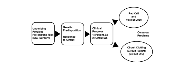
Figure 1. Patient response to extracorporeal life support. DIC = disseminated intravascular coagulation, Δs = changes.

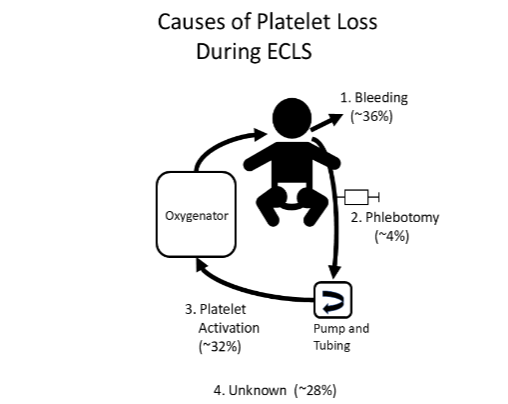
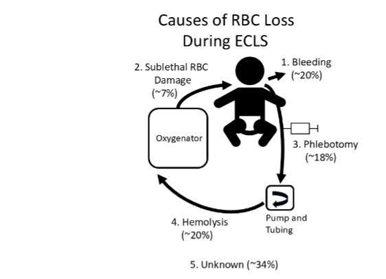
To understand how a patient responds to ECLS we must separate what is happening in the patient versus what is happening in the ECLS circuit. This review will focus on hemostatic, hematologic and thrombotic aspects of the patient response to ECLS including effects on the coagulation system, platelets, red cells, endothelium and leukocytes, and what is known about different mechanisms of blood loss including bleeding, phlebotomy, hemolysis, cell activation and sublethal cell damage. The goal is to summarize how hematologic systems respond to ECLS, what factors affect this response and what future research is needed to minimize ECLS side effects and improve outcomes.

Patient clinical condition

The first factor affecting the patient’s response to ECLS is their pre-existing clinical status including: 1) patient’s age and size, 2) underlying problem leading to ECLS and 3) clinical condition. As blood flows through the ECLS circuit, cells and other humoral factors in the blood may be activated and damaged. As blood returns from the circuit to the patient these activation and damage products must be cleared by the patient’s liver, spleen and kidneys. In an adult patient or large child, the 500–600 mL of blood from the ECLS circuit is diluted 5 to 10-fold in the 3 to 6 L of blood in the patient. In contrast, in a neonate with a 250 mL blood volume, the 500 mL ECLS circuit may hold twice the blood volume of the patient. Even if the amount of damage to blood cells by the circuit is the same, the relative amount returning to the neonate is 20 times higher than that returning to an adult or large child. In addition, compared to adults or older children, neonates have lower levels of coagulation factors making them more vulnerable to coagulation factor related bleeding during ECLS.⁹

The patient’s underlying problem and clinical condition prior to ECLS also play a role in how the patient will respond. Pre-ECLS risk factors include post-operative issues, infection/sepsis, hypoxia, ischemia, shock, disseminated intravascular coagulation (DIC) and prior thrombosis. A patient with a clearly reversible problem in otherwise good clinical condition presents a very different starting point from a patient with a more severe problem and poor clinical condition at the beginning of ECLS. For example, in a child in relatively good condition with treatable infection-related respiratory distress the ECLS run may be uneventful while the patient’s lungs recover. In contrast, a neonate with septic shock that arrives for ECLS in DIC is at increased risk of platelet loss, bleeding and transfusion.¹

Risk factors associated with increased blood loss during ECLS include pre-ECLS surgery (44% higher platelet loss and 82% higher red cell loss) and pre-ECLS DIC (104% higher platelet loss and 65% higher red cell loss).¹⁰,¹¹ Patients may develop DIC prior to starting ECLS for a variety of reasons including hypoxia, shock, trauma and sepsis. Disseminated intravascular coagulation prior to starting ECLS in adults is associated with an increased risk of thrombocytopenia during ECLS and poor outcome.¹²,¹³ A history of venous thrombosis or bacterial infection prior to starting ECLS is associated with an increased risk of venous oxygenator membrane surface thrombosis, oxygenator failure and replacement.¹⁴

Coagulation

Under normal conditions, the hemostatic system is not activated until blood comes into contact with exposed tissue factor and collagen in a wound.¹⁵,¹⁶ Initiation of coagulation requires two factors, an activator for the coagulation system and a procoagulant phospholipid surface for the activated coagulation factors to assemble on. The primary activator for coagulation in a wound is tissue factor, which is present in perivascular tissues. Platelets activated by thrombin bind to the wound and expose procoagulant phospholipids further enhancing coagulation activation, fibrin generation and development of a hemostatic plug that stops bleeding. Thus, in wounds, perivascular tissue factor is the primary coagulation activator while activated platelets provide the procoagulant phospholipid surface.

Instituting extracorporeal circulation exposes the patient’s blood to artificial surfaces and high shear in cannulas, tubing and pumps. Exposure of blood to the artificial surfaces in the ECLS circuit induces a systemic inflammatory response,¹⁷ including cytokine release and activation of the complement, fibrinolytic and coagulation systems, with peak activation of these humoral systems typically occurring during the first few hours of support.¹⁸–²⁴

The contact system of proteins is an ancient form of innate immunity in blood. While still present in humans, its function has been superseded by more complex humoral and cellular immune systems. The contact system plays no role in normal hemostasis in humans, patients deficient in the contact factors

prekallikrein, high molecular weight kininogen and factor XII do not bleed.¹⁵ As blood flows over the ECLS circuit, plasma proteins bind to the artificial surfaces of the pumps, tubing and oxygenator.¹⁸ Contact system factor XII binds to circuit surfaces where it auto-activates to factor XIIa,²⁵ which in turn activates coagulation factor XI. Activated coagulation factor XIa initiates thrombin generation, coagulation activation and thrombus formation on circuit surfaces.¹⁹,²⁶,²⁷ Activation of the contact system peaks during the first 24 hours of ECLS. Initial contact activation is followed by progressive increases over the following days in thrombin generation and circuit fibrin deposition.¹⁸,²⁸–³⁰

In addition to contact activation of coagulation, in vitro studies show that ECLS circuits activate leukocytes stimulating the expression of tissue factor on their surface.³⁰ In vivo studies found that 100% of ECLS patients show evidence of contact system activation of coagulation while about 7% show evidence of circulating tissue factor activation of coagulation.²⁶ Coagulation activation in the circuit consumes coagulation factors in the blood and stimulates thrombus formation on the artificial surfaces of the circuit. Activated blood with reduced levels of coagulation factors flows back into the patient suppressing normal hemostasis at the site of wounds, enhancing the risk of bleeding. This is worse in neonates where the circuit blood volume is twice the blood volume in the patient. Reducing the risk of circuit thrombosis often requires anticoagulation (particularly in children) further increasing the risk of bleeding in the patient.

Fibrinolysis

Under normal conditions, the fibrinolytic system prevents hemostatic clots from growing too large and occluding the vessel. Fibrinolysis is activated by the release of tissue plasminogen activator (tPA) from healthy vascular endothelial cells. tPA activates plasminogen to plasmin which in turn removes excess vascular fibrin preventing thrombus formation and vessel occlusion. The level of tPA in blood is regulated by an inhibitor, plasminogen activator inhibitor type 1 (PAI-1).

Exposure of blood to artificial surfaces during extracorporeal circulation activates the contact system producing factor XIIa and bradykinin. When bradykinin generated in the circuit flows back into the patient it stimulates tPA release increasing fibrin degradation.¹⁹,³¹ tPA release is highest soon after initiation of extracorporeal circulation. Early increased tPA secretion during extracorporeal circulation is typically followed hours later by a systemic inflammatory response which stimulates increased secretion of PAI-1 which suppresses fibrinolysis.

Whether a patient shows more bleeding or more circuit thrombosis while on ECLS is complex combination of their underlying problem, their response to the circuit and their clinical progression. A patient with enhanced fibrinolysis, high platelet consumption and low coagulation factors is more likely to show bleeding while a patient with inflammatory suppression of fibrinolysis, strong coagulation and platelet activation may present with more circuit thrombosis.³¹

Platelets

Extracorporeal life support has multiple adverse effects on platelets including platelet activation leading to circuit thrombus formation and consumption of platelets and platelet damage leading to platelet dysfunction, reduced survival of patient and transfused platelets and an increased risk of bleeding (Figure 2).²⁹,³⁰,³²–³⁵ Platelets are the best example of blood cell loss due to activation by the ECLS circuit. As platelets flow through the ECLS circuit they are activated by a combination of circulating agonists like thrombin and mechanical forces including shear and turbulence in the cannula, pump and tubing. Once activated, platelets are rapidly cleared from the blood through binding to wounds and circuit thrombi and through clearance by the liver and spleen.

Figure 2. Causes of red cell loss during extracorporeal life support (ECLS). Adapted from references 40,41.

Figure 2. Causes of platelet loss during extracorporeal life support (ECLS). Adapted from reference ¹¹.

Some patients show little to no platelet activation, limited platelet loss and receive no platelet transfusions while others with a similar clinical history and circuit dynamics show rapid platelet activation and loss. This suggests there may be a genetic contribution to a patient’s response to the circuit.⁸,¹¹ Genetic variation in platelet activation by the circuit surfaces and by humoral systems like coagulation and complement may play a role in how patients respond to ECLS.

Platelet activation is worse during the first 24 hours on ECLS and is associated with early declines in platelet count.⁸,¹¹,³⁴ Platelet activation is on average 6-fold higher than normal during ECLS and accounts for about 32% of platelet loss during ECLS but this varies widely from less than 5% to more than 90% of platelet loss.⁸ When platelets, red cells, endothelial cells and leukocytes are activated during ECLS, they release small fragments of their outer membrane that are known as microvesicles or extracellular vesicles (EV, Table 1).⁸,²⁶,²⁹,³⁰,³⁶,³⁷

Table 1. Extracellular vesicle levels during ECLS*

ECLS Reference Range p-value**
Platelet Activation – PEV1 76,000/uL 33,000/uL <0.0001
Platelet Damage – PEV2 90,000/uL 30,000/uL <0.0001
Red Cell Activation and Damage – REV1 77,000/uL 11,000/uL <0.0001
Red Cell Activation and Damage – REV2 205,000/uL 39,000/uL <0.0001
Endothelial Activation and Damage – EEV 1,800/uL 700/uL <0.001

Median levels, ECLS = extracorporeal life support, PEV1 = platelet extracellular vesicle type 1, PEV2 = platelet extracellular vesicle type 2, REV1 = red cell extracellular vesicle type 1, REV2 = red cell extracellular vesicle type 2. EEV = endothelial extracellular vesicle. Table adapted from ⁸,¹⁰,¹¹.

The platelet extracellular vesicles that are released show phosphatidylserine on their surface, a procoagulant phospholipid that promotes coagulation activation (termed platelet extracellular vesicle type 1 or PEV1).⁸ Levels of PEV1 in blood can be used to estimate the number of platelets that have been activated and in turn lost due to activation.¹¹ The patient’s pre-existing condition also affects platelet activation and loss. When patients with a history of pre-existing DIC are placed on ECLS, the combination of platelet activation in the circuit and platelet activation in the patient due to their DIC results in a vicious circle that produces up to 100-fold increases in platelet activation and about twice the rate of platelet loss compared to patients without DIC.¹¹–¹³ Platelet and red cell extracellular vesicles circulating in the blood provide a procoagulant phospholipid surface that accelerates coagulation activation during ECLS. Platelet and red cell EV counts correlate with the level of thrombin generation in patients on ECLS.²⁶

In addition to platelet loss due to platelet activation, patients can also lose platelets due to diagnostic phlebotomy and bleeding. On average phlebotomy accounts for about 4% of platelet loss.¹¹ While not all patients show bleeding during ECLS, in those that do, bleeding can be a major source of platelet loss and recurrent transfusions.³⁸ On average bleeding accounts for about 36% of platelet loss during ECLS.¹¹

While about two-thirds of the platelet loss during ECLS population can be quantified, the cause of the remaining platelet loss remains unknown. Platelet exposure to ECLS is associated with a 9-fold increase in platelet extracellular vesicles associated with platelet damage (PEV2).⁸ Platelet interaction with the ECLS circuit may lead to shortened platelet survival, possibly due to removal of altered platelets by the spleen.³⁴ Platelet activation and damage in the ECLS circuit reduces platelet counts and platelet function. Thrombocytopenia results in an increased risk of bleeding and the need for recurrent platelet transfusions during ECLS. Currently there is no way to directly measure platelet loss due to sublethal damage. In contrast, markers are available to measure sublethal damage to red cells.

Red cells

There are multiple reasons for red cell loss during pediatric ECLS including procedural and spontaneous bleeding,²⁴ intravascular hemolysis,²³,³⁹,⁴⁰ diagnostic phlebotomy,²³ and sublethal red cell activation and injury leading to extravascular clearance.¹⁰,¹¹,⁴¹,⁴² Red cells are the best example of blood cell loss due to cell damage during ECLS. Approximately 60% of pediatric patients show evidence of sublethal RBC damage prior to starting ECLS, suggesting that part of the red cell damage seen during ECLS may be due to their underlying condition.⁴¹ Red cell damage during ECLS can be detected in several ways, the most obvious is when red cell destruction occurs resulting in hemoglobin release.

Plasma hemoglobin measurements show that moderate to severe intravascular hemolysis occurs in 20% to 67% of pediatric patients on ECLS.³⁹,⁴³ Severe hemolysis on ECLS results in 100-fold increases in red cell extracellular vesicle (REV) production.⁸ Patients with hemolysis require more red cell transfusions, have an increased risk of renal failure and may have lower survival.³⁹,⁴³ On average intravascular hemolysis accounts for about 20% of red cell loss, with some patients showing up to 68% of red cell loss due to hemolysis (Figure 3).¹⁰ Rapid onset severe hemolysis can occur when fragments of thrombus from the venous tubing break off and embolize to the ECLS pump rotor, resulting in cavitation in the pump.¹⁴ The best monitor for hemolysis is daily plasma hemoglobin measurements. Spectroscopic plasma hemoglobin assays are easy and fast to perform, and can be available stat with turnaround times of less than 60 minutes.

Figure 3. Red cell activation and damage—newly formed thrombi.

Figure 3. Causes of red cell loss during extracorporeal life support (ECLS). Adapted from references¹⁰,⁴¹.

In addition to hemolysis, red cells suffer repeated sublethal damage as they flow through the ECLS circuit as indicated by decreased red cell deformability, increased fragility, increased exposure of phosphatidylserine on their surface and red cell extracellular vesicle release.⁸,¹⁰,⁴¹,⁴⁴ Sublethal red cell injury does not lyse red cells, but marks them for clearance by the liver and spleen resulting in extravascular hemolysis and post-ECLS hyperbilirubinemia.²⁴,⁴⁴,⁴⁷ One marker of sublethal red cell injury is phosphatidylserine exposure on the red cell surface. Red cell phosphatidylserine is increased 6-fold compared to normal during pediatric ECLS, marking these cells for extravascular clearance with a half-life of ~15 hours and accounting for ~7% of red cell loss (range 1% to 60%).¹⁰,⁴¹ Another marker of sublethal red cell injury is red cell extracellular vesicle release which is increased 7 to 9-fold during pediatric ECLS. Increasing levels of red cell damage is a poor prognostic sign that is associated with evidence of vascular damage, DIC and poor outcome.⁸,⁴¹

Other causes of red cell loss during ECLS include bleeding and phlebotomy. Bleeding during ECLS is common with 38% to 70% of pediatric ECLS patients having an episode of bleeding.²⁵,³⁸ Bleeding during ECLS is associated with vascular access and recent surgery sites, disordered hemostasis, platelet dysfunction, loss of high molecular weight von Willebrand factor multimers and enhanced fibrinolysis. Pediatric ECLS patients with chest tubes lose on average 123 mL of blood/kg, with an association between increased chest tube bleeding and decreased survival.⁴⁸ Bleeding accounts for on average about 20% of red cell loss with about half of ECLS patients having major bleeding.¹⁰

On average about 18% of red cell loss during pediatric ECLS is due to phlebotomy, but can account for up to 86% of red cell loss in some patients.¹⁰ Phlebotomy accounts for 10% to 26% of average daily transfusion requirements,³ and is the sole contributor for at least one transfusion in 42% of pediatric ECLS subjects.² Blood loss due to phlebotomy can be reduced by limiting the number of blood draws for anticoagulation monitoring.⁴⁹

While about two-thirds of red cell loss in ECLS can be accounted for, the cause of the remaining one-third is currently unknown. It is likely that sublethal red cell damage and extravascular clearance accounts for more than the 7% of red cell loss estimated based on measurement of phosphatidylserine tagged red cells.⁴¹

Endothelium

Vascular endothelial cells line all normal blood vessels, very few can be found in the circulation and only small numbers of endothelial extracellular vesicles (EEV) are detected in blood.⁸ When endothelium are activated or damaged they release increased numbers of EEV. High levels of EEV in blood are an indication of vascular injury in a variety of cardiovascular disorders.⁵⁰ On average EEV are increased about 2 to 3-fold during ECLS indicating some level of endothelial activation and vascular injury.⁸ Some patients show massive release of red cell and endothelial extracellular vesicles, a bad prognostic sign associated with severe red cell damage, vascular injury, and low survival.⁸

Leukocytes

As blood flows through the ECLS circuit it is exposed to artificial surfaces and to shearing and turbulence in the cannulas, tubing, pump and oxygenator resulting in activation of multiple systems and cells including the contact system, coagulation, complement, platelets and leukocytes.¹⁸–²¹ Activation of the complement system and exposure of the blood to the artificial ECLS surface is associated with leukocyte activation, tissue factor expression on leukocytes, leukocyte extracellular vesicle release and leukocyte deposition on the circuit surfaces.²⁹,³⁰,³⁶,³⁷,⁵¹,⁵²

Circuit thrombosis

Circuit thrombosis continues to be a common complication during ECLS and is associated with multiple clinical problems including arterial thromboembolism, hemolysis, circuit failure, and need for circuit replacement.² Formation of thrombi in ECLS circuits starts with coagulation activation and thrombin generation which in turn stimulates platelet activation which exposes procoagulant phospholipids on the platelet surface and on the surface of the extracellular vesicles they release, further accelerating thrombin generation, fibrin formation and thrombus deposition in the circuit.⁸,¹⁴,²⁶,³⁶,³⁷,⁵³

Thrombi have been reported in 80% to 90% of circuits examined after removal (Table 2).¹⁴,⁵⁴ Currently, detection of circuit thrombosis is based on visual inspection while the circuit is running, which underestimates the true rate and location of thrombi compared to circuit inspection after circuit removal.⁵⁴–⁵⁷

Table 2. ECLS Circuit Thrombi

% of ECLS Circuits Adverse Outcome
Oxygenator Thrombus
Inflow oxygenator membrane surface thrombus 11% Oxygenator failure
Oxygenator deep membrane thrombus 15% Minimal
Inflow oxygenator inner flow distributor thrombus 8% Minimal
Tubing Thrombus
Arterial tubing thrombus 30% Arterial embolus
Venous tubing thrombus 26% Pump embolus
Connector thrombus 26% Minimal
Pump Thrombus
Tubing thrombus embolized to pump 11% Hemolysis
Pump axle thrombus 45% Minimal

Thrombi are most common in tubing and connectors, affecting ~40% of circuits. Fibrin is initially deposited on the tubing, followed by layers of platelets, red blood cells, white blood cells and von Willebrand factor. Thrombi in the arterial side of the circuit increase the risk of arterial thromboembolism in the patient.

Another clinically important circuit problem is the development of oxygenator membrane surface thrombus which can occlude the oxygenator, resulting in rapid increases in circuit delta pressures, oxygenator failure and emergent circuit replacement.¹⁴,⁵⁷ Oxygenator surface membrane thrombi have been reported in 10% of adult oxygenators and 11% of pediatric oxygenators, occurring typically over 1 to 2 days, but at times in only a few hours. Thrombi were not apparent visually when the circuit was running, they were only detected by examination of the circuits after removal. On examination, these oxygenators showed a consistent pattern of adherent occlusive thrombi on the inflow side of the oxygenator membrane. The thrombus does not extend deep into oxygenator membrane, suggesting the surface formation was driven by factors other than exposure of blood to the oxygenator membrane itself. Thrombi can form deep within the oxygenator membrane, but there is little correlation with plasma hemoglobin levels or oxygenator function.¹⁴,⁵⁸ Oxygen membrane surface thrombosis is transient and specific to the patient’s condition, it does not reappear on the new circuit following circuit change. Oxygenator membrane surface thrombi are more common in patients with pre-existing venous thrombosis (odds ratio 39) and bacterial infections (odds ratio 11), suggesting that something circulating in these patients binds to the oxygenator membrane surface stimulating thrombus formation. Anti-Xa heparin levels are similar in patients with and without oxygenator membrane surface thrombosis.

Two kinds of thrombi occur in ECLS circuit pumps: 1) embolic clots trapped in the rotor blades and 2) thrombi that form directly on pump blades or pump axles.¹⁴,⁵⁵,⁵⁶,⁵⁹ If venous tubing thrombi become too large, they can break off and embolize to the pump, where they are trapped in the pump rotor, resulting in cavitation and hemolysis.¹⁴,⁶⁰ Daily plasma hemoglobin measurements are an effective way to screen for hemolysis secondary to pump thromboembolus; however, the only way to definitively determine whether the cause of increased hemolysis was pump thromboembolism is by examining the circuit after removal. Reports of thrombi forming directly on the pump rotor are decreasing in recent studies, likely due to improved rotor designs.

Use of antifibrinolytics in patients on ECLS reduces postoperative bleeding, but is also associated with an increased risk of patient and circuit thrombosis.¹⁴,⁶¹ In the largest retrospective review, use of antifibrinolytics during ECLS reduced surgical site bleeding, but not overall blood loss or transfusion requirements.⁶¹ Antifibrinolytic therapy did not increase patient thrombotic complications, but was associated with an increased number of circuit changes on ECLS. Antifibrinolytics slow the rate of fibrin removal from ECLS circuits which leads to an increased rate of fibrin deposition and enhanced formation of tubing thrombi.

Transfusion Therapy

Most pediatric ECLS patients experience a persistent loss of red cells and platelets that leads to repeated transfusions.³⁶,³⁴,³⁸,⁶² Decreasing blood cell loss during pediatric ECLS requires an understanding of the causes of blood cell loss and a method for quantitating these losses so that we can accurately measure the effect of future interventions. One approach is to track red cell and platelet losses.¹⁰,¹¹ First the total number of red cells and platelets in the patient+circuit blood volume is estimated based on hematocrit and platelet count. Then, over the course of ECLS, changes in red cell and platelet numbers are tracked along with cells added by transfusion, producing an overall estimate of cells lost per liter of total blood volume per hour.¹¹ These estimates of cell loss are termed the platelet loss index and red cell loss index.

index of 1 mL/L/hr is equivalent to a 2.4 decrease in hematocrit every 24 hours. The median red cell loss index during pediatric ECLS was 1.9 mL/L/hr, more than 10-fold higher than the average normal rate and equivalent to a 4.6-point drop in hematocrit per day. A platelet loss index of 1×10⁹ platelets per liter of total blood volume (patient+circuit) per hour (10⁹/L/hr) is equivalent to a platelet count drop of 24,000/µL per day. The median platelet loss index in children on ECLS is 2.8×10⁹/L/hr, equivalent to a 67,000/µL decrease in platelet count/day.

Red cell transfusion rates on ECLS typically average about 15 to 30 mL/kg/day with some centers reporting rates up to 40 mL/kg/day.³,¹⁰,⁶³–⁶⁵ ECLS results in more red cell loss and more transfusions than ventricular assist devices.⁶² Higher RBC transfusion levels are associated with worse outcome.³,⁶⁶ A recent multicenter cohort demonstrated an increase in mortality for each additional 10 mL/kg/day of red cells transfused.³ The red cell loss index is strongly correlated with red cell transfusion (r² = 0.71), but red cell transfusion rates over-estimate red cell loss by 52% in small neonates where patient weight does not accurately account for the expanded total blood volume due to both circuit and patient blood volume.

Due to the high levels of platelet loss during pediatric ECLS, frequent platelet transfusions are also common with average platelet transfusion rates of approximately 10 to 25 mL/kg/day and platelet transfusions occurring on up to 2 out of 3 days.¹¹,³⁸,⁶² The platelet loss index is highly correlated with platelet transfusion (r² = 0.51), but using platelet transfusion rates as a surrogate for platelet loss overestimates platelet loss by 74%.¹¹

Pediatric ECLS patients start with coagulation factor levels that on average are lower than normal due both to the young age of most pediatric ECLS patients (neonates have low coagulation factor levels compared to adults) and the patient’s underlying condition.⁹ Like platelets and red cells, multiple processes contribute to coagulation factor loss during ECLS including bleeding, factor consumption and phlebotomy.⁹,²⁶,³⁸ Low coagulation factor levels are associated with an increased risk of bleeding during ECLS.²⁷ On average pediatric ECLS patients receive plasma transfusions about every third day.³⁸ The median daily plasma dose was 13–16 mL/kg.⁷,³⁸ The best clinical test for evaluating average coagulation factor levels during ECLS is the prothrombin time (PT).⁷,⁶⁸ The PT shows a strong correlation with coagulation factor levels, but minimal change due to the presence of heparin or changes in antithrombin or factor XII. PT international normalized ratio (INR) was the most common test used to guide plasma transfusion.⁷

Conclusion

The patient response to ECLS is highly variable with no single outcome. Some patients require few transfusions, have no circuit problems and need minimal anticoagulation. Others suffer from bleeding, hemolysis and circuit thrombosis, requiring daily transfusions, renal support, close anticoagulant monitoring and circuit changes. Pre-existing conditions including DIC and recent surgery can worsen platelet loss and bleeding during ECLS. Further research is needed on how the patient’s underlying clinical problem and clinical status affect the patient response to ECLS. Evidence showing that patients with similar age and clinical presentation can show dramatically different responses to ECLS suggests that genetic variation may play a role in patient response to ECLS. Research is needed to understand how genetic variation may lead to difference responses on ECLS. Platelet activation during ECLS is one of the major causes of platelet loss and platelet transfusion. Research is needed to better understand the mechanisms of ECLS induced platelet activation. Red cells show damage during ECLS from mechanical trauma, biochemical injury and storage lesion in transfused cells. Red cell damage can lead to red cell loss due to hemolysis and sublethal injury that stimulates red cell removal by the liver and spleen. Phosphatidylserine exposure on the surface of red cells can be used as one marker of sublethal damage, but further work is needed to track other markers of red cell damage during ECLS. to estimate the total amount of red cell loss due to sublethal injury.⁴¹,⁴⁴,⁶⁹ Red cells and platelets are also lost due to bleeding and phlebotomy. Reducing red cell and platelet transfusions for patients on ECLS will require understanding both the causes of cellular damage and loss and determining optimal transfusion thresholds.³,⁶,³⁸,⁷⁰ Circuit thrombosis is a common problem during ECLS that is underestimated unless circuits are carefully examined after removal. Research is needed into new antithrombotic agents like factor XIIa inhibitor that show promise in reducing thrombosis while minimizing bleeding. When contact factor XIIa is inhibited during experimental ECLS, coagulation activation is reduced.⁷¹

Conflict of interest statement:

The authors have no conflicts of interest to declare.

References

1. Thomas J, Kostousov V, Teruya J. Bleeding and Thrombotic Complications in the Use of Extracorporeal Membrane Oxygenation. Semin Thromb Hemost. 2018;44:20-29.

2. Dalton HJ, Reeder R, Garcia-Filion P, et al. Factors Associated with Bleeding and Thrombosis in Children Receiving Extracorporeal Membrane Oxygenation. Am J Respir Crit Care Med. 2017; 196:762-771.

3. Muszynski JA, Reeder RW, Hall MW, et al. RBC Transfusion Practice in Pediatric Extracorporeal Membrane Oxygenation Support. Crit Care Med. 2018;46:e552-e559.

4. Aubron C, Cheng AC, Pilcher D, et al. Factors associated with outcomes of patients on extracorporeal membrane oxygenation support: a 5-year cohort study. Crit Care. 2013;17:R73.

5. Dalton HJ, Garcia-Filion P, Holubkov R, et al. Association of bleeding and thrombosis with outcome in extracorporeal life support. Pediatr Crit Care Med. 2015;16:167-174.

6. Nellis ME, Saini A, Spinella PC, et al. Pediatric Plasma and Platelet Transfusions on Extracorporeal Membrane Oxygenation: A Subgroup Analysis of Two Large International Point-Prevalence Studies and the Role of Local Guidelines. Pediatr Crit Care Med. 2020;21:267-275.

7. Karam O, Nellis ME. Transfusion management for children supported by extracorporeal membrane oxygenation. Transfusion. 2021;61:660-664.

8. Chandler WL. Platelet, Red Cell, and Endothelial Activation and Injury During Extracorporeal Membrane Oxygenation. Asaio j. 2021;67:935-942.

9. Andrew M, Paes B, Milner R, et al. Development of the human coagulation system in the full-term infant. Blood. 1987;70:165-172.

10. Bilodeau KS, Saifee NH, Chandler WL. Causes of red blood cell loss during extracorporeal membrane oxygenation. Transfusion. 2023.

11. Atchison C, Widdershins A, Chandler WL. Causes of platelet loss during extracorporeal life support. Artif Organs. 2022.

12. Abrams D, Baldwin MR, Champion M, et al. Thrombocytopenia and extracorporeal membrane oxygenation in adults with acute respiratory failure: a cohort study. Intensive Care Med. 2016;42:844-852.

13. Kim HS, Cheon DY, Ha SO, et al. Early changes in coagulation profiles and lactate levels in patients with septic shock undergoing extracorporeal membrane oxygenation. J Thorac Dis. 2018;10: 1418-1430.

14. Figueroa Villalba CA, McMullan DM, Reed RC, Chandler WL. Thrombosis in Extracorporeal Membrane Oxygenation (ECMO) Circuits. Asaio j. 2022;68:1083-1092.

15. Smith SA, Travers RJ, Morrissey JH. How it all starts: Initiation of the clotting cascade. Crit Rev Biochem Mol Biol. 2015;50:326-336.

16. Negrier C, Shima M, Hoffman M. The central role of thrombin in bleeding disorders. Blood Rev. 2019;38:100582.

17. Al-Fares A, Pettenuzzo T, Del Sorbo L. Extracorporeal life support and systemic inflammation. Intensive Care Med Exp. 2019;7:46.

18. Plotz FB, van Oeveren W, Bartlett RH, Wildevuur CR. Blood activation during neonatal extracorporeal life support. J Thorac Cardiovasc Surg. 1993;105:823-832.

19. Sniecinski RM, Chandler WL. Activation of the hemostatic system during cardiopulmonary bypass. Anesth Analg. 2011;113:1319-1333.

20. Gorbet MB, Sefton MV. Biomaterial-associated thrombosis: roles of coagulation factors, complement, platelets and leukocytes. Biomaterials. 2004;25: 5681-5703.

21. Doyle AJ, Hunt BJ. Current Understanding of How Extracorporeal Membrane Oxygenators Activate Haemostasis and Other Blood Components. Front Med (Lausanne). 2018;5:352.

22. Chandler WL, Velan T. Secretion of tissue plasminogen activator and plasminogen activator inhibitor 1 during cardiopulmonary bypass. Thromb Res. 2003;112:185-192.

23. Chandler WL, Velan T. Plasmin generation and D-dimer formation during cardiopulmonary bypass. Blood Coagul Fibrinolysis. 2004;15:583-591.

24. Graulich J, Sonntag J, Marcinkowski M, et al. Complement activation by in vivo neonatal and in vitro extracorporeal membrane oxygenation. Mediators Inflamm. 2002;11:69-73.

25. Zhuo R, Siedlecki CA, Vogler EA. Autoactivation of blood factor XII at hydrophilic and hydrophobic surfaces. Biomaterials. 2006;27:4325-4332.

26. Chandler WL. Coagulation activation during extracorporeal membrane oxygenation (ECMO). Thromb Res. 2022;211:154-160.

27. Chandler WL, Velan T. Estimating the rate of thrombin and fibrin generation in vivo during cardiopulmonary bypass. Blood. 2003;101:4355-4362.

28. Hékimian G, Masi P, Lejeune M, et al. Extracorporeal Membrane Oxygenation Induces Early Alterations in Coagulation and Fibrinolysis Profiles in COVID-19 Patients with Acute Respiratory Distress Syndrome. Thromb Haemost. 2021;121: 1031-1042.

29. Meyer AD, Gelfond JA, Wiles AA, Freishtat RJ, Rais-Bahrami K. Platelet-derived microparticles generated by neonatal extracorporeal membrane oxygenation systems. Asaio j. 2015;61:37-42.

30. Meyer AD, Rishmawi AR, Kamucheka R, et al. Effect of blood flow on platelets, leukocytes, and extracellular vesicles in thrombosis of simulated neonatal extracorporeal circulation. J Thromb Haemost. 2020;18:399-410.

31. Helms J, Curtiaud A, Severac F, et al. Fibrinolysis as a Causative Mechanism for Bleeding Complications on Extracorporeal Membrane Oxygenation: A Pilot Observational Prospective Study. Anesthesiology. 2024;141:75-86.

32. Cheung PY, Sawicki G, Salas E, Etches PC, Schulz R, Radomski MW. The mechanisms of platelet dysfunction during extracorporeal membrane oxygenation in critically ill neonates. Crit Care Med. 2000;28:2584-2590.

33. Bleilevens C, Grottke O, Tillmann S, et al. Twelve Hours In Vitro Biocompatibility Testing of Membrane Oxygenators. Asaio j. 2015;61:548-555.

34. Robinson TM, Kickler TS, Walker LK, Ness P, Bell W. Effect of extracorporeal membrane oxygenation on platelets in newborns. Crit Care Med. 1993;21: 1029-1034.

35. Mutlak H, Reyher C, Meybohm P, et al. Multiple electrode aggregometry for the assessment of acquired platelet dysfunctions during extracorporeal circulation. Thorac Cardiovasc Surg. 2015;63:21-27.

36. Siegel PM, Bender I, Chalupsky J, et al. Extracellular Vesicles Are Associated With Outcome in Veno-Arterial Extracorporeal Membrane Oxygenation and Myocardial Infarction. Front Cardiovasc Med. 2021;8:747453.

37. Siegel PM, Hentschel D, Bojti I, et al. Annexin V positive microvesicles are elevated and correlate with flow rate in patients receiving veno-arterial extracorporeal membrane oxygenation. Interact Cardiovasc Thorac Surg. 2020;31:884-891.

38. Karam O, Goel R, Dalton H, Nellis ME. Epidemiology of Hemostatic Transfusions in Children Supported by Extracorporeal Membrane Oxygenation. Crit Care Med. 2020;48:e698-e705.

39. Lou S, MacLaren G, Best D, Delzoppo C, Butt W. Hemolysis in pediatric patients receiving centrifugal-pump extracorporeal membrane oxygenation: prevalence, risk factors, and outcomes. Crit Care Med. 2014;42:1213-1220.

40. Gbadegesin R, Zhao S, Charpie J, Brophy PD, Smoyer WE, Lin JJ. Significance of hemolysis on extracorporeal life support after cardiac surgery in children. Pediatr Nephrol. 2009;24:589-595.

41. Figueroa Villalba CA, Saifee NH, Chandler WL. Red Cell Damage During Extracorporeal Life Support. ASAIO J. 2024.

42. Lutz HU, Bogdanova A. Mechanisms tagging senescent red blood cells for clearance in healthy humans. Front Physiol. 2013;4:387.

43. Dalton HJ, Cashen K, Reeder RW, et al. Hemolysis During Pediatric Extracorporeal Membrane Oxygenation: Associations With Circuitry, Complications, and Mortality. Pediatr Crit Care Med. 2018;19:1067-1076.

44. Olia SE, Maul TM, Antaki JF, Kameneva MV. Mechanical blood trauma in assisted circulation: sublethal RBC damage preceding hemolysis. Int J Artif Organs. 2016;39:150-159.

45. Walsh-Sukys MC, Cornell DJ, Stork EK. The natural history of direct hyperbilirubinemia associated with extracorporeal membrane oxygenation. Am J Dis Child. 1992;146:1176-1180.

46. Kameneva MV, Antaki JF, Borovetz HS, et al. Mechanisms of red blood cell trauma in assisted circulation. Rheologic similarities of red blood cell transformations due to natural aging and mechanical stress. Asaio j. 1995;41:M457-460.

47. Sandza JG, Jr., Clark RE, Weldon CS, Sutera SP. Subhemolytic trauma of erythrocytes: recognition and sequestration by the spleen as a function of shear. Trans Am Soc Artif Intern Organs. 1974;20 b:457-462.

48. Nellis ME, Dalton H, Karam O. Quantifiable Bleeding in Children Supported by Extracorporeal Membrane Oxygenation and Outcome. Crit Care Med. 2019;47:e886-e892.

49. Figueroa Villalba CA, Brogan TV, McMullan DM, Yalon L, Jordan DI, Chandler WL. Conversion From Activated Clotting Time to Anti-Xa Heparin Activity Assay for Heparin Monitoring During Extracorporeal Membrane Oxygenation. Crit Care Med. 2020;48:e1179-e1184.

50. Zhang J. Biomarkers of endothelial activation and dysfunction in cardiovascular diseases. Rev Cardiovasc Med. 2022;23:73.

51. Kappelmayer J, Bernabei A, Edmunds LH, Jr., Edgington TS, Colman RW. Tissue factor is expressed on monocytes during simulated extracorporeal circulation. Circ Res. 1993;72:1075-1081.

52. Fischer M, Sperling C, Tengvall P, Werner C. The ability of surface characteristics of materials to trigger leukocyte tissue factor expression. Biomaterials. 2010;31:2498-2507.

53. Hundalani SG, Nguyen KT, Soundar E, et al. Age-based difference in activation markers of coagulation and fibrinolysis in extracorporeal membrane oxygenation. Pediatr Crit Care Med. 2014;15:e198-205.

54. Hastings SM, Ku DN, Wagoner S, Maher KO, Deshpande S. Sources of Circuit Thrombosis in Pediatric Extracorporeal Membrane Oxygenation. Asaio j. 2017;63:86-92.

55. Lehle K, Philipp A, Zeman F, et al. Technical-Induced Hemolysis in Patients with Respiratory Failure Supported with Veno-Venous ECMO – Prevalence and Risk Factors. PLoS One. 2015;10: e0143527.

56. Hastings SM, Deshpande SR, Wagoner S, Maher K, Ku DN. Thrombosis in centrifugal pumps: location and composition in clinical and in vitro circuits. Int J Artif Organs. 2016;39:200-204.

57. Lehle K, Philipp A, Gleich O, et al. Efficiency in extracorporeal membrane oxygenation-cellular deposits on polymethylpentene membranes increase resistance to blood flow and reduce gas exchange capacity. Asaio j. 2008;54:612-617.

58. Panigada M, L’Acqua C, Passamonti SM, et al. Comparison between clinical indicators of transmembrane oxygenator thrombosis and multidetector computed tomographic analysis. J Crit Care. 2015;30:441.e447-413.

59. Lubnow M, Philipp A, Foltan M, et al. Technical complications during veno-venous extracorporeal membrane oxygenation and their relevance predicting a system-exchange–retrospective analysis of 265 cases. PLoS One. 2014;9:e112316.

60. Wilson JM, Bower LK, Fackler JC, Beals DA, Bergus BO, Kevy SV. Aminocaproic acid decreases the incidence of intracranial hemorrhage and other hemorrhagic complications of ECMO. J Pediatr Surg. 1993;28:536-540; discussion 540-531.

61. Downard CD, Betit P, Chang RW, Garza JJ, Arnold JH, Wilson JM. Impact of AMICAR on hemorrhagic complications of ECMO: a ten-year review. J Pediatr Surg. 2003;38:1212-1216.

62. Stiller B, Lemmer J, Merkle F, et al. Consumption of blood products during mechanical circulatory support in children: comparison between ECMO and a pulsatile ventricular assist device. Intensive Care Med. 2004;30:1814-1820.

63. Henríquez-Henríquez M, Kattan J, Chang M, et al. Blood component usage during extracorporeal membrane oxygenation: experience in 98 patients at a Latin-American tertiary hospital. Int J Artif Organs. 2014;37:233-240.

64. Jackson HT, Oyetunji TA, Thomas A, et al. The
impact of leukoreduced red blood cell transfusion on mortality of neonates undergoing extracorporeal membrane oxygenation. J Surg Res. 2014;192:6-11.

65. Smith A, Hardison D, Bridges B, Pietsch J. Red blood cell transfusion volume and mortality among patients receiving extracorporeal membrane oxygenation. Perfusion. 2013;28:54-60.

66. Cashen K, Dalton H, Reeder RW, et al. Platelet Transfusion Practice and Related Outcomes in Pediatric Extracorporeal Membrane Oxygenation. Pediatr Crit Care Med. 2020;21:178-185.

67. Saifee NH, Brogan TV, McMullan DM, et al. Monitoring Hemostasis During Extracorporeal Life Support. Asaio j. 2020;66:230-237.

68. Yuan S, Ferrell C, Chandler WL. Comparing the prothrombin time INR versus the APTT to evaluate the coagulopathy of acute trauma. Thromb Res. 2007;120:29-37.

69. Shimono T, Makinouchi K, Yada I, Nosé Y. New method of evaluating sublethal damage to erythrocytes by blood pumps. Artif Organs. 1996; 20:568-571.

70. MacLaren G, Monagle P. Platelet Transfusion During Extracorporeal Membrane Oxygenation: Possible Harm, Ongoing Uncertainty. Pediatr Crit Care Med. 2020;21:208-209.

71. Wallisch M, Lorentz CU, Lakshmanan HHS, et al. Antibody inhibition of contact factor XII reduces platelet deposition in a model of extracorporeal membrane oxygenator perfusion in nonhuman primates. Res Pract Thromb Haemost. 2020;4:205-216.

Interested in publishing your own research?
ESMED members can publish their research for free in our peer-reviewed journal.
Learn About Membership

Call for papers

Have a manuscript to publish in the society's journal?