


Ferroptosis Induced by Iron Sucrose in HCC Ascites Treatment
Ferroptosis Induced Following Iron Sucrose Injection into Ascites of Hepatocellular Carcinoma
Baofa Yu, MD 1,2,3,4,5, Hongxi Zhang 1, Peng Jing, MD 1, Feng Gao, MD 1, Guoqin Zheng, Ms.1
- TaiMei Baofa Cancer hospital, Dong ping, Shandong, China, 271500
- Jinan Baofa Cancer Hospital, Jinan, Shandong, China, 250000
- Beijing Baofa Cancer Hospital, Beijing, China, 100010
- South China Hospital of Shenzhen University, Shenzhen, China, 518055
- Immune Oncology Systems Inc, San Diego, CA, USA, 92102
OPEN ACCESS
PUBLISHED: 30 April 2025
CITATION: Yu, B., Zhang, H., et al., 2025. Ferroptosis Induced Following Iron Sucrose Injection into Ascites of Hepatocellular Carcinoma. Medical Research Archives, [online] 13(4).
https://doi.org/10.18103/mra.v13i4.6472
COPYRIGHT: © 2025 European Society of Medicine. This is an open-access article distributed under the terms of the Creative Commons Attribution License, which permits unrestricted use, distribution, and reproduction in any medium, provided the original author and source are credited.
DOI https://doi.org/10.18103/mra.v13i4.6472
ISSN 2375-1924
Abstract
Most of advantage stages of HCC is going into cachexia with asictes which is lacking an effective treatment. Ferroptosis-Enhanced Immunotherapy is used for treating ascites of HCC by an chitosan hydrochloride and oxidized dextran (CH-OD) designed to load sulfasalazine (SSZ). Since iron sucrose (IS) commonly used for treating iron deficiency by IV injection and iron is a critical component of many cellular functions. However, excess iron can be highly toxic, resulting in oxidative DNA damage to induce cancer cell death which is called a ferroptosis. Our current study has indicated that the interaction between mesothelial cells, MPs, neutrophils, TandNK and B cells are induced by IS local fusion treatment ascites of HCC, which promotes the expression of a large number of genes and leads to upregulation of immune response and feroptosis. Our evidence of study has approved local delivery IS can be used as ferroptosis inducer for ferronan immune adjuvant in treatment of cancer ascites of HCC.
Keywords: iron sucrose; ferroptosis; immunotherapy with iron sucrose; local immune inducer; local immune booster.
Introduction
Ascites in advanced hepatocellular carcinoma (HCC) is a complex clinical problem and lacks effective treatment. Their weight and immunity decline, and sometimes lose the ability to take care of themselves, in this condition, any chemotherapy treatment will lead to worsening of the disease, and even lead to death. Therefore, the exploration of non-chemotherapy drugs is useful for take care of ascites.
Iron sucrose (IS) is commonly IV injected for treatment of iron deficiency. Iron is a critical component of any cellular functions including DNA replication and repair, and it is essential for cell vitality. However, excess iron can be highly toxic, resulting in oxidative DNA damage to induce cancer cell death. Ferroptosis-Enhanced Immunotherapy is used for treating ascites of HCC by a chitosan hydrochloride and oxidized dextran (CH-OD) designed to load sulfasalazine (SSZ). To enable growth of cancer cells, they have an increased demand for iron compared to normal non-cancer cells. This dependence on iron can make cancer cells more vulnerable to iron-catalyzed necrosis, known as ferroptosis. The identification of an FDA-approved ferroptosis inducer has raised high expectations for ferroptosis as a promising new way to kill treatment-resistant cancers. There is not report about relation between IS and ferroptosis.
In this study, IS was used for explore of treatment ascites of HCC by local injection replaced IV injection. Tolerance of a single dose for adults and seniors is 10ml (200mg iron) of this product by injection for at least 10 minutes. Safety and efficacy of iron sucrose in patients sensitive to iron dextran: North American clinical trial have showed that it can be administered without a test dose by IV push or infusion; and they concluded that iron sucrose injection administered as 1,000 mg in 10 divided doses by IV push without a prior test dose is safe and effective for the treatment of iron deficiency in patients with dialysis-associated anemia. Doctors have reason to believe that IS is safe and common when used locally, usually off-label. IS was injected with single drug of 10ml (200mg) into ascites of three patients, samples of ascites were taken before and day 1 and day 5 after IS injection. We used single-cell RNA sequencing (scRNA-Seq) to obtain transcriptome profiles of a total of 30909 cells including Mesothelial Cells (UPK3B, WT1, KRT19), MPs (CD14,LYZ,C1QC), Neutrophils (CSF3R,S1-9A,FCGR3B), TandNK (CD3D, CD3E, CD3G) and B cells (CD79A, MS4A1, CD19), and the proportion of changes in immune cells before and after treatment.
Materials and Methods
ETHICAL STATEMENT
All procedures and protocols in the study have been reviewed and approved by the Ethical Committee of the Shandong Baofa Cancer Institute (TMBF 0010, 2021). All informed consent forms for three patients have been signed prior to the start of the study. This experimental treatment was approved by the hospital ethics committee.
CLINICAL SPECIMENS
200 mg of iron sucrose was injected into ascites of HCC, and no adverse reactions such and bleeding were observed. Ascites samples (25-30 ml) were taken before the treatment as control and after treatment at day 1 and day 5 for scRNA-Seq analysis.
TISSUE DISASSOCIATION AND CELLS COLLECTION
The fresh cells samples were immediately stored in the sCelLiVE® Tissue Preservation Solution (Singleron) on ice. Finally, the single cell suspension was collected after re-suspension with PBS, and trypan blue (Sigma) staining was used to calculate cell activity and cell count under a microscope.
SINGLE-CELL RNA SEQUENCING
Briefly, the scRNA-seq library was constructed using the GEXSCOPE® Single Cell RNA Library Kits (Singleron). The library was lastly sequenced with 150 bp that was diluted to 4nM and paired-end reads on the IlluminaHiSeq X platform following an established protocol. Sequencing data processing and quality control was performed as described in previous publications.
DATA PROCESSING AND ANALYSIS
To identify differentially expressed genes (DEGs), genes expressed in more than 10% of the cells were selected in both groups of cells and with an average log (fold changes) value greater than 1 as DEGs. The InferCNV package was used to detect the CNAs in malignant cells. Non-malignant cells (T and NK cells) were used as control references to estimate the CNVs of malignant cells. Genes expressed in more than 20 cells were sorted based on their loci on each chromosome. To investigate the potential functions of DEGs between clusters, the Gene Ontology (GO) and Kyoto Encyclopedia of Genes and Genomes (KEGG) analysis were used with the “clusterProfiler” R package 3.16.1. Cell-cell interaction (CCI) between B cells, Epithelial cells, Fibroblasts, Mononuclear phagocytes, Mast cells, Neutrophils, T and NK cells were predicted based on known ligand–receptor pairs by Cellphone DB v2.1.0.
Result:
CLINICAL BENEFIT
After IS treatment, patient felt a relive from uncomfortable, ascites of HCC is stable for a long time without pumping out of ascites, no more side effect like chemotherapy and extend her life for 6 months, a low-grade fever is the only sensation and may be related to an immune response.
LANDSCAPE OF SINGLE CELL TRANSCRIPTOME SEQUENCING BEFORE AND AFTER TREATMENT
The three samples had a total of 30,909 cells, annotated as 5 cell types, mesothelial Cells (UPK3B, WT1, KRT19), MPs (CD14, LYZ, C1QC), neutrophils (CSF3R, S1-9A, FCGR3B), TandNK (CD3D, CD3E, CD3G) and B, respectively cells (CD79A, MS4A1, CD19) (Fig. 1a/b). The proportion of TandNK decreased at 1 day after treatment, but increased at 5 days after treatment. The proportion of neutrophils increased 5 days after treatment. MPs increased 1 day after treatment and decreased 5 days after treatment.
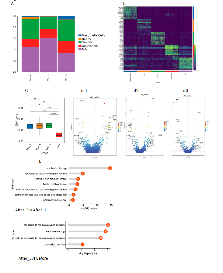
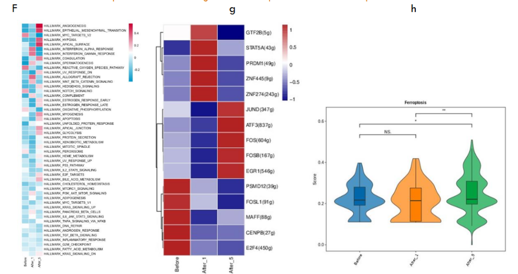
CHANGES OF MESOTHELIAL CELLS BEFORE AND AFTER TREATMENT: IRON DEATH GENE SCORE
The inferCNV analysis of mesothelial Cells against MPs showed deletion on chromosomes 1, 6, 19 and 21, and significant amplification on chromosomes 2, 3, 4, 7, 8, 12, 16 and 18. The results of CNVscore showed that significant changes occurred before treatment at 1 day and 5 day, but there was no significant difference among the three patients. The expression of immune-related genes such as CCL8, VSIG4, MARCO, C1QC, C1QB and CTSD was up-regulated 1 day after treatment compared with that before treatment. The expressions of IGFBP3, LOX, HILPDA, MT-TL1, TPT1, FOS, ID1, ATF3, FOSB, SLC2A1 and other genes were up-regulated 5 days after treatment compared with those before treatment and mesenchymal cell enrichment analysis after 5 days of drug treatment revealed activation of ROS related biological processes and molecular functions (such as reactive oxygen species response, cadherin binding, and antioxidant activity pathways).
The function of each cluster was identified based on GSVA analysis (hallmark database). The After_1 group mesenchymal cells enriched IFN-α and IFN-γ signaling pathways, and the After_5 group mesenchymal cells enriched epithelial mesenchymal transformation and heme metabolism.

CHANGES OF TANDNK CELLS AFTER TREATMENT
There are 8587 TandNK cells in total, which are proliferating T, NK, NaiveT, Teff, CD8MAIT respectively after marker gene annotation. The proportion of Teff cells increased successively. The proportion of NK cells at 5 day increased significantly after treatment. Proliferating T ratio increases on 1 day after treatment, and declines on 5 day after treatment. T cell receptor signaling pathway expression increases in Proliferating T, NK, NaiveT, Teff, and CD8MAIT 5 days after treatment, and Th1, Th17, and Th2 cells differentiate in NK cells and decline 5 days after treatment. The inflammatory effect of T cells was increased at 1 and 5 days after NK, NaiveT, Teff and CD8MAIT treatment. Toxic effects in Teff, NaiveT, NK, Proliferating T, and CD8MAIT increase sequently 1 day after treatment and 5 days after treatment.
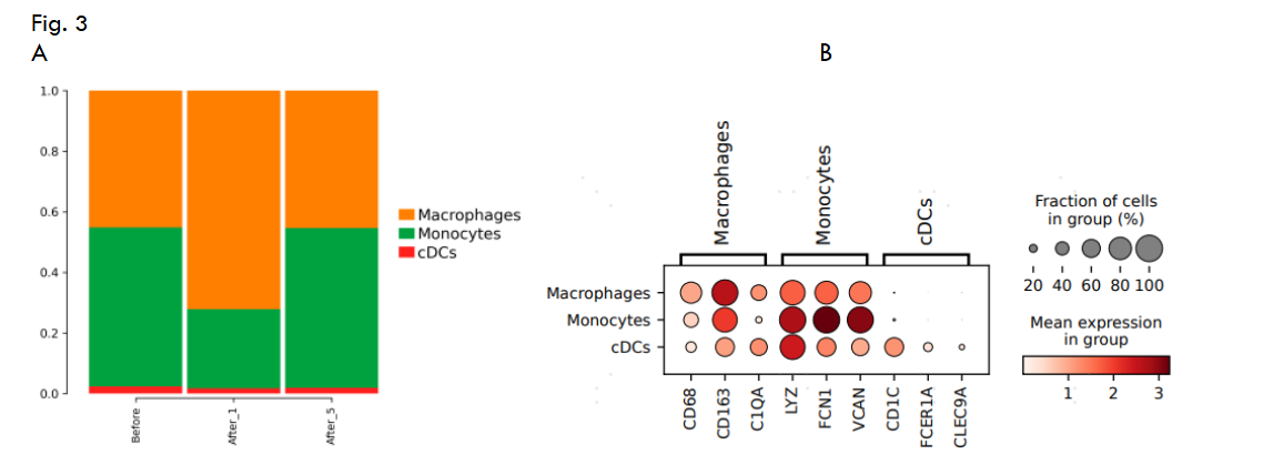
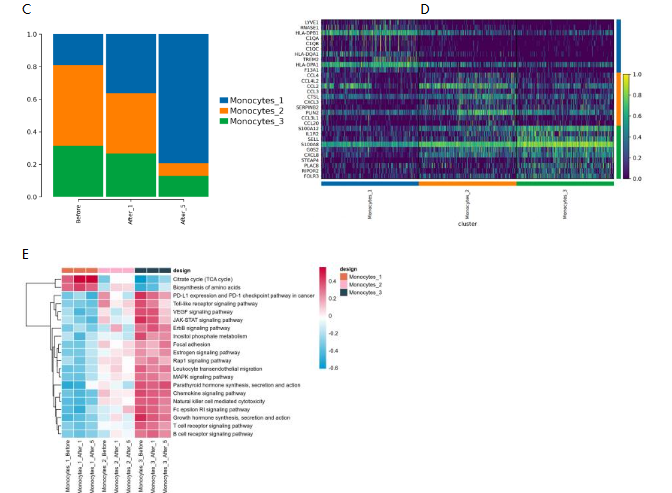
CHANGES OF MONOCYTES AFTER TREATMENT
MPs cells are divided into Macrophages, Monocytes, and cDCs. Macrophages increased in 1 day and decreased in 5 day post treatment. The proportion of monocytes decreased 1 day after treatment and increased 5 days after treatment. A total of 5109 monocytes were divided into 3 subpopulations after unsupervised clustering, Monocytes_1, Monocytes_2, and Monocytes_3. The cell proportion of Monocytes_1 showed an increasing trend after treatment, while the cell proportion of Monocytes_2 and Monocytes_3 showed a decreasing trend after treatment. Monocytes_1 highly expressed C1QA, C1QB, C1QC and other immune-related genes; Monocytes_2 highly expressed CCL2, CCL3, CCL4 and other chemokine-related genes; Monocytes_3 highly expressed FOLR3 and S100A12 extracellular adhesion molecules. Monocytes_3 mainly has immune-related functions but no significant changes after treatment.
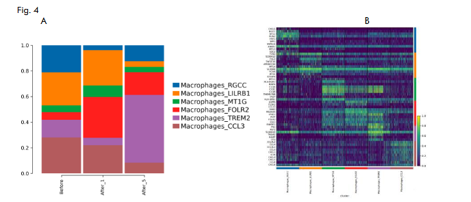
CHANGES OF MACROPHAGES AFTER TREATMENT
Macrophages consist of 7776 cells divided into 6 subpopulations Macrophages_CCL3, new Macrophages_FOLR2, new ges_lilrb1, new ges_mt1g, new ges_rgcc and new ges_TREM2. Macrophages_RGCC is decreased 1 day and increased 5 days after treatment. Macrophages_LILRB1 has no significant change in 1 day post-treatment, but significantly decreased 5 day post-treatment. Macrophages_FOLR2 increased in 1 day, but decreased in 5 day. The proportion of Macrophages_TREM2 increases significantly after 5 days, while the subgroup of Macrophages_CCL3 gradually decreases.
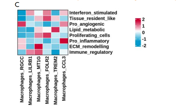
CHANGES IN NEUTROPHILS AFTER TREATMENT
There were 5117 Neutrophils in total, which were divided into 3 subpopulations by unsupervised clustering, namely Neutrophils_1, Neutrophils_2, and Neutrophils_3. The proportion of Neutrophils_1 increased on day 1 and decreased on 5 days after treatment. The proportion of Neutrophils_2 decreased at 1 day and increased at 5 day. However, Neutrophils_3 did not change significantly at 1 day and 5 days after treatment. Neutrophils_3 highly expressed IFIT1 and IFIT2, indicating that this group was related to interferon response. Glycolysis, oxidative phosphorylation, and iron death increased 1 day after Neutrophils_1 treatment compared with before treatment, but decreased 5 days after treatment. Immune-related pathways were elevated at 1 and 5 days after Neutrophils_2 treatment.
Discussion
Iron sucrose (IS) was commonly used for treatment of iron deficiency and iron is important for cancer patient’s nutrition. Ferroptosis-Enhanced Immunotherapy is reported for treating ascites of HCC by a chitosan hydrochloride and oxidized dextran (CH-OD) designed to load sulfasalazine (SSZ) and induces higher levels of immunogenic ferroptosis, then combined with anti-PD-1 immunotherapy to control ascites of HCC. In this study, we used IS local injection in to ascite of HCC to explore if treat ascites of HCC and induce ferroptosis related with immune response including MesothelialCells (UPK3B, WT1, KRT19), MPs (CD14, LYZ, C1QC), Neutrophils (CSF3R, S1-9A, FCGR3B), TandNK (CD3D, CD3E, CD3G) and B cells, respectively cells (CD79A, MS4A1, CD19).
Changes of mesothelial Cells before and after treatment has indicated iron death gene score, The inferCNV analysis of Mesothelial Cells against MPs showed deletion on chromosomes 1, 6, 19 and 21, and significant amplification on chromosomes 2, 3, 4, 7, 8, 12, 16 and 18. The results of CNVscore showed that significant changes occurred before treatment, 1 day after treatment and 5 day after treatment. Induction of HCC cells TGF – beta 1 / BMP – 7 pathways imbalances by increasing differentiation inhibitor 1 (ID1) expression significantly promote HCC cells and dye. The function of each cluster was identified based on GSVA analysis (hallmark database). The After_1 group mesenchymal cells enriched IFN-α and IFN-γ signaling pathways, and the After_5 group mesenchymal cells enriched epithelial mesenchymal transformation and heme metabolism, among which: Has a tendency to literature reported death iron exists a strong correlation between EMT and death may have a promoting effect on EMT of iron. A high propensity for iron death and EMT may result in poor prognosis and non-response to immunotherapy. Have other literature reports after large amounts of iron is input mitochondria, used for synthesis of hemoglobin and hemoglobin disorders has recently been shown to induce cell death of iron. Local high dosage of IS has still induced cell death of iron and increased TandNK highly expressed in proliferating cells, and the expression of amino acid biosynthesis, citric acid cycle, thermogenesis and other pathways decreased at 5 days after treatment. According to the characteristic gene set score of TandNK cells, immunosuppressor effect will significantly increase on ProliferatingT, NK, NaiveT, and Teff 5 days after treatment, and will also significantly increase on proliferatingt, NK, Naivet, and TEFF 5 days after treatment compared with 1 day after treatment.
MPs cells are divided into Macrophages, Monocytes, and cDCs. Macrophages increased in 1 day and decreased in 5 day post treatment. Multiple tumor studies have reported that tumor macrophages expressed by SPP1 and MMP9 are associated with tumor progression, metastasis, and poor prognosis in the remodeled ECM. These macrophages play an immune surveillance and pro-inflammatory role and may actively recruit and regulate immune cells during tumor-related inflammatory responses.
In summary, our current study indicated that the interaction between mesothelial cells, MPs, neutrophils, TandNK and B cells are induced by IS treatment ascites of HCC, which promotes the expression of a large number of genes and leads to upregulation of immune response and feroptosis. There is still need to explore this study in large number of ascites of HCC for providing an strong evidence: IS can used for ferroptosis and immune adjuvant in treatment ascites of HCC and regulate the immune cell function.
Credit author statement
Baofa, Yu, MD conceived the concept and provided overall supervision of all experiments and write. Hongxi Zhang, help to write English. Feng Gao, MD helped the clinical sample collection. Peng Jing, MD, He helped the clinical data collection. Guoqin Zheng, MS conducted and communicated with single cell company and data analysis.
Conflict of Interest Statement
All of authors do not have any conflict interest for the study.
Translational relevance to the manuscript
This research is valuable for medical practice. The study provides evidence to support possible precision immunotherapy with iron sucrose, a old drug for treating iron deficiency, as off label use for inducing ferroptosis and local immune booster that can potentially be applied to treatment of ascites fluid of hepatocellular carcinoma.
Ethical Statement
All procedures and protocols in the study have been reviewed and approved by the Ethical Committee of the Beijing Baofa Cancer Hospital (TMBF 0010, 2015). All informed consent forms from patients have been signed prior to the start of the study.
Ethical Approval
approved by the Ethical Committee of the Beijing Baofa Cancer Hospital (TMBF 0010, 2015).
Data Availability Statement
The data that support the findings of this study are available from [third party name] but restrictions apply to the availability of these data, which were used under license for the current study, and so are not publicly available. Data are however available from the authors upon reasonable request and with permission of [third party name].
Funding
There is not outside funding for this research.
Financial support
This study was sponsored in part by Tai Mei Baofa Cancer Hospital, Dongping, Shandong Province, China 271500.
Clinical trial number
Not applicable.
References:
- Macdougall, I. C., White,C., Anker, S.D., et al. Intravenous Iron in Patients Undergoing Maintenance Hemodialysis. N Engl J Med. 2019 Jan 31;380(5):447-458. doi: 10.1056/NEJMoa1810742. Epub 2018 Oct 26.
- Rich, N. E., Phen, S., Desai, N., et al. Cachexia is Prevalent in Patients With Hepatocellular Carcinoma and Associated With Worse Prognosis. Clin Gastroenterol Hepatol. 2022 May;20(5):e1157-e1169. doi: 10.1016/j.cgh.2021.09.022. Epub 2021 Sep 20.
- Macdougall, I.C., Josep Comin-Colet, J., Breymann, C., et al. Iron Sucrose: A Wealth of Experience in Treating Iron Deficiency. Adv Ther. 2020 May;37(5):1960-2002. doi: 10.1007/s12325-020-01323-z. Epub 2020 Apr 15.
- D B Van Wyck, G Cavallo, B S Spinowitz, et al. Safety and efficacy of iron sucrose in patients sensitive to iron dextran: North American clinical trial. Am J Kidney Dis. 2000 Jul;36(1):88-97. doi: 10.1053/ajkd.2000.8276.
- Meng, J., Yang, X., Huang, J., et al. Ferroptosis-Enhanced Immunotherapy with an Injectable Dextran-Chitosan Hydrogel for the Treatment of Malignant Ascites in Hepatocellular Carcinoma. Adv Sci (Weinh). 2023 Jul;10(20):e2300517. doi: 10.1002/advs.202300517. Epub 2023 May 3.
- Hassannia B., Vandenabeele P., Berghe T. V. Targeting Ferroptosis to Iron Out Cancer. Cancer Cell. 2019 Jun 10;35(6):830-849. doi: 10.1016/j.ccell.2019.04.002. Epub 2019 May.
- C Charytan, N Levin, M Al-Saloum, et al. Efficacy and safety of iron sucrose for iron deficiency in patients with dialysis-associated anemia: North American clinical trial. Am J Kidney Dis. 2001 Feb;37(2):300-7. doi: 10.1053/ajkd.2001.21293.
- Morales, M., Xiang Xue,X. Targeting iron metabolism in cancer therapy. Theranostics. 2021 Jul 25;11(17):8412-8429. doi: 10.7150/thno.59092. eCollection 2021.
- Chen, Z., et al., Dissecting the single-cell transcriptome network underlying esophagus non-malignant tissues and esophageal squamous cell carcinoma. EBioMedicine, 2021. 69: p. 103459.
- Kechin, A., et al., cutPrimers: A New Tool for Accurate Cutting of Primers from Reads of Targeted Next Generation Sequencing. J Comput Biol, 2017. 24(11): p. 1138-1143.
- Dobin, A., et al., STAR: ultrafast universal RNA-seq aligner. Bioinformatics, 2013. 29(1): p. 15-21.
- Yu, G., et al., clusterProfiler: an R package for comparing biological themes among gene clusters. Omics, 2012. 16(5): p. 284-7.
- Efremova, M., et al., CellPhoneDB: inferring cell-cell communication from combined expression of multi-subunit ligand-receptor complexes. Nat Protoc, 2020. 15(4): p. 1484-1506.
- Yao, J., Zhang, Y., Li, M., et al. Single-Cell RNA-Seq Reveals the Promoting Role of Ferroptosis Tendency During Lung Adenocarcinoma EMT Progression. Frontiers in Cell and Developmental Biology. 2022 Jan:(9). doi: 10.3389/fcell.2021.822315.
- Ma, M., Ruixia Wang, R., Xu, M., et al. Thorium (IV) triggers ferroptosis through disrupting iron homeostasis and heme metabolism in the liver following oral ingestion. Journal of Hazardous Materials. 452 (2023) 131217. DO1:10.1016 / j.jhazmat.2023.131217.
- Qiu, X., et al., Single-cell mRNA quantification and differential analysis with Census. Nat Methods, 2017. 14(3): p. 309-315.
- Ning, J., Ye, Y., Dechao Bu, D., et al. Imbalance of TGF-β1/BMP-7 pathways induced by M2-polarized macrophages promotes hepatocellular carcinoma aggressiveness. Mol Ther. 2021 Jun 2;29(6):2067-2087. doi: 10.1016/j.ymthe.2021.02.016. Epub 2021 Feb 15.
- Pittet, M. J., Michielin, O., Denis Migliorini, D. Clinical relevance of tumour-associated macrophages. Nat Rev Clin Oncol. 2022 Jun;19(6):402-421. doi: 10.1038/s41571-022-00620-6. Epub 2022 Mar 30.