

Human Cells: Self-Oxygenation Mechanisms Explained
The unsuspected ability of the human cell to oxygenate itself. Applications in cell biology
Arturo Solís Herrera, MD., PhD. 1, María del Carmen Arias Esparza, MD, MSc. 1
- Human Photosynthesis™ Research Center. Aguascalientes 20000, México
OPEN ACCESS
PUBLISHED: 31 March 2025
CITATION: Herrera, AS., and Arias Esparza, Md., 2025. The unsuspected ability of the human cell to oxygenate itself. Applications in cell biology. Medical Research Archives, [online] 13(3). https://doi.org/10.18103/mra.v13i3.6344
COPYRIGHT: © 2025 European Society of Medicine. This is an open-access article distributed under the terms of the Creative Commons Attribution License, which permits unrestricted use, distribution, and reproduction in any medium, provided the original author and source are credited.
DOI https://doi.org/10.18103/mra.v13i3.6344
ISSN 2375-1924
ABSTRACT
The transport of water across epithelia has remained an enigma ever since it was discovered over 200 years ago that water was transported across the isolated small intestine in the absence of osmotic and hydrostatic pressure gradients. While it is accepted that water transport is linked to solute transport, the actual mechanisms are not well understood. The movement of water and small molecules across the selectively permeable membranes of mammalian cells is a fundamental concept of physiology, which has not been resolved.
Cell membranes are complexes multicomponent structures, related to many basic cellular processes, such as substance transporting, energy conversion, signal transduction, mechano-sensing, cell adhesion and so on. However, cell membranes have long been difficult to study at a single-molecule level due to their complex and dynamic properties. Cell membranes are highly permeable to water, and water follows osmotic gradients. Osmotic gradients are generated when the concentration of solutes, such as sodium, is higher on one side of the membrane than the other.
But the mechanisms described are too mild to explain the control of the dynamics of the enormous amount of water that passes through the cell every minute. And since the control of the movement of water as well as solutes is fundamental for life, our observation about the presence of molecules capable of transforming the power of light into chemical energy, through the dissociation of intracellular water, as in plants, and this inside eukaryotic cells, marks a before and after in the knowledge about the mysteries of water movement and solutes inside cells.
Keywords: Cell, energy, hydrogen, intestine, ions, oxygen, water dissociation.
Introduction
All cells are surrounded by cell membrane, which separates a cell from the environment, creating and maintaining the fundamental differences between the cytosol and the extracellular environment 1. serve as many crucial biological functions, such as forming barriers between extracellular and intracellular environments, regulating the transport of substances 2, mediating the communications between cells 3, identifying and transmitting electrical/chemical signals through protein receptors 4.
And like many things in biology, the cell membrane is not understood, given the dynamic and fluid nature of its structure. This implies significant challenges in being able to define its chemical composition. And a composition of chemical elements that explain its multiple properties and functions, which to date has not been fully achieved. Ideally, it would be best to study it by touching it as little as possible. And the cell membrane and the cell as a whole function whose functioning stops or at least changes as soon as it is touched or opened. Hence the enormous difficulties we face in studying not only the cell membrane but the cell in general. Due to the extraordinary technical difficulties in studying the components of cells in vivo, or in situ, there is no particular methodology that will allow us to advance quickly or at least take firm steps in this regard.
The primary function of the cell membrane is to modulate the interplay between cells from their surroundings. All substances need across the cell membrane to enter the cells cytoplasm 5.
And despite the remarkable techniques developed in the past 100 years, such as detergent extraction, centrifugation, and cholesterol depletion by methyl-beta-cyclodextrin 6, and the recent development of super resolution imaging that further confirmed the existence of lipid rafts 7 to date, there are unsolved enigmas. Other techniques have broken through the optical diffraction limit and greatly improved the spatial resolution to tens of nanometers during the past decades 8. These methods provide a powerful tool for studying the distributions and functions of proteins and carbohydrates on cell membranes 9.
In general terms, the techniques described and other even more recent ones such as single molecule biophysical techniques 10, have been designed and developed with the aim of deepening knowledge of the structure and function of proteins, lipids, and carbohydrates that are important components of cell membranes and undertake primary functions for cell membranes. Exploring the composition and distribution of proteins and carbohydrates on cell membranes is essential to explain their functions 11.
As a highly heterogeneous and dynamically organized entity in the cells, the plasma membrane actively participates in numerous cellular functions 12, because the cell works as a whole, with all its components acting simultaneously and in coordination in different but very well defined roles, as well as very exact, amazingly exact, hence the enormous difficulty of studying them separately, because as soon as the cell is touched, it significantly modifies its functioning, or worse, stops completely. Deciphering the dynamics of cell membranes on different times and spatial scales and knowing how much force is needed for cellular transport has become the key to understanding biological functions of membranes 13. Single Molecule techniques have now become well-established tools for investigating the complex behaviors of different molecules on living cell membranes 14. These techniques can detect the structure, dynamics, and functions of single molecules, thus revealing their information that may be lost in ensemble averages.
But understanding the dynamics of water is a different challenge, as it is considered, so far, to be a rather passive role, as it easily crosses cell membranes, apparently following the laws of osmosis and simple diffusion. In the mammalian cell membrane, the phospholipid bilayer alone is permeable to some substances such as oxygen, a small nonpolar molecule, and partially permeable to water, but some substances such as charged ions and glucose are impermeant without the additional presence of protein channels and transporters in the membrane. The combined properties of the phospholipid and proteins has resulted in the use of the term the “selectively permeable” membrane 15. The extent to which solutes can cross the cell membrane dictates the tonicity of extracellular fluids and, therefore, the size and shape of cells from the resultant osmotic water movement 16.
Cell’s volume
Maintenance of a constant volume in the face of extracellular and intracellular osmotic perturbations is a critical problem faced by all cells. Most cells respond to swelling or shrinkage by activating specific membrane transport and/or metabolic processes that serve to return cell volume to its normal resting state. Water is effectively in thermodynamic equilibrium across the plasma membrane. The osmotic concentration of cytoplasmic (πi) and extracellular (π °) fluids are equal under steady-state conditions. Changes in intracellular or extracellular solute content generate a transmembrane osmotic gradient (∆π). Because cell membranes are freely permeable to water, any such gradient results in the immediate flow of water into or out of the cell until equilibrium is again achieved. All membranes have finite solute permeabilities. While many biologically relevant solutes have permeabilities substantially lower than water and behave as though they were effectively impermeable, some solutes have permeabilities approaching that of water. These high-permeability solutes diffuse across the membrane down their concentration gradient. As they do so, the osmotic pressure driving water flow is reduced. If the movement of solute is fast enough, the concentrations of the solute on the two sides of the membrane can become equalized before significant osmotic water flow occurs.
Osmolarity and tonicity are often used interchangeably by students, but they are not the same. Tonicity refers to the effect a solution has on cell volume because of the permeability of the membrane to that solute. Tonicity is, therefore, determined by the osmolarity and whether the solute can cross the cell membrane; it is the concentration of the impermeant solutes alone that determines tonicity. When comparing fluid concentrations to that of extracellular body fluid, the terms isotonic, hypertonic, and hypotonic are used rather than osmolarity, as they describe the effect the solution has on cell volume, which is of physiological significance. The tonicity will result in the following: no net movement of water (isotonic), net flow of water out of a cell (hypertonic), or net flow of water into a cell (hypotonic). Two solutions that are isosmotic may not be isotonic. A key example is isosmotic urea and isosmotic NaCl.
Both urea and NaCl have the same osmolarity, having the same total number of osmolyte particles; however, the membrane is permeable to urea, which will freely diffuse across the cell membrane, and impermeable to NaCl. An isosmotic urea is, therefore, hypotonic compared with an isosmotic and isotonic solution of the impermeant NaCl. As a result, the volume of a cell is determined by the solution in which it is being bathed and whether the cell’s membrane is permeable to the solute. If a membrane is not equally permeable to all solutes, then a difference in water movement will be observed that is not explained by osmolarity alone, and, hence, an additional term, tonicity, is required. Hypotonic solutions lead to cell swelling and eventual rupture or lysis if the resultant osmotic movement of water is great enough. In the case of red blood cells, this is referred to as hemolysis 17.
Thereby, it was thought that since animal cells, its membranes, or organelles are unable to generate or sustain significant hydrostatic pressure gradients, water flow can cause cell swelling or shrinkage.
Cell volume regulation
So far, it is believed that cells respond to volume perturbations by activating poorly defined volume regulatory mechanisms. The processes by which swollen and shrunken cells return to normal volume are collectively termed regulatory volume decrease and regulatory volume increase, respectively. Cell volume can only be regulated by the gain or loss of osmotically active solutes, primarily inorganic ions such as Na+1, K+1, Cl±1.357, or small organic molecules termed organic osmolytes, that could be compatible or non-perturbing and perturbing or counteracting solutes 18.
Volume regulatory electrolyte loss and gain it is believed that are mediated almost exclusively by membrane transport processes 19 since it is the only mechanism that is already known, although it is not well understood. In most animal cells, regulatory volume decrease occurs through loss of KCl via activation of separate K+1 and Cl ±1.357 channels or by activation of the K-Cl co-transporter. Regulatory volume increase occurs by uptake of both KCl and NaCl. Accumulation of these salts is brought about by activation of Na+1/H+1 and Cl-1.357/HCO3 -1 exchangers or the Na-K-2Cl co-transporter.
Elevated electrolyte levels and intracellular ionic strength can denature or precipitate cell macromolecules. Even smaller changes in cellular inorganic ion levels can alter resting membrane potential, the rates of enzymatically catalyzed reactions and membrane solute transport that is coupled to ion gradients. Accumulation of organic osmolytes is mediated either by energy-dependent transport from the external medium or by changes in the rates of osmolyte synthesis and degradation 20.
Volume regulatory organic osmolyte accumulation is typically a quite slow process relative to electrolyte uptake and requires many hours after initial activation to reach completion. This slow time course is observed because activation of organic osmolyte accumulation pathways usually requires transcription and translation of genes coding for organic osmolyte transporters and synthesis enzymes. Opposite, they are the extra-dynamic fast and exact mechanisms of water dissociation, which fluctuate in the nano and picosecond range.
Volume sensing mechanisms of the cell
They appear to be extremely sensitive. Studies on the renal proximal tubule have demonstrated that these cells can sense and respond to volume changes of 3% 21. However, our understanding of the mechanisms by which cells sense volume perturbations and transduce those changes into regulatory responses is rudimentary.
Several possible volume signals have been postulated including swelling- and shrinkage-induced changes in membrane tension, cytoskeletal architecture, cellular ion concentrations and the concentration of cytoplasmic macromolecules 22. All these hypotheses have their strengths and weaknesses. At present, it appears that no one signaling mechanism can account for the volume sensitivity of the various genes and membrane transport pathways that are activated or inactivated in response to cell volume perturbations. To further complicate the picture, recent evidence suggests that cells can detect more than simple swelling or shrinkage. Cells most likely possess a wide array of volume detectors and effector mechanisms that respond selectively to both the magnitude and nature of the volume perturbation 23. Such functionally distinct sensors and effector pathways may afford the cell simultaneous control over a variety of parameters (e.g., intracellular pH and ionic composition) in addition to volume.
The volume of water and intracellular solutes are within very narrow ranges
The ability to tightly control solute and water balance during osmotic challenge is an essential prerequisite for cellular life. Cellular osmotic homeostasis is maintained by the regulated accumulation and loss of inorganic ions and small organic solutes termed organic osmolytes. Organic osmolytes are “compatible” or “nonperturbing” solutes and are typically found in concentrations of tens to hundreds of milli-molars in the cytosol of all organisms from bacteria to humans. The effector mechanisms responsible for osmoregulatory solute accumulation and loss in animal cells are generally well understood.
However, major gaps exist in our understanding of the signals and signaling pathways by which animal cells detect volume perturbations and activate volume regulatory mechanisms. Elucidation of volume sensing mechanisms and signaling pathways represents the most pressing and significant challenge in the field and is essential for understanding fully cell volume control and related cellular processes.
Water pump
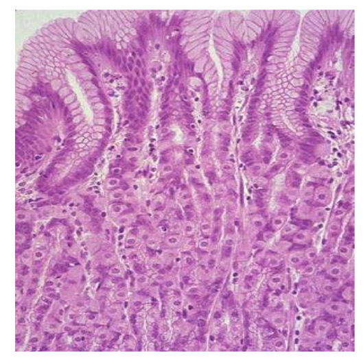
Current dogma holds that active ion transport sets up local osmotic gradients in the spaces between epithelial cells, the lateral intercellular spaces, and this in turn drives water transport by local osmosis. In the case of the small intestine, which in humans absorbs about 8 L of water a day, there is no direct evidence for either local osmosis or aquaporin gene expression in enterocytes. Intestinal water absorption is greatly enhanced by glucose, and this is the basis for oral rehydration therapy in patients with secretory diarrhea.
The question of how water is transported across epithelial cells in the absence of external driving forces has intrigued physiologists for over a century 24, when it was demonstrated that water is transported across the intestine in the absence of transepithelial osmotic and hydrostatic pressure differences.
In the intestine there is little, if any, expression of aquaporins in enterocytes and this raises the possibility that the high-water permeability of the brush border and basolateral membranes is either due to a high intrinsic water permeability of the lipid bilayer or the presence of other water channels. Yet another possibility is that local osmosis does not account for water transport across the intestine. There is ample evidence that intestinal water absorption, 8 L a day in man, is intimately linked to ‘active’ solute absorption and that glucose in the gut lumen greatly enhances salt and water absorption.
Although the membrane potential, the external Na+ and sugar concentrations, and temperature 25 plays an important role. But on the other hand, we have the unknown of the origin of the energy, which in the case of the activation energy for Na+–glucose cotransport is identical (25–30 kcal mol-1) to that for coupled-water transport 26. Plus, the conflicting experimental data that Coupled-water transport is independent of the osmotic gradient and even occurs against an osmotic gradient 27. Therefore, seems to be a close link between Na+–glucose cotransport and the initial rate of water transport.
However, when the intracellular fluid becomes hypertonic as transport proceeds, this is expected to draw more water into the cell by osmosis. The small size of the water molecule lends itself to accommodating it in the theoretical processes of cotransporters, since the volume occupied by 200–400 water molecules (6000–12 000 Å3) only amounts to 7% of the volume of the co-transporter (100 000 Å3).
The human intestine absorbs about 1 mol of D-glucose and 8 L of water per day and given that the stoichiometry of hSGLT1 is 2 Na+ : 1 glucose : 264 water, this indicates that co-transport alone can account for ~4 L of water transport. Since the co-transported fluid is hypertonic this will result in an additional component due to osmosis, but the exact amount is difficult to estimate due to uncertainties about the magnitude of the osmotic gradient and the water permeability of the membrane.
Thereby, the mystery remains about how water is transported across the basolateral membrane of the enterocyte in the absence of aquaporins and hypertonic fluid in the lateral intercellular spaces? One theoretical possibility is that basolateral membrane transport proteins act as water pumps and channels.
The Na+–glucose cotransporter behaves as a low conductance water channel, In the steady state, direct sugar-coupled water flow constitutes one-third of the total water transport in isotonic water flow, the remaining being osmotic water flow. Under the assumption of extremely low intracellular mobilities 28 and based on steady-state flows alone, some researchers concluded that water flow was linked to Na+–glucose cotransport by simple osmosis. They concluded that 65% of the steady-state water flow was osmotic.
That water cotransport accounts for 35% of the steady-state flow is still controversial. The measurement of the thermodynamics of Na+, sugar and water transport is especially difficult, since the osmotic gradients required to influence Na+–sugar cotransport are large. On the other hand, the use of the Gibbs equation predicts that osmotic gradients of 100 mosmol L-1 will shift the reversal potential by only about 5 mV.
It is interesting that the problems raised since the nineteenth century have not been solved to date. Researcher E. WAYMOUTH REID, M.B. CAMB., from the department of Physiology in University College. Dundee; St. Andrews; University, concludes his article with the following: “The facts I have observed, then, seem to point to the conclusion that it is possible to have a true ‘absorptive process taking place without the aid of ordinary. osmotic action. That intestinal absorption is governed by other laws than those of ordinary osmosis has long been known.” But the redox properties of melanin, which originate in its hitherto unknown intrinsic property of dissociating the molecules of water, will largely explain the accurate observations of Dr. Reid, which date from 1892.
Current dogmas cannot be able to solve the mysteries about water dynamics because it is still wrongly believed that water is pushed into the cell, despite the remarkable permeability of the cell membrane to water, but the laws of simple diffusion or even by hydrostatic gradients or osmosis, do not explain the passage of isotonic water through the intestinal walls, as Dr. Reid experimentally demonstrated since 1892.
The unsuspected ability of eukaryotic and prokaryotic cells to dissociate water molecules by means of molecules normally present in their cytoplasm, such as chlorophyll in plants, opens new paths to understand both normal and diseased cells.
Thereby, isotonic water is not pushed from the extracellular space into the intracellular space but is attracted into the cell through water dissociation mechanisms is explained as follows.
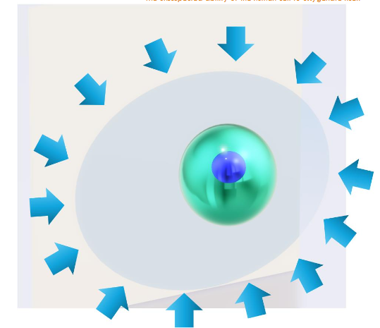
The intensity and direction of water currents resulting from constant negative pressure mainly around the cell nucleus, resulting from water dissociation; they affect both the cell cytoplasm and the nucleus of the cell, and undoubtedly reach the immediate environment of the living thing.
The transformation of the power of light into chemical energy that can be used by the cell is the beginning of the explanation of the functioning of the cell nucleus, apparently without mitochondria or ATP. Then, this source of oxygen and hydrogen, which is the universal carrier of energy, would explain the presence of energy inside the cell nucleus.
So, to analyze the physiology of the process of isotonic water absorption, we must now start by including the intrinsic property of melanin to dissociate water molecules at the intracellular level. This water dissociation creates significant negative pressure, which is the main basis for the absorption of water and food throughout the gastrointestinal tract. The hitherto inexplicable constants and very rapid variations observed in the flow of isotonic water through cell membranes are largely because the turnover rate of water dissociation is under constantly changing, as it is not linear at all but seems to adapt very quickly to the changing conditions of the intracellular environment.
So, the cell does not have a passive role with respect to the absorption of isotonic water, but the process that gives rise to life, that is: the dissociation of water, also generates a zone of negative pressure that attracts water from the interstitial space, and this negative pressure changes according to the conditions of the immediate environment. For example, the addition of glucose to water increases the degree of absorption of water, which can be explained by the obstacle that glucose molecules represent to the passage of isotonic water through the aquaporins of the cell membrane, and by decreasing this flow, melanin responds by increasing the rate of turnover of the dissociation of isotonic water molecules.
We can consider that the other processes described in the literature, which intervene in the flow of isotonic water into the cell, are rather accessory, since they are a complement to the main mechanism in this regard, referring to the vacuum that is generated by the constant dissociation of water molecules.
The redox properties of Melanin
Melanin’s redox properties seem especially important to its biological function, and there have been several attempts to investigate these redox properties 30. Melanin can exist in different redox states helps to explain how melanin properties can depend on context. Specifically, depending on its redox-state, melanin can serve as either a reductant or an oxidant. However, context also depends on the environment 31. It seems the context undoubtedly can determine the action of melanin.
The first two reactions of life are the dissociation and further reforming of water molecules. The dissociation of water that necessarily occurs inside melanin is a highly endergonic process (blue color), as it absorbs and requires a lot of energy to happen properly, and can be described as follows:
2H2O (Liq) → 2H2(gas) + O2(gas) → 2H2O (liq) + 4e–
The second part of the process (green color) happens inside and outside the melanin molecule and is an exergonic process that releases energy. For every two water molecules that are re-formed, 4 high-energy electrons are generated 32.
The rate of turnover between one process and the other (dissociation/reforming) is the basis that establishes the redox properties of melanin molecules at a given time. Melanin is reported to offer important biological and technological properties that include antimicrobial, 33 antioxidant 34 and anti-cancer activities 35; radiation protective 36 and photothermal energy conversion properties 37; and heavy metal binding abilities 38.
Because of melanin’s structural complexity, it has been difficult to apply conventional bottom-up molecular structure-based approaches to understand these various properties.
The suction of isotonic water through the aquaporins of the cell membrane brings the glucose molecules closer to the cell membrane and from there the glucose molecule is transported into cells via its transporter 39. It takes time (10–40 ms) and force (~40 pN) to transport the single glucose molecule into cells via the glucose transporter.
And it is not ruled out that the molecular hydrogen that is formed when water dissociates and that is the molecule that transports energy in living beings but in the entire universe, is the same one that provides the energy for the glucose transporter to carry out its function.
Characterization of the multi-molecular structures of cell membranes is an essential step towards understanding their processes and functions, which could potentially lead to alternate applications in medicine and biotechnology 40. All cells are threatened by possible isosmotic swelling or shrinkage. Under steady-state conditions, intracellular solute levels are held constant by a precise balance between solute influx and efflux across the plasma membrane, and by the metabolic production and removal of osmotically active substances. A variety of physiological and pathophysiological conditions, however, can disrupt this balance 41. For example, the cell swelling that occurs in the brain after a stroke or head trauma is an example of isosmotic volume increase and is due to intracellular accumulation of NaCl and other solutes. But the main impact is on the redox properties of melanin, since when these are altered, the effect is diffuse because all cell processes are impacted since they depend directly or indirectly on the very first reaction of life, which is the dissociation of the molecule from water.
The mediators with a chemical of interest and we observed that 42 some redox-active environmental toxins (i.e., paraquat) 43 drugs (e.g., clozapine or acetaminophen can undergo redox cycling with melanin. While such in vitro MEP measurements cannot prove any in vivo activity, it has revealed a previously unknown mechanism of potential biological relevance to 51 chemical toxicities and drug activities.
Conclusion
The principle of the origin of life and therefore of cellular function is the dissociation of the water molecules that the cell itself contains inside, which could not be otherwise since the cell handles this gaseous element in a very precise way. Thus, as CO2 is generated inside the cells, and then passes into the bloodstream, and from there to the lungs to be expelled into the atmosphere, oxygen is also generated inside the cells.
The oxygen and hydrogen requirements of each cell are very precise and demanding, and if the cell can provide both molecules to itself, the efficiency of the organism as a whole is greater, since each cell can oxygenate by itself, in the best way and under various circumstances, since the redox properties of melanin are surprising. What we can describe as auto-oxygenation solves at least two basic problems: first: that the amount of oxygen inside the cells is adequate in most of the ever-changing situations that cells face throughout life, and the second is that oxygen toxicity is kept at low or minimal levels in this way, because the excess of O2 that may be present, is quickly eliminated by the same cell by adding a carbon atom to each oxygen molecule, generating the most oxidized form of carbon which is carbon dioxide. And the cells, tissues, organs or systems have quickly handled this toxic gas as they have done since the beginning of time, and the process is so efficient that it is almost identical in all living beings.
The approximately 900 grams of CO2 that are eliminated on average every 24 hours by an average human being is irrefutable proof of the efficiency of an elimination process that Nature developed over eons of years of evolution. And surprisingly, the functioning of the CO2 biological cycle inside the cell and the body depends entirely on the integrity of the dissociation process of the water molecules found inside the cells. And as long as the process of dissociation and reformation of water happens properly inside the melanosomes that each of the cells that make us up contains, the body will function well because it is very well made. For the health of our body to be affected either in its form or function, it is necessary to unbalance the fundamental process of life (dissociation of water at the intracellular level), otherwise the health of the person is not significantly affected.
The resilience of the human body to different diseases is a manifestation of the surprising resilience of melanin’s redox properties.
Acknowledgment
This work was supported by an unrestricted grant from Human Photosynthesis™ Research Center, Aguascalientes 20000, México.
Conflict of interest: None
References
- Raposo G, Stoorvogel W (2013) Extracellular vesicles: exosomes, microvesicles, and friends. J Cell Biol 200: 373−383
- Jiang Y, Lee A, Chen J, Ruta V, Cadene M, Chait BT, MacKinnon R. (2003) X-ray structure of a voltage-dependent K+ channel. Nature 423: 33−41
- Lin JC, Duell K, Konopka JB (2004) A microdomain formed by the extracellular ends of the transmembrane domains promotes activation of the G protein-coupled α-factor receptor. Mol Cell Biol 24: 2041−2051
- Rivière S, Challet L, Fluegge D, Spehr M, Rodriguez I (2009) Formyl peptide receptor-like proteins are a novel family of vomeronasal chemosensors. Nature 459: 574−577
- Aderem A, Underhill DM (1999) Mechanisms of phagocytosis in macrophages. Annu Rev Immun 17: 593−623
- Brown DA, Rose JK (1992) Sorting of GPI-anchored proteins to glycolipid-enriched membrane subdomains during transport to the apical cell surface. Cell 68: 533−544
- Lillemeier BF, Mörtelmaier MA, Forstner MB, Huppa JB, Groves JT, Davis MM (2010) TCR and Lat are expressed on separate protein islands on T cell membranes and concatenate during activation. Nat Immunol 11: 90−96
- Huang B, Babcock H, Zhuang X (2010) Breaking the diffraction barrier: super-resolution imaging of cells. Cell 143: 1047−1058
- Jacobson K, Liu P, Lagerholm BC (2019) The lateral organization and mobility of plasma membrane components. Cell 177:806−819
- Zhang, Qingrong. Li, Siying. Yang, Yu, Shan, Yuping. Wang, Hongda. Studying structure and functions of cell membranes by single molecule biophysical techniques Biophys Rep 2021, 7(5):384−398 https://doi.org/10.52601/bpr.2021.210018
- Bretscher MS, Raff MC (1975) Mammalian plasma membranes. Nature 258: 43−49
- Garcia-Parajo MF, Cambi A, Torreno-Pina JA, Thompson N, Jacobson K (2014) Nanoclustering as a dominant feature of plasma membrane organization. J Cell Sci 127: 4995−5005
- Lesniak A, Salvati A, Santos-Martinez MJ, Radomski MW, Dawson KA, Åberg C (2013) Nanoparticle adhesion to the cell membrane and its effect on nanoparticle uptake efficiency. J Am Chem Soc 135: 1438−1444
- Xie XS, Yu J, Yang WY (2006) Living cells as test tubes. Science 312:228−230
- Cooper GM. The Cell: A Molecular Approach (6th Ed.). Sunderland, MA: Sinauer, 2013.
- Strange K. Cellular volume homeostasis. Adv Physiol Educ 28: 155–159, 2004. doi:10.1152/advan.00034.2004.
- Dourmashkin RR, Rosse WF. Morphologic changes in the membranes of red blood cells undergoing hemolysis. Am J Med 41: 699–710, 1966. doi:10.1016/0002-9343(66)90031-3.
- Yancey PH. Compatible and counteracting solutes. In: Cellular and Molecular Physiology of Cell Volume Regulation, edited by Strange K.Boca Raton, FL: CRC, 1994, p. 81–109.
- Lang F, Busch GL, Ritter M, Volkl H, Waldegger S, Gulbins E, and Haussinger D. Functional significance of cell volume regulatory mechanisms. Physiol Rev 78: 247–306, 1998.
- Chamberlin ME and Strange K. Anisosmotic volume regulation: a comparative view. Am J Physiol Cell Physiol 257: C159–C173, 1989.
- Lohr JW and Grantham JJ. Isovolumetric regulation of isolated S2 proximal tubules in anisotonic media. J Clin Invest 78: 1165–1172, 1986
- O’Neill WC. Physiological significance of volume-regulatory transporters. Am J Physiol Cell Physiol 276: C995–C1011, 1999.
- Emma F, McManus M, and Strange K. Intracellular electrolytes regulate the volume set point of the organic osmolyte/anion channel VSOAC. Am J Physiol Cell Physiol 272: C1766–C1775, 1997
- REID, E. W. (1892). Preliminary report on experiments upon intestinal absorption without osmosis. British Medical Journal 2, 1133–1134.
- MEINILD, A.-K., HIRAYAMA, B. A., WRIGHT, E. M. & LOO, D. D. F. (2002). Fluorescence studies of ligand-induced conformational changes of the Na+/glucose cotransporter. Biochemistry 41, 1250–1258
- LOO, D. D. F., HIRAYAMA, B. A., MEINILD, A.-K., CHANDY, G., ZEUTHEN, T. & WRIGHT, E. M. (1999). Passive water and ion transport by cotransporters. Journal of Physiology 518, 195–202.
- MEINILD, A.-K., KLAERKE, D. A., LOO, D. D. F., WRIGHT, E. M. & ZEUTHEN, T. (1998). The human Na+–glucose cotransporter is a molecular water pump. Journal of Physiology 508, 15–21.
- ZEUTHEN, T. (2002). General models for water transport across leaky epithelia. International Review of Cytology 215, 285–319.
- REID, E. W. (1892). Preliminary report on experiments upon intestinal absorption without osmosis. British Medical Journal 2, 1133–1134
- M. Rózanowska , T. Sarna , E. J. Land and T. G. Truscott , Free Radical Biol. Med., 1999, 26 , 518 —525
- Kim, Eunkyoung , Wang, Zheng. Jun Wei Phua, . Bentley, William E. Dadachova, Ekaterina, Napolitano, Alessandra, and Payne, Gregory F. Enlisting electrochemistry to reveal melanin’s redox-related properties.Mat. Adv. Royal Society of Chemistry (2024) https://doi.org/10.1039/D3MA01161E
- Herrera, A.S. (2015) The Biological Pigments in Plants Physiology. Agricultural Sciences, 6, 1262-1271. http://dx.doi.org/10.4236/as.2015.610121
- I. Ben Tahar, M. Kus-Lis´kiewicz, Y. Lara, E. Javaux and P. Fickers, Biotechnol. Prog., 2020, 36, e2912.
- T. Rahmani Eliato, J. T. Smith, Z. Tian, E. S. Kim, W. Hwang, C. P. Andam and Y. J. Kim, J. Mater. Chem. B, 2021, 9, 1536–1545
- O. Al-Obeed, A. S. El-Obeid, S. Matou-Nasri, M. A. Vaali-Mohammed, Y. Alhaidan, M. Elwatidy, H. Al Dosary, Z. Alehaideb, K. Alkhayal, A. Haseeb, J. McKerrow, R. Ahmad and M. H. Abdulla, Cancer Cell Int., 2020, 20, 1–17.
- W. Song, H. Yang, S. Liu, H. Yu, D. Li, P. Li and R. Xing, J. Mater. Chem. B, 2023, 11, 7528–7543.
- H. Liu, Y. Yang, Y. Liu, J. Pan, J. Wang, F. Man, W. Zhang and G. Liu, Adv. Sci., 2020, 7, 1903129
- A. S. ElObeid, A. Kamal-Eldin, M. A. K. Abdelhalim and A. M. Haseeb, Basic Clin. Pharmacol. Toxicol., 2017, 120, 515–522.
- Pan YG, Zhang FX, Zhang LY, Liu SH, Cai MJ, Shan YP, Wang XQ, Wang HZ, Wang HD (2017) The process of wrapping virus revealed by a force tracing technique and simulations. Adv Sci4: 1600489. https://doi.org/10.1002/advs.201600489
- Escribá PV, González-Ros JM, Goñi FM, Kinnunen PKJ, Vigh L, Sánchez-Magraner L, Fernández AM, Busquets X, Horváth I, Barceló-Coblijn G (2008) Membranes: a meeting point for lipids, proteins and therapies. J Cell Mol Med 12: 829−875
- McManus ML and Churchwell KB. Clinical significance of cellular osmoregulation. In: Cellular and Molecular Physiology of Cell Volume Regulation, edited by Strange K. Boca Raton, FL: CRC, 1993, p. 63–77.
- E. Kim, W. T. Leverage, Y. Liu, L. Panzella, M. L. Alfieri, A. Napolitano, W. E. Bentley and G. F. Payne, ACS Chem. Neurosci., 2016, 7, 1057–1067.
- Z. Temocin, E. Kim, J. Li, L. Panzella, M. L. M. L. Alfieri, A. Napolitano, D. L. D. L. Kelly, W. E. W. E. Bentley and G. F. G. F. Payne, ACS Chem. Neurosci., 2017, 8, 2766–2777.