


Transmission of Pneumococcal Pneumonia: Key Insights
What do you get? Transmission of pneumococcal pneumonia.
Tom Boom¹, Anna Budińská¹, Júlia Šeligová¹, Ger T. Rijkers¹*
- Department of Science and Engineering, University College Roosevelt, P.O. Box 94, 4330 AB Middelburg, The Netherlands.
OPEN ACCESS
PUBLISHED: 31 August 2024
CITATION: Boom, T., Budinska, A., et al., 2024.
What do you get? Transmission of pneumococcal pneumonia. Medical Research Archives, [online] 12(8).
https://doi.org/10.18103/mra.v12i8.5612
COPYRIGHT: © 2024 European Society of Medicine. This is an open-access article distributed under the terms of the Creative Commons Attribution License, which permits unrestricted use, distribution, and reproduction in any medium, provided the original author and source are credited.
DOI https://doi.org/10.18103/mra.v12i8.5612
ISSN 2375-1924
ABSTRACT
Streptococcus pneumoniae is a polysaccharide encapsulated bacterium responsible for the majority of cases of community acquired pneumonia. The upper respiratory tract of children becomes colonized with pneumococci early in life, from the first weeks of life up to 18 months. Factors which influence timing of colonization include geographical localization, socio-economic circumstances, and household conditions.
Pneumococcal pneumonia to a degree is a seasonal disease, peaking in the winter months. One of the epidemiological determinants is that viral infections (in particular influenza) predispose for pneumococcal pneumonia. The infectious nature of pneumococcal pneumonia was underscored during the COVID-19 pandemic. Due to societal restrictions imposed (face masks, social distancing) the incidence of invasive pneumococcal disease (IPD) dropped, and returned to pre-COVID-19 numbers after lifting of the restrictions.
There are 100 different S. pneumoniae serotypes, based on differences in the capsular polysaccharide. Introduction of protein conjugated polysaccharide vaccines (PCV) has reduced the incidence of IPD in children. Because children are the reservoir for other risk groups, in particular the elderly, introduction of PCV has indirectly also reduced the burden of IPD in latter risk group. The first generation of PCV consisted of the seven most prevalent pneumococcal serotypes. After implementation, replacement colonization of the upper respiratory tract with non-vaccine serotypes was observed and also replacement disease. Upcoming 24-valent PCVs will have a much broader coverage, but whether replacement disease now has been eliminated remains to be seen.
Introduction
“What do you get when you kiss a girl?” is a line from the Burt Bacharach song I’ll never fall in love again. The answer to this open question is in the next line of the song: “You get enough germs to catch pneumonia”. What makes this song unique in the context of pneumonia is that it is the only song which emphasizes the infectious nature of the disease. In 1968, the opening night of the musical Promises promises was already advertised, but it still lacked a catchy song to end the first episode before the interval. The producer pushed Bacharach, who was admitted to the hospital with pneumonia, to finish the song in a hurry, which he did. Apart from I’ll never fall in love again, all other popular music songs about pneumonia infer that you can get pneumonia by standing in the rain and/or cold.
While pneumonia is a respiratory infectious disease, there is no direct evidence that it can be transmitted by kissing. Streptococcus pneumoniae, the bacterium responsible for the majority of cases of community acquired pneumonia, can easily be detected in saliva, either by molecular techniques or conventional bacterial cultures²,³. The major risk groups for community acquired pneumonia are the young (infants and toddlers up to the age of 2 years) and the old (from which age is debated, but surely from 65 years onwards). The natural reservoir for S. pneumoniae is the upper respiratory tract of young children, from which the bacteria can be transmitted to others, including the elderly⁴. Apart from the “what do you get” therefore also the “how do you get” is an important question. These questions, as well as the impact of vaccination, will be addressed in this review on transmission of pneumococcal pneumonia.
Methods
A weighted linear regression analysis (from week 1, 2018 to week 52, 2022) was performed to assess the effect of season as well as non-pharmaceutical interventions (i.e. the societal restrictions implemented throughout the SARS-CoV2 pandemic) on the incidence of pneumococcal diseases. Analysis was restricted to the United States. Data about invasive pneumococcal disease was available from the American Centers for Disease Control and Prevention and was used ranging from 2019 to 2024 (https://www.cdc.gov/pneumococcal/php/surveillance/index.html; Assessed June 2024). Data about the stringency index was acquired from the Oxford Coronavirus Government Response Tracker (OxCGRT) from 2020 to 2022 (https://ourworldindata.org/covid-stringency-index; Assessed June 2024). The OxCGRT project used nine metrics (school closures; workplace closures; cancellation of public events; restrictions on public gatherings; closures of public transport; stay-at-home requirements; public information campaigns; restrictions on internal movements; and international travel controls) to calculate the stringency index of multiple countries across 2020-2023.
For statistical analysis, data were tested for linearity, normal distribution of error and multicollinearity. Heteroscedasticity was present based on the results of a Breusch-Pagan test (p < .001). Therefore a weighted least squares analysis was conducted. The weighted data showed homoscedasticity (Breusch-Pagan test p = 0.8805).
Children are colonized with S. pneumoniae early in life
Clinical trials using conventional nasopharyngeal swabs and subsequent culturing on blood agar determined that infants 0-2 years of age are colonized with S. pneumoniae early in life. This age of first colonization varies among different parts of the world (Figure 1). In North America, the age of colonization ranged from 4 days to 18 months, with a mean age of acquisition of 6 months⁵. A study in Finland found that colonization by 6 months was rare and that most infants were colonized at 18 months⁶. In a different study in Finland, 9% of infants were colonized at 2 months, and a majority (43%) at 24 months⁷. In the Netherlands, 8% of infants were colonized at age 1.5 months, 31% at 6 months, and 45% at 14 months⁸. In other parts of the world, the age of first acquisition was much lower for most infants. In Papua New Guinea, acquisition occurred as early as day 1 after birth, more than 60% of infants were colonized by 15 days, the mean age of acquisition was 17.1 days, and all were colonized within 3 months⁹. A study in the Thailand-Myanmar border region found that 76% of infants were colonized at 3 months, and 97% by 6 months¹⁰. In South India, 54% of children were colonized at 2 months of age, 64% at 4 months and 70% at 6 months, respectively¹¹. In Gambia, 30% were colonized by week 1, 60% were colonized by 15 days, 90% by 15 weeks, and all the infants by 4 months¹². Another Gambian study found similar early acquisition rates, as the mean age at first acquisition was 33 days, respectively¹³. A study in Kenya found that the earliest acquisition was on day 1, 63% were colonized by 3 months, and the median age of colonization was 38.5 days¹⁴. In South Africa, 3% were colonized by 2 weeks, 64% at 6 months, 95 % by 260 days (8.7 months), and 98% by the first year of life¹⁵. The overarching trend is that colonization is positively correlated with age⁵,⁸,¹². However, in low-income countries, it occurs much sooner as compared to medium to high-income countries. Please note that the data cited above are national averages, and that regional or population differences may exist. Furthermore, sampling and microbiological techniques may also vary between studies.
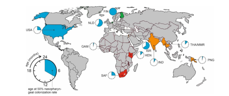
Figure 1. Time of acquisition of pneumococci in the upper respiratory tract. Shown is the age (in months) at which at least 50% of the children are colonized with pneumococci. Countries are indicated by their alpha-3 codes (https://www.iban.com/country-codes).
Factors influencing early colonization
A number of factors contribute to earlier colonization by S. pneumoniae. Crowding is the most significant factor. Living in a larger family with older siblings is associated with an earlier age of first acquisition as S. pneumoniae is contagious within families⁵,⁶,¹⁶,¹⁷. Older siblings attend daycare and come into contact with other children and people, which contributes to earlier colonization⁸,¹⁶,¹⁷. Colonization could also be due to exposure of shared toys and surfaces, as pneumococci can be readily isolated from fomites such as plush toys recently manipulated by colonized children, and do not undergo desiccation on surfaces for many days¹⁸ due to biofilm formation and catabolism of capsular polysaccharide as a carbon source¹⁹.
Moreover, first-born children in a family have higher age at first acquisition and they acquired fewer serotypes⁵. In low-income countries, several additional socioeconomic and environmental factors could play a role in the earlier acquisition compared to their counterparts in high income regions. These factors include maternal and secondary (household) smoking¹⁰,¹¹,¹⁴,¹⁷, exposure to cooking fumes¹⁴, and home delivery¹⁰. Moreover, earlier age of acquisition was associated with very low educational levels (<1 year) of the mother¹¹.
Prevalence of different pneumococcal serotypes and carriage
There are 100 serotypes of S. pneumoniae documented as of 2020²⁰. The serotype frequency of both colonization as well as pneumococcal disease varies in time, between age groups, geographic locations and socio-economic circumstances. Gray et al. showed that serotypes 6, 14, 19, and 23, known as the pediatric serotypes, were the most prevalent ones, and they were acquired earlier and carried for longer periods (mean of 4.2 months) than other serotypes⁵. They are often carried by infants, regardless of their socio-economic circumstances⁷. The occurrence of serotypes causing infection was proportional to the rate of carriage – with serotypes 6, 14, 19, and 23 causing most infections⁹. A trend was observed in the carriage sequence – each consecutive strain was carried for a shorter period than the previous one. On average, the acquisition of a second strain took place six months following the initial one, and the acquisition of a third strain occurred three months later. Additionally, some children acquired more than one serotype⁹. In a trial by Leino et al. in Finland, serotypes 23, 19, and 6 were most common (even in family members > 5 years old). Overall, serotypes 4, 6, 9, 14, 18, 19, 23 comprised 64% of all present serotypes⁶. A Dutch study by Bogaert et al. showed that the most common serotypes were 6B, 19F, 23F, 6A, 3, 11, and 14²¹. This finding is consistent with studies from other European countries. A different Dutch study suggests that repeated pneumococcal carriage is not limited to particular serotypes, as the serotype distribution among two sampling occasions closely resembled that of those at a single sampling time⁸.
As indicated above, the serotype distribution of pneumococci may vary in different geographical regions. This variability would imply the need to consider regional factors when assessing pneumococcal carriage dynamics and vaccine effectiveness. In Papua New Guinea, serotypes 6, 19, and 23 were the most prevalent. The observed pattern of carriage was that serotypes 6, 13, 15, 19, 20, 21, 22, and 29 lasted longer than 100 days⁹. Infants on the Thailand-Myanmar border showed a similar pattern, and 19F, 23F, and 6B were the dominant serotypes¹⁰. In Southern India, the most prevalent serotypes were 6, 9, 10, 11, 14, 15, 19, 23 and 33, which entailed 76.7% of all isolated serotypes¹¹. In infants in Israel, the carriage trend showed that there was a lower risk of acquisition of types 6A, 14, and 23F following prior exposure to a homologous serotype²². In Gambia, serotypes 4, 6A, 6B, 19F, 23F, and 35B were most prevalent, and serotypes 4, 23F, and 35B were carried for a shorter period¹³. A study of infants in Turkey found that the carriage rate and serotype prevalence are the same as in other developed countries, but the acquisition is earlier (first two months)²³. However, in other parts of the world, serotype prevalence in infants differed. For example, a trial in Kenya has shown that 50% of identified serotypes were not included in any current vaccine¹⁴. In Bangladeshi children, the predominant identified serotypes were 2, 1, 6B, 14, and 5²⁴. This distribution differed from the global analysis of serotype distribution because of high frequency of serotypes 12A, 45, 18F that are not covered by any PCV vaccine²⁴.
Viral infections predisposing for pneumococcal infection
Coinfections play a significant role in both community-acquired and hospital-acquired pneumonia. Children under 5 and adults over 65 are particularly vulnerable. Evidence suggests that influenza alters the immune response and allows pneumococci to thrive in virus-modified environments, leading to severe lung infections²⁵. In coinfections, pneumococci utilize sialic acids provided by influenza, facilitating colonization and pneumonia development through aspiration²⁵. Viral infections such as influenza further promote pneumococcal colonization²⁵. This is because during influenza infection, desialylation of airway epithelial cells enhances pneumococcal adhesion through galectin binding²⁶.
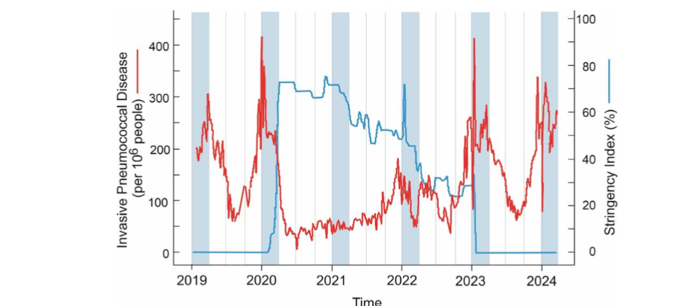
Figure 2. Seasonality of invasive pneumococcal disease and the impact of societal restrictions during COVID-19 restrictions in the USA. The incidence of invasive pneumococcal disease is shown as a red line. The stringency index (as a measure for societal restrictions) is indicated in blue. Time scale from week 1 of 2019 to week 10 of 2024. Winter periods (January-March) are indicated in light blue.
Seasonality of pneumococcal disease
Like many other respiratory infectious diseases, pneumococcal disease shows a clear seasonal pattern. The data in Figure 2 clearly shows a sharp increase of the frequency of invasive pneumococcal diseases during the winter period. Epidemiological factors associated with this pattern are lower temperatures and higher relative humidity during the winter, with people staying indoors more. Because, as discussed above, viral infections can predispose for pneumococcal infections, the seasonality of pneumococcal disease could closely follow that of influenza²⁶. It is also suggested that the immune system would function suboptimal during the winter, potentially related to vitamin D levels.
Apart from environmental conditions, also a number of public holidays with close transgenerational family contacts (Thanksgiving, Christmas, New Year) take place during the winter season on the Northern hemisphere. However, in the Southern hemisphere pneumococcal infections peak in June-August, not during the November-December holiday season²⁷.
Societal restrictions during COVID-19 had impact on seasonality and incidence of pneumococcal infections.
A unique opportunity to study the importance of physical contact for the transfer of S. pneumoniae arose when societal restrictions were implemented during the COVID-19 pandemic. These interventions included the closing of schools, wearing of face masks and social distancing, all of which reduce person-to-person contact²⁸. Specifically the closure of schools highlights, again, the importance of children in the spread of communicable diseases. Epidemiological observations made throughout the time in which societal restrictions were implemented showed a general worldwide decrease in both COVID-19. But for other communicable viral and bacterial respiratory diseases including S. pneumoniae those historical data (i.e. from the period before societal restrictions) were available and also showed a clear-cut decline in the incidence of respiratory infections²⁸–³³. Figure 2 shows that implementation of societal restrictions in the USA, aimed at controlling the spread of SARS-CoV-2, also reduced the incidence of invasive pneumococcal disease. Lifting of the restrictions lead to recurrence of the seasonal increase of IPD²⁸. A weighted least squares regression was used to test if the stringency index significantly predicted the number of IPD cases. The overall regression was statistically significant (R² = 0.4636, F(1, 207) = 178.9, p < .001. It should be noted that the direct effect of societal restrictions cannot be determined due to the multitude of factors that may contribute towards such a decline, it is likely that they played a major role in the reduction of IPD and other respiratory diseases nonetheless³⁴. Thus, these observation underline the pivotal role of close physical contact in the direct or indirect transmission of S. pneumoniae.
Impact of pneumococcal vaccination.
The best way to manage infectious diseases is through prevention. Vaccines are the most clinical and economically efficient method for preventing illness and death attributable to pneumococcal disease³⁵. The first available vaccines against pneumococcal pneumonia were based on capsular polysaccharides which trigger antibody production and opsonophagocytosis³⁶ (Table 1). The polysaccharide-based vaccine resulted in prevention of invasive pneumococcal infections in adult immunocompetent individuals but not in those that were immunocompromised³⁷. The PPV23 vaccine showed to be less effective in the elderly population³⁸,³⁹. Moreover, the response to the PPV23 in children younger than 2 years is poor and does not confer clinical protection against disease. The reason is that vaccines purely based on capsular polysaccharides do not induce an immune response in children and elderly population³⁶. The vaccine’s poor effectiveness in children is attributed to the antigenic nature of the polysaccharide antigen. PS antigens are classified as type 2 T-cell independent (TI-2) antigens, which stimulate mature B cells without requiring assistance from T cells⁴⁰. In newborns, B cells typically do not respond to most PS antigens, and responsiveness develops gradually during the early years of life⁴¹. Furthermore, TI-2 antigens fail to induce immunologic memory. Anti-PS antibodies generated by the vaccine exhibit low avidity, and limited isotype switching, even after repeated immunizations⁴².
Table 1. Composition of pneumococcal polysaccharide and pneumococcal conjugate vaccines
| Vaccine | Year of release | Pneumococcal serotypes included |
|---|---|---|
| PPV23 | 1983 | 1, 2, 3, 4, 5, 6B, 7F, 8, 9N, 9V, 10A, 11A, 12F, 14, 15B, 17F, 18C, 19A, 19F, 20, 22F, 23F, 33F |
| PCV7 | 2000 | 4, 6B, 9V, 14, 18C, 19F, 23F |
| PCV10 | 2009 | PCV7 + 1, 5, 7F |
| PCV13 | 2009 | PCV10 + 3, 6A, 19A |
| PCV15 | 2021 | PCV13 + 22F, 33F |
| PCV20 | 2021 | PCV15 + 8, 10A, 11A, 12F,15B |
| PCV24 | 2024 | PCV20 + 2, 9N, 17F, 20B |
PPV = pneumococcal polysaccharide vaccine; PCV = pneumococcal conjugate vaccine. The numbers indicate the number of different serotypes included in the vaccine.
By coupling the polysaccharide molecule to a protein, the issue of low immunogenic response in children can be overcome. The principle of increasing the immunogenicity by conjugation of a polysaccharide with a protein was already discovered by Oswald Avery, almost 100 years ago⁴³. Conjugate vaccines result in a stronger immune response as polysaccharides are co-expressed with carrier proteins, allowing for their interaction with the major histocompatibility complex. Consequently, this facilitates the provision of cognate CD4⁺ T-cell help for the activation of polysaccharide-specific B-cells³⁶. The first conjugate vaccine was the seven-valent conjugate vaccine (PCV7). The PCV7 showed to be highly effective in reducing pneumococcal disease caused by vaccine-included serotypes in young children⁴⁴–⁴⁶ and in elderly population⁴⁷. Vaccination with PCV7 also resulted in a decrease in pneumococcal meningitis⁴⁸ and otitis media⁴⁴. Furthermore, PCV7 provided cross-protection for serotype 6A infection, but not for serotypes 6C and 6D cross-protection⁴⁹.
In 2009, PCV13 was released which contained the serotype 19A and 5 other additional serotypes, as well as those included in PCV7. PCV13 provided optimal and larger coverage against pneumococcal disease than PCV7 in children (Table 1)⁵⁰,⁵¹. PCV13 was also effective in pneumococcal disease prevention in the elderly population⁵². The development of PCVs incorporating more serotypes has continued and currently a 24-valent (PCV24) vaccine is in the stage of clinical testing in humans. The PCV24 showed to achieve higher serotype valency and enhanced immune response compared to the PCV20 in adults⁵³ as well as in toddlers⁵⁴. While PCV24 has a much broader serotype coverage, for parts of the world such as China the coverage of PCV24 would still only be 64%⁵⁵.
Transmission of S. pneumoniae from infants to elderly and impact of vaccination
The introduction of PCV10 and PCV15 in national childhood vaccination programs has reduced the incidence of mucosal pneumococcal as well as invasive pneumococcal disease (IPD). Childhood vaccination also had an indirect effect on pneumococcal disease in the elderly. Many studies have shown the connection between introduction of pediatric PCVs and the reduction of pneumococcal disease in the elderly population¹⁴,⁴⁷,⁵⁶–⁶⁰. This is explained by the fact, as discussed above, that children are generally the ecological niche of S. pneumoniae and thus an integral part of the transmission route of pneumococci to the elderly population⁴,²⁹. It should be noted that, in most of these studies, only cases of IPD were included. This means that mucosal pneumococcal diseases such as CAP would not be included when assessing the benefit for the elderly population from pediatric vaccination programs. However, a study by van Werkhoven et al. in 2016 showed that IPD data sufficiently represented CAP cases when it came to assessing the involvement of elderly⁶². Therefore, it can be assumed that studies that used IPD as a measure can be considered to be representative of other pneumococcal cases too, therefore increasing the representativeness of these studies when it comes to the prevalence of S. pneumoniae as a whole.
Serotype replacement and replacement disease
Many studies have shown a shift in nasopharyngeal colonization patterns of S. pneumoniae serotypes as a result of the introduction of PCVs within a population⁶³–⁷¹. All of these studies show a decrease in vaccine-type serotypes with a concomitant increase of non-vaccine serotypes which will occupy the newly available ecological niche. Such a change in colonization pattern is of importance because pneumococcal carriage precedes disease⁷¹, and thus may result in so-called “replacement disease”, pneumococcal disease caused by non-vaccine serotypes⁷². The pattern of recolonization can be of interest as it might be able to predict which pneumococcal serotypes could cause future replacement disease, and thus could be candidate serotypes to be included in next generation conjugate vaccines.
Unfortunately, the replacement serotypes are not necessarily the ones originally “next in line” when compared to serotype prevalence pre-vaccination but rather increase in prevalence based on other factors⁶⁴,⁶⁵,⁷⁰. These could include niche preference, antibiotic susceptibility and others²⁹.
Interesting is that, after the introduction of PCV7, serotype 19A increased in prevalence within all age groups worldwide⁴⁷,⁷³,⁷⁴. Serotype 19A became one of the most predominant serotypes for IPD in children and elderly adults⁷³. The replacement disease of serotype 19A was remarkable because serotype 19F was included in PCV7. This is in contrast with the concept of “cross-protection” which states that serotype-specific antibodies can offer protection against serotypes within the same serogroup, although the efficacy of these cross-reacting antibodies may differ⁴¹. However, at the same time, a decrease in serotype 6A was observed following introduction of PCV7 (which included 6B)⁶⁹. These observations highlight the existence of differential cross-protection between serogroups, with some serotypes offering more protection to other serotypes within the serogroup they belong to than other serotypes might. Cross-protection, or the lack thereof, and replacement disease therefore are still aspects of the “what do you get” and “how do you get” questions on transmission of pneumococcal pneumonia that still are not fully answered.
Conclusions
So what do you get with a 24-valent PCV? You will get immunity against the 24 most prevalent pneumococcal serotypes. This will provide protection against the prevalent serotypes in many parts of the world, not just Northern America and Western Europe, but probably still incomplete in (parts of) Asia. Vaccination will also prevent colonization of the upper respiratory tract with these 24 vaccine serotypes. The indirect effect of reduced colonization of young children is that transmission to the vulnerable elderly population will be reduced as well. In young, vaccinated, children, the ecological niches of the upper respiratory tract can be (and will be) repopulated with representatives of the 76 non-vaccine pneumococcal serotypes. Which serotypes that will be cannot be predicted. Neither can it be predicted whether replacement colonization would lead to replacement disease (as was the case with 19A following introduction of PCV7) in the young, and potentially also the elderly population. Close monitoring of pneumococcal carriage and disease therefore remains warranted.
Conflict of Interest Statement:
The authors have no conflicts of interest to declare.
Funding Statement:
The authors received no external funding for the work discussed in the manuscript.
Acknowledgements:
None.
References
1. Rijkers GT, Rodriguez Gomez M. Rocking pneumonia. Pneumonia (Nathan). 2017;9:18. doi:10.1186/s41479-017-0043-0
2. Wyllie AL, Chu ML, Schellens MH, et al. Streptococcus pneumoniae in saliva of Dutch primary school children. PLoS One. 2014;9(7):e 102045. doi:10.1371/journal.pone.0102045
3. Gundel M, Okura. Untersuchungen über das gleichzeitige Vorkommen mehrerer Pneumokokkentypen bei Gesunden und ihre Bedeutung für die Epidemiologie [in German]. Zeitschrift für Hyg und Infekt 1933;114: 678–704.
4. Althouse BM, Hammitt LL, Grant L, et al. Identifying transmission routes of Streptococcus pneumoniae and sources of acquisitions in high transmission communities. Epidemiol Infect. 2017; 145(13):2750-2758. doi:10.1017/S095026881700125X
5. Gray BM, Converse GM 3rd, Dillon HC Jr. Epidemiologic studies of Streptococcus pneumoniae in infants: acquisition, carriage, and infection during the first 24 months of life. J Infect Dis. 1980;142(6):923-933. doi:10.1093/infdis/142.6.923
6. Leino T, Auranen K, Jokinen J, Leinonen M, Tervonen P, Takala AK. Pneumococcal carriage in children during their first two years: important role of family exposure. Pediatr Infect Dis J. 2001;20(11) :1022-1027. doi:10.1097/00006454-200111000-00004
7. Syrjänen RK, Kilpi TM, Kaijalainen TH, Herva EE, Takala AK. Nasopharyngeal carriage of Streptococcus pneumoniae in Finnish children younger than 2 years old. J Infect Dis. 2001;184(4):451-459. doi: 10.1086/322048
8. Labout JA, Duijts L, Arends LR, et al. Factors associated with pneumococcal carriage in healthy Dutch infants: the generation R study. J Pediatr. 2008;153(6):771-776. doi:10.1016/j.jpeds.2008.05.061
9. Gratten M, Gratten H, Poli A, Carrad E, Raymer M, Koki G. Colonisation of Haemophilus influenzae and Streptococcus pneumoniae in the upper respiratory tract of neonates in Papua New Guinea: primary acquisition, duration of carriage, and relationship to carriage in mothers. Biol Neonate. 1986;50(2):114-120. doi:10.1159/000242576
10. Turner P, Turner C, Jankhot A, et al. A longitudinal study of Streptococcus pneumoniae carriage in a cohort of infants and their mothers on the Thailand-Myanmar border. PLoS One. 2012;7 (5):e38271. doi:10.1371/journal.pone.0038271
11. Coles CL, Kanungo R, Rahmathullah L, et al. Pneumococcal nasopharyngeal colonization in young South Indian infants. Pediatr Infect Dis J. 2001;20(3):289-295. doi:10.1097/00006454-20010 3000-00014
12. Kwambana BA, Barer MR, Bottomley C, Adegbola RA, Antonio M. Early acquisition and high nasopharyngeal co-colonisation by Streptococcus pneumoniae and three respiratory pathogens amongst Gambian new-borns and infants. BMC Infect Dis. 2011;11:175. doi:10.1186/ 1471-2334-11-175
13. Hill PC, Cheung YB, Akisanya A, et al. Nasopharyngeal carriage of Streptococcus pneumoniae in Gambian infants: a longitudinal study. Clin Infect Dis. 2008;46(6):807-814. doi:10. 1086/528688
14. Tigoi CC, Gatakaa H, Karani A, et al. Rates of acquisition of pneumococcal colonization and transmission probabilities, by serotype, among newborn infants in Kilifi District, Kenya. Clin Infect Dis. 2012;55(2):180-188. doi:10.1093/cid/cis371
15. Dube FS, Ramjith J, Gardner-Lubbe S, et al. Longitudinal characterization of nasopharyngeal colonization with Streptococcus pneumoniae in a South African birth cohort post 13-valent pneumococcal conjugate vaccine implementation. Sci Rep. 2018;8(1):12497. doi:10.1038/s41598-018 -30345-5
16. Otsuka T, Chang B, Shirai T, et al. Individual risk factors associated with nasopharyngeal colonization with Streptococcus pneumoniae and Haemophilus influenzae: a Japanese birth cohort study. Pediatr Infect Dis J. 2013;32(7):709-714. doi:10.1097/INF.0b013e31828701ea
17. Koliou MG, Andreou K, Lamnisos D, et al. Risk factors for carriage of Streptococcus pneumoniae in children. BMC Pediatr. 2018;18(1):144. Published 2018 Apr 26. doi:10.1186/s12887-018-1119-6
18. Weiser JN, Ferreira DM, Paton JC. Streptococcus pneumoniae: transmission, colonization and invasion. Nat Rev Microbiol. 2018;16(6):355-367. doi:10.1038/s41579-018-0001-8
19. Morimura A, Hamaguchi S, Akeda Y, Tomono K. Mechanisms Underlying Pneumococcal Transmission and Factors Influencing Host-Pneumococcus Interaction: A Review. Front Cell Infect Microbiol. 2021;11:639450. doi:10.3389/fci mb.2021.639450
20. Ganaie F, Saad JS, McGee L, et al. A New Pneumococcal Capsule Type, 10D, is the 100th Serotype and Has a Large cps Fragment from an Oral Streptococcus. mBio. 2020;11(3):e00937-20. doi:10.1128/mBio.00937-20
21. Bogaert D, Sluijter M, Toom NL, et al. Dynamics of pneumococcal colonization in healthy Dutch children. Microbiology (Reading). 2006;152 (Pt 2):377-385. doi:10.1099/mic.0.28394-0
22. Weinberger DM, Dagan R, Givon-Lavi N, Regev-Yochay G, Malley R, Lipsitch M. Epidemiologic evidence for serotype-specific acquired immunity to pneumococcal carriage. J Infect Dis. 2008;19 7(11):1511-1518. doi:10.1086/587941
23. Ozdemir B, Beyazova U, Camurdan AD, Sultan N, Ozkan S, Sahin F. Nasopharyngeal carriage of Streptococcus pneumoniae in healthy Turkish infants. J Infect. 2008;56(5):332-339. doi:10.1016/ j.jinf.2008.02.010
24. Saha SK, Hossain B, Islam M, et al. Epidemiology of Invasive Pneumococcal Disease in Bangladeshi Children Before Introduction of Pneumococcal Conjugate Vaccine. Pediatr Infect Dis J. 2016;35(6):655-661. doi:10.1097/INF.00000 00000001037
25. Sender V, Hentrich K, Henriques-Normark B. Virus-Induced Changes of the Respiratory Tract Environment Promote Secondary Infections With Streptococcus pneumoniae. Front Cell Infect Microbiol. 2021;11:643326. doi:10.3389/fcimb.20 21.643326
26. Rijkers G, Croon S, Nguyen TA. Rocking Pneumonia and the Boogie Woogie Flu. Eur Medical J. 2019; 48–54. doi.org/10.33590/emj/10311819
27. Silva MEP, Oliveira JR, Carvalho AG, et al. Colonization by Streptococcus pneumoniae among children in Porto Velho, Rondônia, Western Brazilian Amazon. Braz J Biol. 2022;82:e260617. doi:10.1590/1519-6984.260617
28. Chow EJ, Uyeki TM, Chu HY. The effects of the COVID-19 pandemic on community respiratory virus activity. Nat Rev Microbiol. 2023;21(3):195-210. doi:10.1038/s41579-022-00807-9
29. Brueggemann AB, Jansen van Rensburg MJ, Shaw D, et al. Changes in the incidence of invasive disease due to Streptococcus pneumoniae, Haemophilus influenzae, and Neisseria meningitidis during the COVID-19 pandemic in 26 countries and territories in the Invasive Respiratory Infection Surveillance Initiative: a prospective analysis of surveillance data. Lancet Digit Health. 2021;3(6):e360-e370. doi:10.1016/S2589-7500(21) 00077-7
30. Principi N, Autore G, Ramundo G, Esposito S. Epidemiology of Respiratory Infections during the COVID-19 Pandemic. Viruses. 2023;15(5). doi:10.3 390/v15051160
31. Achangwa C, Park H, Ryu S, Lee MS. Collateral Impact of Public Health and Social Measures on Respiratory Virus Activity during the COVID-19 Pandemic 2020–2021. Viruses. 2022;14(5). doi:10. 3390/v14051071
32. Tanislav C, Kostev K. Fewer non-COVID-19 respiratory tract infections and gastrointestinal infections during the COVID-19 pandemic. J Med Virol. 2022;94(1):298-302. Doi:10.1002/jmv.27321
33. RIVM. Epidemiological Impact and Effectiveness of COVID-19 Measures.; 2024. https://open.overheid.nl/documenten/2c1c4e77-1b0b-4808-9098-2b47b331369d/file (assessed June 26, 2024)
34. Feldman C, Anderson R. Recent advances in the epidemiology and prevention of Streptococcus pneumoniae infections. F1000Res. 2020;9. doi:10. 12688/f1000research.22341.1
35. Briles DE, Paton JC, Mukerji R, Swiatlo E, Crain MJ. Pneumococcal Vaccines. Microbiol Spectr. 2019;7(6):10.1128/microbiolspec.gpp3-0028-2018. doi:10.1128/microbiolspec.GPP3-0028-2018
36. Berical AC, Harris D, Dela Cruz CS, Possick JD. Pneumococcal Vaccination Strategies. An Update and Perspective. Ann Am Thorac Soc. 2016;13(6): 933-944. doi:10.1513/AnnalsATS.201511-778FR
37. Shapiro ED, Berg AT, Austrian R, et al. The protective efficacy of polyvalent pneumococcal polysaccharide vaccine. N Engl J Med. 1991;325(2 1):1453-1460. doi:10.1056/NEJM199111213252101
38. Ortqvist A, Hedlund J, Burman LA, et al. Randomised trial of 23-valent pneumococcal capsular polysaccharide vaccine in prevention of pneumonia in middle-aged and elderly people. Swedish Pneumococcal Vaccination Study Group. Lancet. 1998;351(9100):399-403. doi:10.1016/s01 40-6736(97)07358-3
39. Honkanen PO, Keistinen T, Miettinen L, et al. Incremental effectiveness of pneumococcal vaccine on simultaneously administered influenza vaccine in preventing pneumonia and pneumococcal pneumonia among persons aged 65 years or older. Vaccine. 1999;17(20-21):2493-2500. doi:10.1016/s 0264-410x(99)00069-9
40. Rijkers GT, Mosier DE. Pneumococcal polysaccharides induce antibody formation by human B lymphocytes in vitro. J Immunol. 1985;135(1):1-4.
41. Griffioen AW, Sanders L, Rijkers GT, Zegers BJ. Cell biology of B lymphocyte activation by polysaccharides. J Infect Dis. 1992;165 Suppl 1:S71-S73. doi:10.1093/infdis/165-supplement_1-s71
42. Käyhty H, Eskola J. New vaccines for the prevention of pneumococcal infections. Emerg Infect Dis. 1996;2(4):289-298. doi:10.3201/eid0204 .960404
43. Avery OT, Goebel WF, Babers FH. Chemo-immunological studies on conjugated carbohydrate-proteins: VII. Immunological specificity of antigens prepared by combining alpha- and beta-glucosides of glucose with proteins. J Exp Med. 1932 Apr 30;55(5):769-80. doi: 10.1084/jem. 55.5.769
44. Black S, Shinefield H, Fireman B, et al. Efficacy, safety and immunogenicity of heptavalent pneumococcal conjugate vaccine in children. Northern California Kaiser Permanente Vaccine Study Center Group. Pediatr Infect Dis J. 2000;19(3 ):187-195. doi:10.1097/00006454-200003000-00003
45. Black SB, Shinefield HR, Ling S, et al. Effectiveness of heptavalent pneumococcal conjugate vaccine in children younger than five years of age for prevention of pneumonia. Pediatr Infect Dis J. 2002;21(9):810-815. doi:10.1097/000 06454-200209000-00005
46. Whitney CG, Pilishvili T, Farley MM, et al. Effectiveness of seven-valent pneumococcal conjugate vaccine against invasive pneumococcal disease: a matched case-control study. Lancet. 2006;368(9546):1495-1502. doi:10.1016/S0140-67 36(06)69637-2
47. Pilishvili T, Lexau C, Farley MM, et al. Sustained reductions in invasive pneumococcal disease in the era of conjugate vaccine. J Infect Dis. 2010;201(1):32-41. doi:10.1086/648593
48. Hsu HE, Shutt KA, Moore MR, et al. Effect of pneumococcal conjugate vaccine on pneumococcal meningitis. N Engl J Med. 2009;360 (3):244-256. doi:10.1056/NEJMoa0800836
49. McEllistrem MC, Nahm MH. Novel pneumococcal serotypes 6C and 6D: anomaly or harbinger. Clin Infect Dis. 2012;55(10):1379-1386. doi:10.1093/cid/cis691 World Health Organization. Pneumococcal conjugate vaccines in infants and children under 5 years of age: WHO position paper. Weekly epidemiological record 2019; 8, 94: 85–104
50. Esposito S, Principi N. Impacts of the 13-Valent Pneumococcal Conjugate Vaccine in Children. J Immunol Res. 2015;2015:591580. doi:10.1155/201 5/591580
51. Bonten MJ, Huijts SM, Bolkenbaas M, et al. Polysaccharide conjugate vaccine against pneumococcal pneumonia in adults. N Engl J Med. 2015;372(12):1114-1125. doi:10.1056/NEJMoa1408544
52. Wassil J, Sisti M, Fairman J, et al. Evaluating the safety, tolerability, and immunogenicity of a 24-valent pneumococcal conjugate vaccine (VAX-24) in healthy adults aged 18 to 64 years: a phase 1/2, double-masked, dose-finding, active-controlled, randomised clinical trial. Lancet Infect Dis. 2024;24 (3):308-318. doi:10.1016/S1473-3099(23)00572-8
53. Borys D, Rupp R, Smulders R, et al. Safety, tolerability and immunogenicity of a novel 24-valent pneumococcal vaccine in toddlers: A phase 1 randomized controlled trial. Vaccine. 2024;42(10) :2560-2571. doi:10.1016/j.vaccine.2024.02.001
54. Miao C, Yan Z, Chen C, et al. Serotype, antibiotic susceptibility and whole-genome characterizatio of Streptococcus pneumoniae in all age groups living in Southwest China during 2018-2022. Front Microbiol. 2024;15:1342839. doi:10.3 389/fmicb.2024.1342839
55. Dirmesropian S, Wood JG, MacIntyre CR, Newall AT. A review of economic evaluations of 13-valent pneumococcal conjugate vaccine (PCV13) in adults and the elderly. Hum Vaccin Immunother. 2015;11(4):818-825. doi:10.1080/21645515.2015.1011954
56. Lexau CA, Lynfield R, Danila R, et al. Changing epidemiology of invasive pneumococcal disease among older adults in the era of pediatric pneumococcal conjugate vaccine. JAMA. 2005;29 4(16):2043-2051. doi:10.1001/jama.294.16.2043
57. Vila-Córcoles A. Advances in pneumococcal vaccines: what are the advantages for the elderly?. Drugs Aging. 2007;24(10):791-800. doi:10.2165/0 0002512-200724100-00001
58. Tsaban G, Ben-Shimol S. Indirect (herd) protection, following pneumococcal conjugated vaccines introduction: A systematic review of the literature. Vaccine. 2017;35(22):2882-2891. doi:10. 1016/j.vaccine.2017.04.032
59. Sando E, Suzuki M, Furumoto A, et al. Impact of the pediatric 13-valent pneumococcal conjugate vaccine on serotype distribution and clinical characteristics of pneumococcal pneumonia in adults: The Japan Pneumococcal Vaccine Effectiveness Study (J-PAVE) Vaccine. 2019;37(20) :2687-2693. doi:10.1016/j.vaccine.2019.04.009
60. Hanquet G, Krizova P, Valentiner-Branth P, et al. Effect of childhood pneumococcal conjugate vaccination on invasive disease in older adults of 10 European countries: implications for adult vaccination. Thorax. 2019;74(5):473-482. doi:10.11 36/thoraxjnl-2018-211767
61. van Werkhoven CH, Hollingsworth RC, Huijts SM, et al. Pneumococcal conjugate vaccine herd effects on non-invasive pneumococcal pneumonia in elderly. Vaccine. 2016;34(28):3275-3282. doi:10. 1016/j.vaccine.2016.05.002
62. Desmet S, Wouters I, Heirstraeten L Van, et al. In-depth analysis of pneumococcal serotypes in Belgian children (2015–2018): Diversity, invasive disease potential, and antimicrobial susceptibility in carriage and disease. Vaccine. 2021;39(2):372-379. Doi:10.1016/j.vaccine.2020.11.044
63. Keck JW, Wenger JD, Bruden DL, et al. PCV7-induced changes in pneumococcal carriage and invasive disease burden in Alaskan children. Vaccine. 2014;32(48):6478-6484. doi:10.1016/j.vac cine.2014.09.037
64. Roca A, Hill PC, Townend J, et al. Effects of community-wide vaccination with PCV-7 on pneumococcal nasopharyngeal carriage in the Gambia: A cluster-randomized trial. PLoS Med. 2011;8(10). doi:10.1371/journal.pmed.1001107
65. Gonzales BE, Mercado EH, Castillo-Tokumori F, et al. Pneumococcal serotypes and antibiotic resistance in healthy carriage children after introduction of PCV13 in Lima, Peru. Vaccine. 2023; 41(28):4106-4113. doi:10.1016/j.vaccine.2023.05.042
66. Collins DA, Hoskins A, Snelling T, et al. Predictors of pneumococcal carriage and the effect of the 13-valent pneumococcal conjugate vaccination in the Western Australian Aboriginal population. Pneumonia. 2017;9(1). doi:10.1186/s4 1479-017-0038-x
67. Bruce MG, Singleton R, Bulkow L, et al. Impact of the 13-valent pneumococcal conjugate vaccine (pcv13) on invasive pneumococcal disease and carriage in Alaska. Vaccine. 2015;33(38):4813-4819. doi:10.1016/j.vaccine.2015.07.080
68. Roca A, Dione MM, Bojang A, et al. Nasopharyngeal Carriage of Pneumococci Four Years after Community-Wide Vaccination with PCV-7 in The Gambia: Long-Term Evaluation of a Cluster Randomized Trial. PLoS One. 2013;8(9). doi:10.1371/journal.pone.0072198
69. Roca A, Bojang A, Bottomley C, et al. Effect on nasopharyngeal pneumococcal carriage of replacing PCV7 with PCV13 in the Expanded Programme of Immunization in The Gambia. Vaccine. 2015;33(51):7144-7151. doi:10.1016/j.vac cine.2015.11.012
70. Gladstone RA, Jefferies JM, Tocheva AS, et al. Five winters of pneumococcal serotype replacement in UK carriage following PCV introduction. Vaccine. 2015;33(17):2015-2021. doi:10.1016/j.vaccine.201 5.03.012
71. Weinberger DM, Malley R, Lipsitch M. Serotype replacement in disease after pneumococcal vaccination. Lancet. 2011;378(9807):1962-1973. doi:10.1016/S0140-6736(10)62225-8
72. Hicks LA, Harrison LH, Flannery B, et al. Incidence of pneumococcal disease due to non-pneumococcal conjugate vaccine (PCV7) serotypes in the United States during the era of widespread PCV7 vaccination, 1998-2004. J Infect Dis. 2007;196(9):1346-1354. doi:10.1086/521626
73. Singleton RJ, Hennessy TW, Bulkow LR, et al. Invasive pneumococcal disease caused by nonvaccine serotypes among Alaska native children with high levels of 7-valent pneumococcal conjugate vaccine coverage. JAMA. 2007;297(16) :1784-1792. doi:10.1001/jama.297.16.1784