


AI Insights into Neurodegenerative Disease Diagnosis
From Pixels to Prognosis: AI-Driven Insights into Neurodegenerative Diseases
OPEN ACCESS
PUBLISHED: June 30, 2024
CITATION: Ganjizadeh, A., et al., 2024. From Pixels to Prognosis: AI-Driven Insights into Neurodegenerative Diseases. Medical Research Archives, [online] 12(6).
https://doi.org/10.18103/mra.v12i6.5512
COPYRIGHT: © 2024 European Society of Medicine. This is an open-access article distributed under the terms of the Creative Commons Attribution License, which permits unrestricted use, distribution, and reproduction in any medium, provided the original author and source are credited.
DOI https://doi.org/10.18103/mra.v12i6.5512
ISSN 2375-1924
Introduction
Neurodegenerative diseases, a group of debilitating conditions characterized by progressive degeneration and death of neurons in the brain and spinal cord, are increasingly prevalent in the aging population worldwide¹. The need for early and accurate diagnosis of neurodegenerative diseases is growing, given the emergence of potential disease-modifying treatments and ongoing clinical trials²–⁵. This alarming trend highlights the important challenge clinicians face in discerning disease states, as they differentiate between early symptoms based solely on clinical information or those discovered by neuropsychological testing, which can be subjective and prone to error. The consequences of misdiagnosis can be severe, leading to inappropriate treatment regimens, delayed access to care, and adverse impacts on patients’ quality of life. Delays in diagnosis often result in prolonged distress, increased healthcare expenses, and missed opportunities for research⁵–⁹.
Although a large number of neurodegenerative diseases have been defined, we target the ones with the highest prevalence and representative of the neurodegenerative spectrum¹⁰. Further, while Alzheimer’s disease (AD) is the most common neurodegenerative disorder globally, several previous reviews have extensively covered the applications of artificial intelligence (AI) in its diagnosis and analysis, so this review will not repeat that. The reader is referred to: Täüjan et al.¹, Yao et al.¹¹, and Borchert et al.¹².
Obtaining an accurate and definitive diagnosis for neurodegenerative diseases is a complex process, with brain imaging playing a crucial role. The analysis of these complex brain images, however, can be subjective and prone to variability among clinicians¹³,¹⁴. This highlights the need for advanced techniques, such as machine learning and deep learning, to enhance diagnostic accuracy and consistency in the interpretation of structural brain MRI data.
Among neurodegenerative Parkinsonian disorders, Progressive Supranuclear Palsy (PSP), Multiple System Atrophy-Parkinsonian subtype (MSA-P), and Dementia with Lewy Bodies (DLB) follow Parkinson’s Disease (PD) in prevalence. Despite advances in medical technology, up to 20% of patients with neurodegenerative diseases may receive an incorrect initial diagnosis¹⁵. Their early diagnosis is complicated by shared clinical and neuropathological traits⁷,¹⁶,¹⁷.
While single-photon emission computed tomography (SPECT) offers differential diagnostic advantages⁶,⁹,¹⁸–²⁰, Magnetic Resonance Imaging (MRI), particularly through longitudinal studies, has superior performance and is preferred due to lower cost and greater accessibility²¹–²³. MRI can also help to identify other disease processes that might initially be confusing. With its superior ability to render high-resolution images of brain anatomical structures, MRI provides precise differentiation between gray and white matter, enabling the detection of subtle changes associated with these conditions²⁴–²⁶. As the disease progresses, MRI can clearly depict the widening and deepening of cerebral sulci and ventricles, which are hallmarks of neurodegeneration²⁷. The Magnetic Resonance Parkinsonism Index (MRPI 2.0) aids in differential diagnosis¹⁸,¹⁹,²⁸–³⁰. Despite the advantages offered by MRI, the interpretation of these complex brain images can be subjective, prone to variability among clinicians¹⁴, and inaccuracies can arise from asymmetry in brainstem atrophy¹⁷,³¹–³⁴. While MRI findings like the ‘hummingbird’, ‘Mickey mouse’, and ‘hot cross-bun’ signs can aid early diagnosis when present, similar early-stage symptoms pose challenges in accurately distinguishing these conditions⁷,³¹,³²,³⁵–³⁷.
This limitation underscores the need for advanced techniques, such as Artificial Intelligence (AI) driven analysis, to enhance diagnostic accuracy and consistency. By leveraging machine learning and deep learning algorithms, researchers aim to develop automated systems capable of analyzing structural brain MRI data, extracting relevant features, and providing objective and reliable diagnostic support³⁴,³⁸–⁴⁰.
In this narrative review, we will explore the cutting-edge applications of machine learning and deep learning techniques in the analysis of brain imaging data for the diagnosis and evaluation of Parkinson’s Disease, Multiple System Atrophy, Dementia with Lewy Bodies, and Progressive Supranuclear Palsy. We will examine the latest research and advancements in these AI-driven approaches, discussing their potential to revolutionize the field of neurodegenerative disease management. By exploring the current state of the art and future directions, we aim to highlight how these sophisticated techniques can enhance diagnostic accuracy, enable early intervention, and ultimately improve patient outcomes. Furthermore, we will address the challenges and limitations associated with implementing these AI methodologies in clinical practice and discuss potential solutions to overcome these obstacles.
Parkinson’s Disease
Deep learning is increasingly recognized for its influence across multiple aspects of Parkinson’s disease (PD) management, encompassing diagnosis, evaluation of neurological impairment, forecasting disease progression, and the detection of subtle motor features.
Recent studies harnessed deep learning approaches to improve the diagnosis and future outlook of PD, utilizing an array of medical imaging techniques such as MRI and positron emission tomography (PET)⁴¹,⁴². Wang et al. developed a PD diagnostic model using T1-weighted MRI images, achieving area under the receiver operating characteristic curve (AUC) scores of 0.901 and 0.845 in internal and external testing cohorts, respectively. Dünnwald et al. employed a 3D-Unet model for the precise localization and segmentation of the locus coeruleus in aging and Parkinson’s disease cohorts, utilizing neuromelanin-sensitive MRI to demonstrate the model’s effectiveness⁴³. Furthermore, Wu et al. demonstrated that a deep learning algorithm applied to 18F-FDG PET images achieved a diagnostic accuracy of 98.6%⁴⁴.
Deep learning has also been utilized for the prediction of PD progression, prognosis in PD patients, and assessment of severity of neurological dysfunction in PD⁴⁵–⁴⁷. As shown by Kaur et al., predicting gait disturbances and classifying tremor types in PD patients have also been advanced by DL methodologies. This study is the first attempt to apply and demonstrate the potential of DL within a multi-view digital camera-based gait analysis framework for neurological gait dysfunction prediction. They presented a comprehensive quantitative comparison of 16 diverse traditional machine and deep learning algorithms. The results indicated that multi-scale residual neural networks achieved the highest levels of accuracy and AUC, at 78.1% and 0.87, respectively⁴⁸.
For instance, Ahn et al. developed an algorithm using fundus photography and deep learning to predict the Hoehn and Yahr (H-Y) scale and Unified Parkinson’s Disease Rating Scale part III (UPDRS-III) score among patients with PD⁴⁹. Leung et al. employed deep learning for the prognosis of PD, showcasing its ability to forecast the course of the disease and contribute to the management of patients⁴⁵.
Collectively, these investigations illuminate the significant role of deep learning in delivering precise and efficient diagnostic and classification systems for PD symptomatology.
Multiple system atrophy
Multiple system atrophy (MSA) is a severe rapid-onset neurodegenerative disease resulting in atrophy of several midbrain structures (Figure 1). Its prevalence is low, and as a consequence, the number of studies using computational approaches for its characterization is limited³²,⁵⁰.
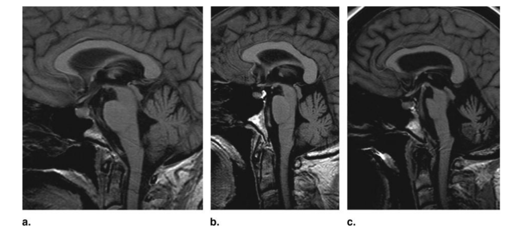
Figure 1. Pontine atrophy, cerebellar atrophy, and enlargement of the fourth ventricle in probable multiple systems atrophy (MSA) on sagittal T1-weighted images. (a) Absent, (b) moderate, and (c) severe⁵¹.
A major challenge in diagnosing MSA is differentiating it from other Parkinsonian syndromes, such as Parkinson’s disease (PD) and progressive supranuclear palsy (PSP). **Duchesne et al.**⁵¹ reported that the initial error rate for clinical diagnosis of idiopathic Parkinson’s disease (IPD) against other Parkinson Plus Syndromes (PPS) could reach up to 35%. To address this issue, they developed an automated technique based on structural, cross-sectional T1-weighted (T1w) magnetic resonance imaging (MRI) to perform differential classification of IPD patients versus those with either PSP or MSA. The system achieved 91% accuracy, 88% specificity, and 93% sensitivity, demonstrating the potential of AI-based approaches in assisting the differential diagnosis of IPD versus PSP and MSA.
Kanatani et al.¹³ examined the validity of MSA diagnosis using a pointwise linear model (deep learning-based method) and identified features associated with disease differentiation. The study used a large dataset of 3,377 registered MSA cases. It estimated the diagnostic probabilities of striatonigral degeneration (SND) to be 0.852 ± 0.107, Shy-Drager syndrome (SDS) to be 0.650 ± 0.235, and olivopontocerebellar atrophy (OPCA) to be 0.858 ± 0.270. The model also identified autonomic dysfunction, respiratory failure, dysphagia, and brain-stem atrophy as characteristic features of SDS, SND, and OPCA, respectively.
Tsuda et al.⁵² aimed to differentiate the Parkinson’s variant of MSA (MSA-P) from PD in the early stages using neural network (NN) analyses before the ‘hot cross-bun’ and putaminal rim imaging features of MSA appeared. The analysis involved data from voxel-based morphometry (VBM) and magnetic resonance spectroscopy (MRS). The study found that MSA-P patients showed atrophy in various brain regions compared to PD patients, and NNs contributed to the clinical diagnosis when using both VBM and MRS data.
Rau et al.⁵³ determined the performance of a Deep Neural Patchwork (DNP) in comparison to the established segmentation algorithms for delineating the putamen in MSA, PD, and healthy controls. The DNP achieved a Dice coefficient of 0.96, significantly outperforming other methods. The DNP-based segmentation was also more capable of differentiating between MSA and PD than other algorithms.
Tan et al.⁵⁴ employed a deep learning approach to differentiate between PD and MSA based on MR and PET/CT images. The study used the nnU-Net network to segment the putamen and caudate nucleus regions in MRI images and convolutional neural networks (CNNs) for classification. The nnU-Net achieved a Dice score of 84.92% in the segmentation task, and the CNNs could distinguish PD from MSA with an accuracy of 89.71% and an AUC of 0.87.
Nemmi et al.⁵⁵ developed a fully data-driven analysis pipeline to discriminate between PD, MSA, and healthy controls (HC) using a MRI protocol that included sagittal 3D T1 magnetization prepared rapid gradient-echo (MPRAGE) and transverse T2 fluid-attenuated inversion recovery (T2-FLAIR). The pipeline combined several feature selection and reduction steps to obtain interpretable models with just a few discriminant features. The study achieved accuracies of 0.78, 0.94, and 0.88 for discriminating between PD and HC, MSA and HC, and PD and MSA, respectively. The results showed that indices derived from resting-state fMRI alone could discriminate between PD and HC, while mean diffusivity in the cerebellum and the putamen alone could discriminate between MSA and HC.
Jucaite et al.⁵⁶ assessed the performance of TSPO imaging as a diagnostic marker for MSA using [^11C]PBR28 binding to TSPO in a multicenter PET study. The study analyzed imaging data of 66 patients with MSA and 24 patients with PD and found a pattern of significantly increased regional glial TSPO binding in patients with MSA. Visual reading discriminated MSA from PD with 100% specificity and 83% sensitivity, while the machine learning approach improved sensitivity to 96%.
Abos et al.⁵⁷ investigated whether the strength of structural connectivity between subcortical structures, measured as the number of streamlines (NOS) derived from tractography, can be used to classify MSA and PD patients at the single-patient level. The study found reduced NOS in MSA compared with controls and PD in connections between the putamen, pallidum, ventral diencephalon, thalamus, and cerebellum. The classification procedure achieved an overall accuracy of 78%, with 71% of the MSA subjects and 86% of the PD patients correctly classified. The results suggest that structural connectivity derived from tractography has the potential to correctly distinguish between MSA and PD patients.
Coll et al.⁵⁸ CNNs to MRI data to predict the course of multiple sclerosis (MS) and identify key disease mechanisms leading to disability accumulation. The study used T1-weighted and T2-FLAIR brain MRI sequences from a cohort of patients prospectively followed after a first demyelinating attack. The CNN model achieved a mean accuracy of 79% and proved to be superior to the equivalent logistic regression model (77%). The attention-map analyses revealed the predominant role of the frontotemporal cortex and cerebellum for CNN decisions, suggesting that the mechanisms leading to disability accrual exceed the mere presence of brain lesions or atrophy.
Kim et al.² developed a fully automated volumetric diagnostic decision tree to facilitate early and accurate differential diagnosis of neurodegenerative movement disorders (NMDs), including MSA, PSP, and PD. The study used 3DT1 MRI from 171 NMD patients and 171 matched healthy subjects and produced decision trees employing substructure volumes and a novel volumetric pons-to-midbrain ratio (3D-PMR). The optimal tree separating NMD from healthy subjects achieved a sensitivity of 84%, specificity of 94%, accuracy of 84%, and kappa of 0.69 in cross-validation. The optimal tree restricted to NMD patients yielded sensitivities/specificities of 94/84% for MSA, 72/96% for PSP, and 73/92% for PD, with 79% accuracy and 0.62 kappa.
Tupe-Waghmare et al.⁵⁹ explored the feasibility of radiomics features extracted from T1-weighted MRI to differentiate PD from atypical parkinsonian syndromes (APS). The study computed radiomics features from T1 images of 65 patients with PD, 61 patients with APS (31: PSP and 30: MSA), and 75 healthy controls (HC). The PD vs. HC classifier illustrated an accuracy of 70%, while the PD vs. APS classifier demonstrated a superior test accuracy of 92%. Moreover, a 3-way PD/MSA/PSP classifier was performed with 96% accuracy.
Bu et al.⁶⁰ investigated the potential of radiomics with multiple parameters from conventional T1-weighted imaging (T1WI) and susceptibility-weighted imaging (SWI) in distinguishing between idiopathic Parkinson’s disease (PD) and multiple system atrophy (MSA). The study used 2,640 radiomic features extracted from both T1WI and SWI scans of 57 patients with PD, 74 with MSA, and 70 healthy control (HC) individuals. The light gradient boosting machine (LGBM) model trained by the features combination of T1WI and SWI exhibited the most outstanding differential performance in both the three-class classification task of MSA vs. PD vs. HC and the binary classification task of MSA vs. PD, with an accuracy of 0.814 and 0.854, and an AUC of 0.904 and 0.881, respectively.
Progressive Supranuclear Palsy
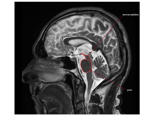
Progressive Supranuclear Palsy (PSP), a devastating neurodegenerative disorder, poses significant diagnostic challenges due to its overlapping clinical presentations with other Parkinsonian syndromes³⁶. Accurate and early diagnosis is crucial for initiating appropriate treatment strategies and improving patient outcomes. In recent years, the advent of artificial intelligence (AI) and machine learning techniques has opened new frontiers in addressing these diagnostic challenges, offering the potential for a more objective and reliable assessment of PSP⁷,¹⁶,¹⁷. While some cases demonstrate a common imaging appearance (Figure 2), not all do, resulting in a diagnostic challenge.
Figure 2: Hummingbird sign, a classic MRI presentation of PSP-RS²⁸ which reflects the atrophy particularly of the pons, resulting in the hummingbird shape.
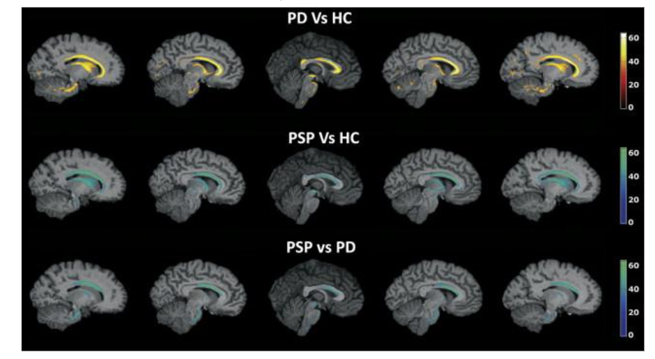
One of the pioneering studies in this field was conducted by Salvatore et al.⁶¹ (Figure 3), who employed a supervised machine learning algorithm to analyze morphological T1-weighted Magnetic Resonance Imaging (MRI) data from patients with Parkinson’s disease (PD), PSP, and healthy controls. By combining Principal Component Analysis for feature extraction and Support Vector Machines (SVMs) for classification, their algorithm achieved remarkable accuracy, sensitivity = 94.4%, and specificity = 91.3% in distinguishing individual cases of PD, PSP, and healthy controls. Notably, the critical brain regions influencing the classification between PD and PSP patients, such as the midbrain, pons, corpus callosum, and thalamus, are known to be heavily involved in the pathophysiological mechanisms of PSP.
Figure 3. Maps of voxel-based pattern distribution of brain structural differences (sagittal view, threshold = 60%). The importance of each voxel in the SVM classification is expressed according to the color scale. A, PD versus Controls; B, PSP versus controls; C, PSP versus PD; PD, Parkinson’s disease; PSP, Progressive Supranuclear Palsy⁶¹.
Building on this approach, Focke et al.⁶² leveraged voxel-based morphometry (VBM) and SVM analysis to enable individual classification of PSP cases from idiopathic Parkinson’s syndrome and healthy controls. Their SVM analysis allowed for reliable classification of individual PSP cases with up to 96.8% accuracy, 90% sensitivity, and 100% specificity. These results demonstrated the potential for translating VBM findings into clinically useful tools for differential diagnosis.
In another study, Singh et al.⁶³ proposed a novel synergetic paradigm integrating Kohonen self-organizing maps (KSOM) and least-squares support vector machines (LS-SVM) for individual-level clinical diagnosis of neurodegenerative diseases, including Parkinson’s disease (PD). Their unsupervised approach involved extracting features from preprocessed brain MRIs using KSOM, which were then fed into LS-SVM for subject classification. Remarkably, they achieved up to 99% classification accuracy for differential diagnosis of PD with 99.9% confidence interval, using 831 T1-weighted MRIs from the Parkinson’s Progression Markers Initiative (PPMI) database. This study highlighted the potential of unsupervised learning techniques, coupled with powerful classifiers, to serve as first-line diagnostic tools for early-stage PD and other neurodegenerative diseases.
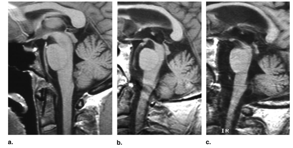
Duchesne et al.⁵¹ took a different approach, employing an automated technique based on structural MRI features in the hindbrain region. Their method achieved 91% accuracy, 88% specificity, and 93% sensitivity in differentiating PSP (Figure 4) and Multiple System Atrophy (MSA) from idiopathic PD. These findings highlighted the potential of quantitative parameters derived from MRI data to assist in the differential diagnosis of Parkinsonian syndromes, thereby reducing initial clinical error rates.
Figure 4. *Mesencephalon atrophy in probable progressive supranuclear palsy (PSP) on sagittal T1-weighted images. (a) Absent, (b) moderate, and (c) Severe.*⁵¹
Recognizing the limitations of relying solely on structural MRI data, researchers have explored the potential of other neuroimaging modalities, such as diffusion tensor imaging (DTI) and susceptibility-weighted imaging (SWI). Haller et al.⁶⁴ employed SVM-based pattern recognition of DTI data, accurately detecting patients with PD among those with suspected PD at an individual level, with accuracies up to 97%. Their subsequent study⁶⁵ using SVM analysis of SWI data demonstrated accurate discrimination of PD among patients with various forms of Parkinsonism despite the absence of visually detectable alterations.
In a more recent study, Illán-Gala et al.⁶⁶ conducted a comprehensive assessment of the diagnostic value of various MRI-based measures of cerebral atrophy in differentiating PSP, Corticobasal Degeneration (CBD), and other neurodegenerative diseases. Their findings demonstrated that a combination of cortical and subcortical atrophy measures achieved excellent diagnostic accuracy, even in participants without the canonical clinical presentations of PSP or CBD at the time of MRI acquisition. This study underscored the potential of structural MRI analysis to support the diagnosis of these tauopathies in diverse clinical scenarios.
Moving beyond structural imaging, Wu et al.⁶⁷ developed deep metabolic imaging indices based on deep learning analysis of 18F-FDG PET data from a large Parkinsonian patient database. Their approach exhibited robust performance in differentiating PD, MSA, and PSP, with high sensitivity and specificity rates, even when applied to an external cohort with different imaging acquisition protocols. This study highlighted the potential of AI-driven metabolic imaging analysis to provide accurate differential diagnosis of parkinsonism, addressing the overlap in clinical presentations among these conditions.
Recognizing the importance of neuropathological assessment in the diagnosis of PSP, Koga et al.⁶⁸ took a unique approach by developing a machine learning-based decision tree classifier for the differential diagnosis of PSP and CBD. By analyzing tau pathology scores in specific brain regions, their decision tree achieved remarkable accuracy in distinguishing PSP and CBD cases, simplifying the neuropathological differential diagnosis process. This study underscored the potential of AI-driven analysis to enhance diagnostic accuracy at the neuropathological level.
These studies collectively highlight the immense potential of AI and machine learning techniques in improving the diagnosis of PSP, a condition that has historically been challenging to diagnose accurately, particularly in its early stages. By leveraging various neuroimaging modalities and advanced computational methods, researchers are paving the way for more reliable and objective diagnostic tools, which could ultimately lead to earlier intervention, personalized treatment strategies, and improved patient outcomes⁶⁹,⁷⁰.
Dementia with Lewy bodies
Dementia with Lewy bodies (DLB) is a prominent neurodegenerative condition marked by the accumulation of Lewy bodies in the brain. Accounting for 25–45% of dementia cases, Lewy body pathology is frequently identified during autopsies, making it one of the leading causes of dementia⁷¹. While DLB presents challenges in diagnosis and management due to its overlap with other dementias and under-detection, efforts are being made to improve diagnostic rates⁷². Nevertheless, recent progress in machine learning and deep learning has been encouraging in improving the diagnosis and distinction of DLB from other dementia forms.
Several studies have highlighted the effectiveness of deep learning algorithms, especially support vector machines and deep learning models, in predicting the onset of AD from its early stages and in differentiating between various forms of dementia, including DLB, PD Dementia, and AD⁷³,⁷⁴. Several models employ imaging techniques such as fluorodeoxyglucose (FDG) PET and structural MRI to estimate discrepancies in brain age and detect metabolic patterns unique to DLB⁷⁵–⁷⁷. Furthermore, machine learning classifiers have been developed to distinguish among prevalent dementia syndromes using FDG PET scans, offering valuable support in differential diagnosis⁷⁴. Additionally, Lizuka et al. showcased the effectiveness of deep learning in pinpointing specific imaging markers, like the cingulate island sign, in DLB patients using PET images. This study employed a deep CNN for autonomous feature extraction, underscoring deep learning’s potential in image classification and recognition).
Furthermore, studies have explored using EEG and quantitative electroencephalogram (qEEG) in diagnosing and differentiating DLB from AD. EEG has been investigated for its role in providing insights into the clinical correlations of DLB and has shown the potential in helping predict the prognosis of the disease. Additionally, deep learning models based on EEG spectrograms have been utilized to identify patterns associated with rapid eye movement behavior disorder, a prodromal stage of α-synucleinopathies that can progress to PD, DLB, or other neurodegenerative diseases⁷⁹–⁸².
In addition to the techniques mentioned, deep learning integration has also been promising in identifying crucial microRNA signatures for various neurodegenerative diseases, including DLB⁸³. Furthermore, machine learning classifiers that analyze speech and language attributes have successfully differentiated between AD and DLB, as evidenced by the work of Yamada et al. Their study achieved impressive accuracy rates: 87.0% in distinguishing AD from HC, 93.2% for DLB versus CN, and 87.4% for AD compared to DLB, illustrating machine learning’s capacity to utilize non-imaging data for precise diagnosis⁸⁴.
Conclusion
In conclusion, the utilization of deep learning in neurodegenerative diseases has made significant progress in early diagnosis, disease subtype classification, and understanding disease mechanisms. As the field of deep learning continues to mature, its applications in neurodegenerative diseases are poised to transition from theoretical explorations to practical clinical tools. Some things that we expect will occur in this field include:
First, since most of the above studies utilized a single data type (e.g. T1-weighted MRI), more efforts should be made to combine imaging, -omics, and clinical data through deep learning to produce multimodal deep learning tools. This integration can create more comprehensive disease models, shedding light on the multifactorial nature of neurodegeneration. By combining CT, MRI, and PET scans with genomic and proteomic data, deep learning models can generate comprehensive analyses that may lead to better diagnosis and treatment plans. This integration could help identify early markers of disease progression or predict individual responses to treatment. Second, interpretability remains a bottleneck in the adoption of AI in healthcare. Research dedicated to unraveling the “black box” of neural networks will be essential.
Future research should focus on improving model interpretability, allowing clinicians to understand and trust AI-assisted tools. In addition, current models need to effectively estimate uncertainty in their predictions. Researchers can focus on how customized uncertainty metrics should be developed and how these uncertainty measures can be communicated to and utilized by healthcare professionals⁸⁵. Finally, with deep learning’s entry into the clinical realm, addressing data bias and ethical considerations will take center stage. Ensuring that AI systems serve diverse populations equitably will require large amounts of high-quality data for training and diligent research oversight. By delving into these areas, the field can move towards more transparent and reliable AI tools for diagnosing and treating neurodegenerative diseases, ultimately improving patient care and outcomes.
Abbreviations:
| Term | Abbreviation |
|---|---|
| Alzheimer’s disease | AD |
| Artificial Intelligence | AI |
| cognitively normal | CN |
| computed tomography | CT |
| convolutional neural networks | CNNs |
| Deep Neural Patchwork | DNP |
| Dementia with Lewy Bodies | DLB |
| diffusion tensor imaging | DTI |
| electroencephalogram | EEG |
| fluorodeoxyglucose | FDG |
| healthy controls | HC |
| Kohonen self-organizing maps | KSOM |
| least-squares support vector machines | LS-SVM |
| Magnetic Resonance Imaging | MRI |
| magnetic resonance spectroscopy | MRS |
| Multiple System Atrophy | MSA |
| Number of streamlines | NOS |
| olivopontocerebellar atrophy | OPCA |
| operating characteristic curve | AUC |
| Parkinson Plus Syndromes | PPS |
| Parkinson’s disease | PD |
| Parkinson’s variant of MSA | MSA-P |
| Positron emission tomography | PET |
| Progressive Supranuclear Palsy | PSP |
| Shy-Drager syndrome | SDS |
| Single-photon emission computed tomography | SPECT |
| Striatonigral degeneration | SND |
| T1-weighted imaging | T1WI |
| Voxel-based morphometry | VBM |
| Translocator Protein | TSPO |
Conflict of Interest:
None
Funding:
None
Acknowledgements:
None
References
1. Tăuţan AM, Ionescu B, Santarnecchi E. Artificial intelligence in neurodegenerative diseases: A review of available tools with a focus on machine learning techniques. Artif Intell Med. 2021;117:102081. doi:10.1016/j. artmed.2021.102081
2. Kim J, Young GS, Willett AS, et al. Toward More Accessible Fully Automated 3D Volumetric MRI Decision Trees for the Differential Diagnosis of Multiple System Atrophy, Related Disorders, and Age-Matched Healthy Subjects. Cerebellum Lond Engl. 2023;22(6): 1098-1108. doi:10.1007/s12311-022-01472-7
3. Höglinger GU, Huppertz HJ, Wagenpfeil S, et al. Tideglusib reduces progression of brain atrophy in progressive supranuclear palsy in a randomized trial. Mov Disord Off J Mov Disord Soc. 2014;29(4):479-487. doi:10.1002/ mds.25815
4. Brusa L, Ponzo V, Mastropasqua C, et al. Theta burst stimulation modulates cerebellar-cortical connectivity in patients with progressive supranuclear palsy. Brain Stimulat. 2014;7(1): 29-35. doi:10.1016/j.brs.2013.07.003
5. Persely A, Beszedics B, Paloczi K, et al. Analysis of Genetic and MRI Changes, Blood Markers, and Risk Factors in a Twin Pair Discordant of Progressive Supranuclear Palsy. Med Kaunas Lith. 2023;59(10). doi:10.3390/ medicina59101696
6. Wiblin L, Durcan R, Galna B, Lee M, Burn D. Clinical Milestones Preceding the Diagnosis of Multiple System Atrophy and Progressive Supranuclear Palsy: A Retrospective Cohort Study. J Mov Disord. 2019;12(3):177-183. doi:10.14802/jmd.19015
7. Calomino C, Quattrone A, Sarica A, et al. Neuroimaging correlates of postural instability in Progressive Supranuclear Palsy. Parkinsonism Relat Disord. 2023;113:105768. doi:10.1016/j.parkreldis.2023.105768
8. Krismer F, Seppi K, Wenning GK, et al. Abnormalities on structural MRI associate with faster disease progression in multiple system atrophy. Parkinsonism Relat Disord. 2019;58: 23-27. doi:10.1016/j.parkreldis.2018.08.004
9. Wenning GK, Stankovic I, Vignatelli L, et al. The Movement Disorder Society Criteria for the Diagnosis of Multiple System Atrophy. Mov Disord Off J Mov Disord Soc. 2022;37(6): 1131-1148. doi:10.1002/mds.29005
10. Przedborski S, Vila M, Jackson-Lewis V. Series Introduction: Neurodegeneration: What is it and where are we? J Clin Invest. 2003;111(1):3-10. doi:10.1172/JCI17522
11. Yao Z, Wang H, Yan W, et al. Artificial intelligence-based diagnosis of Alzheimer’s disease with brain MRI images. Eur J Radiol. 2023;165:110934. doi:10.1016/j.ejrad.2023.110934
12. Borchert RJ, Azevedo T, Badhwar A, et al. Artificial intelligence for diagnostic and prognostic neuroimaging in dementia: A systematic review. Alzheimers Dement. 2023; 19(12):5885-5904. doi:10.1002/alz.13412
13. Kanatani Y, Sato Y, Nemoto S, Ichikawa M, Onodera O. Improving the Accuracy of Diagnosis for Multiple-System Atrophy Using Deep Learning-Based Method. Biology. 2022; 11(7):951. doi:10.3390/biology11070951
14. Smits M. MRI biomarkers in neuro-oncology. Nat Rev Neurol. 2021;17(8):486-500. doi:10.1038/s41582-021-00510-y
15. Fischer CE, Qian W, Schweizer TA, et al. Determining the impact of psychosis on rates of false-positive and false-negative diagnosis in Alzheimer’s disease. Alzheimers Dement Transl Res Clin Interv. 2017;3(3):385-392. doi:10.1016/j.trci.2017.06.001
16. Sekiya H, Koga S, Murakami A, et al. Validation Study of the MDS Criteria for the Diagnosis of Multiple System Atrophy in the Mayo Clinic Brain Bank. Neurology. 2023;101 (24):e2460-e2471. doi:10.1212/WNL.0000000000207905
17. Furuta M, Sato M, Tsukagoshi S, Tsushima Y, Ikeda Y. Criteria-unfulfilled multiple system atrophy at an initial stage exhibits laterality of middle cerebellar peduncles. J Neurol Sci. 2022;438:120281. doi:10.1016/j.jns.2022.120281
18. Miyoshi F, Kanasaki Y, Shinohara Y, et al. Significance of combined use of MRI and perfusion SPECT for evaluation of multiple system atrophy, cerebellar type. Acta Radiol Stockh Swed 1987. 2016;57(6):742-749. doi: 10.1177/0284185115598810
19. Sakamoto F, Shiraishi S, Kitajima M, et al. Diagnostic Performance of (123)I-FPCIT SPECT Specific Binding Ratio in Progressive Supranuclear Palsy: Use of Core Clinical Features and MRI for Comparison. AJR Am J Roentgenol. 2020;215(6):1443-1448. doi:10. 2214/AJR.19.22436
20. Bae YJ, Kim JM, Kim E, et al. Loss of Nigral Hyperintensity on 3 Tesla MRI of Parkinsonism: Comparison With (123) I-FP-CIT SPECT. Mov Disord Off J Mov Disord Soc. 2016;31(5):684-692. doi:10.1002/mds.26584
21. Whitwell JL, Tosakulwong N, Schwarz CG, et al. MRI Outperforms [18F]AV-1451 PET as a Longitudinal Biomarker in Progressive Supranuclear Palsy. Mov Disord Off J Mov Disord Soc. 2019;34(1):105-113. doi:10.1002/ mds.27546
22. Sun F, Lyu J, Jian S, Qin Y, Tang X. Accurate measurement of magnetic resonance parkinsonism index by a fully automatic and deep learning quantification pipeline. Eur Radiol. 2023;33(12):8844-8853. doi:10.1007/s00330-023-09979-1
23. Bateman TM. Advantages and disadvantages of PET and SPECT in a busy clinical practice. J Nucl Cardiol. 2012;19(1):3-11. doi:10.1007/s12350-011-9490-9
24. Martín-Noguerol T, Casado-Verdugo OL, Beltrán LS, Aguilar G, Luna A. Role of advanced MRI techniques for sacroiliitis assessment and quantification. Eur J Radiol. 2023;163:110793. doi:10.1016/j.ejrad.2023.110793
25. Buckner RL, Krienen FM, Yeo BTT. Opportunities and limitations of intrinsic functional connectivity MRI. Nat Neurosci. 2013;16(7):832-837. doi:10.1038/nn.3423
26. Zhang Y, Dong Z, Wang S, Phillips P, Ji G. Prediction of Alzheimer’s disease and mild cognitive impairment based on structural volumetric MR images by 3D discrete wavelet transform and artificial neural network. Alzheimers Dement. 2015;11(7, Supplement): P78-P79. doi:10.1016/j.jalz.2015.06.136
27. Eickhoff SB, Yeo BTT, Genon S. Imaging-based parcellations of the human brain. Nat Rev Neurosci. 2018;19(11):672-686. doi:10.10 38/s41583-018-0071-7
28. Madetko N, Alster P, Kutyłowski M, et al. Is MRPI 2.0 More Useful than MRPI and M/P Ratio in Differential Diagnosis of PSP-P with Other Atypical Parkinsonisms? J Clin Med. 2022;11(10):2701. doi:10.3390/jcm11102701
29. Eraslan C, Acarer A, Guneyli S, et al. MRI evaluation of progressive supranuclear palsy: differentiation from Parkinson’s disease and multiple system atrophy. Neurol Res. 2019;41 (2):110-117. doi:10.1080/01616412.2018.1541115
30. Meijer FJ, Aerts MB, Abdo WF, et al. Contribution of routine brain MRI to the differential diagnosis of parkinsonism: a 3-year prospective follow-up study. J Neurol. 2012;259(5):929-935. doi:10.1007/s00415-01 1-6280-x
31. Constantinides VC, Paraskevas GP, Velonakis G, Toulas P, Stefanis L, Kapaki E. Midbrain morphology in idiopathic normal pressure hydrocephalus: A progressive supranuclear palsy mimic. Acta Neurol Scand. 2020;141(4):328-334. doi:10.1111/ane.13205
32. Heim B, Krismer F, Seppi K. Differentiating PSP from MSA using MR planimetric measurements: a systematic review and meta-analysis. J Neural Transm Vienna Austria 1996. 2021;128(10):1497-1505. doi:10.1007/s00702-021-02362-8
33. Shinde S, Prasad S, Saboo Y, et al. Predictive markers for Parkinson’s disease using deep neural nets on neuromelanin sensitive MRI. NeuroImage Clin. 2019;22: 101748. doi:10.1016/j.nicl.2019.101748
34. Picillo M, Tepedino MF, Abate F, et al. Uncovering clinical and radiological asymmetry in progressive supranuclear palsy-Richardson’s syndrome. Neurol Sci Off J Ital Neurol Soc Ital Soc Clin Neurophysiol. 2022;43(6):3677-3682. doi:10.1007/s10072-022-05919-x
35. Muñoz-Lopetegi A, Berenguer J, Iranzo A, et al. Magnetic resonance imaging abnormalities as a marker of multiple system atrophy in isolated rapid eye movement sleep behavior disorder. Sleep. 2021;44(1):zsaa089. doi:10.1093/sleep/zsaa089
36. Wattjes MP, Huppertz HJ, Mahmoudi N, et al. Brain MRI in Progressive Supranuclear Palsy with Richardson’s Syndrome and Variant Phenotypes. Mov Disord Off J Mov Disord Soc. 2023;38(10):1891-1900. doi:10.1002/md s.29527
37. Page I, Gaillard F. Descriptive neuroradiology: beyond the hummingbird. Pract Neurol. 2020;20(6):463-471. doi:10. 1136/practneurol-2020-002526
38. Krismer F, Péran P, Beliveau V, et al. Progressive Brain Atrophy in Multiple System Atrophy: A Longitudinal, Multicenter, Magnetic Resonance Imaging Study. Mov Disord Off J Mov Disord Soc. 2024;39(1):119-129. doi:10.1002/mds.29633
39. Bang J, Lobach IV, Lang AE, et al. Predicting disease progression in progressive supranuclear palsy in multicenter clinical trials. Parkinsonism Relat Disord. 2016;28:41-48. doi:10.1016/j.parkreldis.2016.04.014
40. Tsai RM, Lobach I, Bang J, et al. Clinical correlates of longitudinal brain atrophy in progressive supranuclear palsy. Parkinsonism Relat Disord. 2016;28:29-35. doi:10.1016/ j.parkreldis.2016.04.006
41. Wang Y, He N, Zhang C, et al. An automatic interpretable deep learning pipeline for accurate Parkinson’s disease diagnosis using quantitative susceptibility mapping and T1-weighted images. Hum Brain Mapp. 2023;44(12):4426-4438. doi:10. 1002/ hbm.26399
42. Zhang J, Zhou C, Xiao X, et al. Magnetic resonance imaging image analysis of the therapeutic effect and neuroprotective effect of deep brain stimulation in Parkinson’s disease based on a deep learning algorithm. Int J Numer Methods Biomed Eng. 2022; 38(11):e3642. doi:10.1002/cnm.3642
43. Dünnwald M, Ernst P, Düzel E, Tönnies K, Betts MJ, Oeltze-Jafra S. Fully automated deep learning-based localization and segmentation of the locus coeruleus in aging and Parkinson’s disease using neuromelanin-sensitive MRI. Int J Comput Assist Radiol Surg. 2021;16(12):2129-2135. doi:10.1007/s11548-021-02528-5
44. Wu P, Zhao Y, Wu J, et al. Differential Diagnosis of Parkinsonism Based on Deep Metabolic Imaging Indices. J Nucl Med Off Publ Soc Nucl Med. 2022;63(11):1741-1747. doi:10.2967/jnumed.121.263029
45. Leung KH, Rowe SP, Pomper MG, Du Y. A three-stage, deep learning, ensemble approach for prognosis in patients with Parkinson’s disease. EJNMMI Res. 2021;11 (1):52. doi:10.1186/s13550-021-00795-6
46. Nilashi M, Abumalloh RA, Minaei-Bidgoli B, et al. Predicting Parkinson’s Disease Progression: Evaluation of Ensemble Methods in Machine Learning. J Healthc Eng. 2022; 2022:2793361. doi:10.1155/2022/2793361
47. Arslan J, Racoceanu D, Benke KK. Deep Learning Using Images of the Retina for Assessment of Severity of Neurological Dysfunction in Parkinson Disease. JAMA Ophthalmol. 2023;141(3):240-241. doi:10. 1001/jamaophthalmol.2022.6036
48. Kaur R, Motl RW, Sowers R, Hernandez ME. A Vision-Based Framework for Predicting Multiple Sclerosis and Parkinson’s Disease Gait Dysfunctions-A Deep Learning Approach. IEEE J Biomed Health Inform. 2023;27(1):190-201. doi:10.1109/JBHI.2022.3208077
49. Ahn S, Shin J, Song SJ, et al. Neurologic Dysfunction Assessment in Parkinson Disease Based on Fundus Photographs Using Deep Learning. JAMA Ophthalmol. 2023;141(3):234-240. doi:10.1001/jamaophthalmol.2022.5928
50. Koga S, Aoki N, Uitti RJ, et al. When DLB, PD, and PSP masquerade as MSA. Neurology. 2015;85(5):404-412. doi:10.1212/WNL.0000000000001807
51. Duchesne S, Rolland Y, Vérin M. Automated computer differential classification in Parkinsonian Syndromes via pattern analysis on MRI. Acad Radiol. 2009;16(1):61-70. doi:10.1016/j.acra.2008.05.024
52. Tsuda M, Asano S, Kato Y, Murai K, Miyazaki M. Differential diagnosis of multiple system atrophy with predominant parkinsonism and Parkinson’s disease using neural networks. J Neurol Sci. 2019;401:19-26. doi:10.1016/j.jns.2019.04.014
53. Rau A, Schröter N, Rijntjes M, et al. Deep learning segmentation results in precise delineation of the putamen in multiple system atrophy. Eur Radiol. 2023;33(10):7160-7167. doi:10.1007/s00330-023-09665-2
54. Tan H, Luo B, Cong C. Using Deep Learning to differentiate between Parkinson’s Disease and Multiple System Atrophy based on PET and MRI images. In: IEEE Computer Society; 2023:3181-3187. doi:10.1109/BIBM5 8861.2023.10385520
55. Nemmi F, Pavy-Le Traon A, Phillips OR, et al. A totally data-driven whole-brain multimodal pipeline for the discrimination of Parkinson’s disease, multiple system atrophy and healthy control. NeuroImage Clin. 2019; 23:101858. doi:10.1016/j.nicl.2019.101858
56. Jucaite A, Cselényi Z, Kreisl WC, et al. Glia Imaging Differentiates Multiple System Atrophy from Parkinson’s Disease: A Positron Emission Tomography Study with [11C]PBR28 and Machine Learning Analysis. Mov Disord. 2022;37(1):119-129. doi:10.1002/mds.28814
57. Abos A, Baggio HC, Segura B, et al. Differentiation of multiple system atrophy from Parkinson’s disease by structural connectivity derived from probabilistic tractography. Sci Rep. 2019;9(1):16488. doi: 10.1038/s41598-019-52829-8
58. Coll L, Pareto D, Carbonell-Mirabent P, et al. Deciphering multiple sclerosis disability with deep learning attention maps on clinical MRI. NeuroImage Clin. 2023;38:103376. doi: 10.1016/j.nicl.2023.103376
59. Tupe-Waghmare P, Rajan A, Prasad S, Saini J, Pal PK, Ingalhalikar M. Radiomics on routine T1-weighted MRI can delineate Parkinson’s disease from multiple system atrophy and progressive supranuclear palsy. Eur Radiol. 2021;31(11):8218-8227. doi:10. 1007/s00330-021-07979-7
60. Bu S, Pang H, Li X, et al. Multi-parametric radiomics of conventional T1 weighted and susceptibility-weighted imaging for differential diagnosis of idiopathic Parkinson’s disease and multiple system atrophy. BMC Med Imaging. 2023;23(1):204. doi:10.1186/s1 2880-023-01169-1
61. Salvatore C, Cerasa A, Castiglioni I, et al. Machine learning on brain MRI data for differential diagnosis of Parkinson’s disease and Progressive Supranuclear Palsy. J Neurosci Methods. 2014;222:230-237. doi:1 0.1016/j.jneumeth.2013.11.016
62. Focke NK, Helms G, Scheewe S, et al. Individual voxel-based subtype prediction can differentiate progressive supranuclear palsy from idiopathic parkinson syndrome and healthy controls. Hum Brain Mapp. 2011;32(1 1):1905-1915. doi:10.1002/hbm.21161
63. Singh G, Samavedham L. Unsupervised learning based feature extraction for differential diagnosis of neurodegenerative diseases: A case study on early-stage diagnosis of Parkinson disease. J Neurosci Methods. 2015;256:30-40. doi:10.1016/j.jneumeth.2015.08.011
64. Haller S, Badoud S, Nguyen D, Garibotto V, Lovblad KO, Burkhard PR. Individual Detection of Patients with Parkinson Disease using Support Vector Machine Analysis of Diffusion Tensor Imaging Data: Initial Results. Am J Neuroradiol. 2012;33(11):2123-2128. doi:10.3174/ajnr.A3126
65. Haller S, Badoud S, Nguyen D, et al. Differentiation between Parkinson disease and other forms of Parkinsonism using support vector machine analysis of susceptibility-weighted imaging (SWI): initial results. Eur Radiol. 2013;23(1):12-19. doi:10.1 007/s00330-012-2579-y
66. Illán-Gala I, Nigro S, VandeVrede L, et al. Diagnostic Accuracy of Magnetic Resonance Imaging Measures of Brain Atrophy Across the Spectrum of Progressive Supranuclear Palsy and Corticobasal Degeneration. JAMA Netw Open. 2022;5(4):e229588. doi:10.1001/ jamanetworkopen.2022.9588
67. Wu P, Zhao Y, Wu J, et al. Differential diagnosis of parkinsonism based on deep metabolic imaging indices. J Nucl Med. Published online April 1, 2022. doi:10.2967/ jnumed.121.263029
68. Koga S, Zhou X, Dickson DW. Machine learning‐based decision tree classifier for the diagnosis of progressive supranuclear palsy and corticobasal degeneration. Neuropathol Appl Neurobiol. 2021;47(7):931-941. doi:10. 1111/nan.12710
69. Erickson BJ. Magician’s corner: 2. Optimizing a simple image classifier. Radiol Artif Intell. 2019;1(5):e190113. doi:10.1148/ ryai.2019190113
70. Erickson BJ, Korfiatis P, Akkus Z, Kline TL. Machine Learning for Medical Imaging. Radiographics. 2017;37(2):505-515. doi:10.11 48/rg.2017160130
71. Combi R, Salsone M, Clara V, Ferini-Strambi L. Genetic Architecture and Molecular, Imaging and Prodromic Markers in Dementia With Lewy Bodies: State of the Art, Opportunities and Challenges. Int J Mol Sci. Published online 2021. doi:10.3390/ijms22083960
72. Suzuki Y, Suzuki M, Shigenobu K, et al. A prospective multicenter validation study of a machine learning algorithm classifier on quantitative electroencephalogram for differentiating between dementia with Lewy bodies and Alzheimer’s dementia. PloS One. 2022;17(3):e0265484. doi:10.1371/journal.pone.0265484
73. Katako A, Shelton P, Goertzen AL, et al. Machine learning identified an Alzheimer’s disease-related FDG-PET pattern which is also expressed in Lewy body dementia and Parkinson’s disease dementia. Sci Rep. 2018;8 (1):13236. doi:10.1038/s41598-018-31653-6
74. Perovnik M, Vo A, Nguyen N, et al. Automated differential diagnosis of dementia syndromes using FDG PET and machine learning. Front Aging Neurosci. 2022;14:1005 731. doi:10.3389/fnagi.2022.1005731
75. Lee J, Burkett BJ, Min HK, et al. Deep learning-based brain age prediction in normal aging and dementia. Nat Aging. 2022;2(5): 412-424. doi:10.1038/s43587-022-00219-7
76. Perovnik M, Tomše P, Jamšek J, Tang CC, Eidelberg D, Trošt M. Metabolic Brain Pattern in Dementia With Lewy Bodies: Relationship to Alzheimer’s Disease Topography. Neuroimage Clin. Published online 2022. doi:10.1016/j.nicl.2022.103080
77. Etsuko Imabayashi, Tsutomu Sawada, Daichi Sone, et al. Validation of the Cingulate Island Sign With Optimized Ratios for Discriminating Dementia With Lewy Bodies From Alzheimer’s Disease Using Brain Perfusion SPECT. Ann Nucl Med. Published online 2017. doi:10.1007/s12149-017-1181-4
78. Iizuka T, Fukasawa M, Kameyama M. Deep-learning-based imaging-classification identified cingulate island sign in dementia with Lewy bodies. Sci Rep. 2019;9(1):8944. doi:10.1038/s41598-019-45415-5
79. Ruffini G, Ibañez D, Castellano M, et al. Deep Learning With EEG Spectrograms in Rapid Eye Movement Behavior Disorder. Front Neurol. 2019;10:806. doi:10.3389/fneur .2019.00806
80. Jeong E. EEG-based Machine Learning Models for the Prediction of Phenoconversion Time and Subtype in Isolated Rapid Eye Movement Sleep Behavior Disorder. Sleep. Published online 2024. doi:10.1093/sleep/zsae031
81. Salman A, Lapidot I, Shufan E, Agbaria AH, Porat Katz BS, Mordechai S. Potential of infrared microscopy to differentiate between dementia with Lewy bodies and Alzheimer’s diseases using peripheral blood samples and machine learning algorithms. J Biomed Opt. 2020;25(4):1-15. doi:10.1117/1.JBO.25.4.046501
82. Dauwan M, van der Zande JJ, van Dellen E, et al. Random forest to differentiate dementia with Lewy bodies from Alzheimer’s disease. Alzheimers Dement Amst Neth. 2016;4:99-106. doi:10.1016/j.dadm.2016.07.003
83. Li Z, Guo W, Ding S, et al. Identifying Key MicroRNA Signatures for Neurodegenerative Diseases With Machine Learning Methods. Front Genet. 2022;13:880997. doi:10.3389/ fgene.2022.880997
84. Yamada Y, Shinkawa K, Nemoto M, Ota M, Nemoto K, Arai T. Speech and language characteristics differentiate Alzheimer’s disease and dementia with Lewy bodies. Alzheimers Dement Amst Neth. 2022;14(1):e1 2364. doi:10.1002/dad2.12364
85. Faghani S, Moassefi M, Rouzrokh P, et al. Quantifying Uncertainty in Deep Learning of Radiologic Images. Radiology. 2023;308(2):e2 22217. doi:10.1148/radiol.222217