

Home > Medical Research Archives > Issue 149 > [articles-from-mrab sid="177"]

Published in the Medical Research Archives
March 2023 Issue
[articles-from-mrab sid="177"]
Published on [articles-from-mradate sid=”177″]
DOI
Abstract
[articles-from-mraabstract sid=”177″]
Author info
[articles-from-mraauthors sid=”3562″]
Introduction
In January 1968, the Clark Panel [1] released its long-awaited review of the President John F. Kennedy (JFK) autopsy. That report described an apparent 6.5 mm cross section of a bullet fragment that lay inside JFK’s right orbit on the anterior-posterior (AP) X-ray (Figures 1 and 2).
Curiously, despite the fact that it was (by far) the largest apparent metal fragment on this X-ray, it had not been described in the autopsy report. Furthermore, it had not been removed during the autopsy, even though the sole point of the autopsy X-rays had been to locate and to collect (for forensic purposes) precisely such objects.
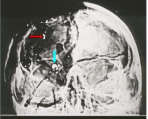
Moreover, this 6.5 mm object had not been cited anywhere in the 1964 Warren Report [13] nor in its accompanying 26 volumes. In fact, the X-ray images had not been introduced to the Warren Commission. The Commission however, did conclude that both the nose and the tail of this (supposedly identical) bullet had been found inside the presidential limousine (Warren Commission Exhibit Numbers 567 and 569). In other words, this 6.5 mm “metal fragment” supposedly represented an internal cross section that had been sliced out of the inside of that same bullet, and then deposited onto the back of the skull (near the supposed entry site at the cowlick area—Figure 2).
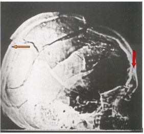
Subsequently (1976-1978) the House Select Committee on Assassinations (HSCA) [10] correlated this image on the AP X-ray to its partner image on the lateral X-ray (Figure 2)—by employing shared anatomic features of the two X-rays.
Paradoxically though, this partner image on the lateral X-ray was only a tiny metal fragment that was a poor match for the large (and very transparent) image on the AP X-ray.
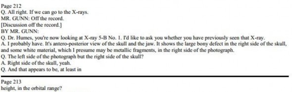
To further confound this mystery, during 1994-1998 each of JFK’s three attending pathologists were asked (under oath) by the Assassination Records Review Board (ARRB) [8] if they had seen this thing during the autopsy. Each one (independently) denied that they had. As an example, see the deposition of the chief autopsy pathologist, James J. Humes in Figure 3.
There is wide agreement that the partner image (on the lateral X-ray) of this mysterious 6.5 mm object must appear at the rear of the skull (near the cowlick area—Figure 2). However, when the forensic radiologist, Dr. John J. Fitzpatrick, reviewed the X-rays (as the premier expert for the ARRB) he remained puzzled by this object, even returning for a second day in an attempt to extract its secrets. Ultimately, however he failed in this task, saying,
No object directly and clearly corresponding to the bright, 6.5 mm wide radio- opaque object in the A-P X-Ray could be identified by the consultant on the lateral skull X-Rays. Although there is a mere trace of some additional density near the fragment bilocation at the vertex of the skull, the consultant did not feel this object was anywhere near the density/brightness required for it to correspond to the bright, radio-opaque object on the A-P X-Ray. After briefly speculating that the small metallic density behind the right eye in the lateral X- Rays might correspond to the bright radio-opaque density in the A-P X-Ray, this idea was abandoned because neither the locations nor the density/brightness of the 2 objects are consistent [2].
For all practical purposes, after this failed attempt (by the most appropriate specialist for the task) this 6.5 mm object become the most curious—and unsolved— mystery in the history of diagnostic radiology.
During the lifetime of the HSCA, Larry Sturdivan served as its ballistics consultant. In his subsequent book [14] he emphasized that he had never, in his entire career, seen a cross section of a bullet deposited in such an odd fashion on a skull. So, totally contrary to all prior government investigations, he concluded that the 6.5 mm object could not be a metal fragment:
I’m not sure just what that 6.5 mm fragment is. One thing I’m sure it is NOT is a cross-section from the interior of a bullet. I have seen literally thousands of bullets, deformed and undeformed, after penetrating tissue and tissue simulants. Some were bent, some torn in two or more pieces, but to have a cross-section sheared out is physically impossible. That fragment has a lot of mystery associated with it. Some have said it was a piece of the jacket, sheared off by the bone and left on the outside of the skull. I’ve never seen a perfectly round piece of bullet jacket in any wound. Furthermore, the fragment seems to have great optical density thin-face on [the frontal X-ray] than it does edgewise [on the lateral X-ray]….The only thing I can think is that it is an artifact (e-mail from Larry Sturdivan to Stuart Wexler on 9 March 1998).
This was a radical statement. After all, the HSCA in particular, had relied on the (metallic) authenticity of this fragment in the most fundamental manner: based on the supposed reality of this 6.5 mm object, the HSCA had concluded that the bullet (from the sole headshot) had deposited this 6.5 mm “metal fragment” near its entry site at the back of the skull. So now, if this was merely an artifact, what was to become of the HSCA’s conclusion?
In 1993 I had two telephone conversations with the (sole) autopsy radiologist, Dr. John Ebersole. On the second occasion, he telephoned me. That call was recorded and later transcribed [7]. After (somewhat reluctantly) discussing the autopsy, I asked him about this 6.5 mm object—and Ebersole instantly stopped the conversation. In fact, that was Ebersole’s final comment to history about the JFK autopsy, as he died shortly afterwards [4].
Materials and Methods
For its unexpected entrance onto the historical stage in 1968, and also for its bizarre properties, a possible explanation occurred to me—perhaps this 6.5 mm object had indeed not been present on the original X-ray, but had been added later (e.g., in the darkroom). To pursue this hypothesis I began (in the early 1990s before the ARRB got underway) to query older radiology technologists. In particular, I asked them: How exactly had they copied X-ray films in the 1960s? With the further assistance of a close colleague (diagnostic radiologist, Dr. John Szabo) that riddle was solved. In particular, I soon discovered a technologist’s handbook [5]1 that contained detailed recipes (p. 56) for converting standard (double-sided) X-ray films into duplicating films. (In that era, Kodak did not make duplicating films—as I was later able to confirm from their inventory lists.) Cahoon even makes this comment: “By variations of the copying time, one may even improve on the original” (p. 55).
Here then was the key to the 6.5 mm mystery: if an X-ray film could be copied (with high fidelity), then it could also be altered. The key step was to add a second image during a second exposure. For example, first the image of the original film would be imprinted onto a duplicating film via a light box in the darkroom (which was how X-rays were then copied). But then, before developing that duplicate film, a second exposure would be made. In particular, a piece of cardboard, with a 6.5 mm hole in it, could be precisely positioned over the duplicate film—and then a second exposure made (using only this mask). I soon showed the feasibility of this approach (using modern duplicating film) by preparing fantastic X-rays with such double exposures (Figures 4 and 5).
References
“1968 Panel Review of Photographs, X-Ray Films, Documents and Other Evidence Pertaining to the Fatal Wounding of President John E Kennedy on November 22, 1963, in Dallas, Texas” [Clark Panel Report] at http://www.jfklancer.com/ClarkPanel.html
“ARRB staff report of observations and opinions of forensic radiologist Dr. John J. Fitzpatrick, after viewing the JFK autopsy photos and X-rays” at
“The [ARRB] deposition of Dr. James J. Humes” at http://jfkassassination.net/russ/testimony/hu mesa.htm
Author, “A Telephone Conversation with Dr. John Ebersole” at http://jfkassassination.net/russ/testimony/h umesa.htm
Cahoon, John B. 1961. Formulating X-ray Techniques (5th edition). Duke University Press, Durham, North Carolina at http://europepmc.org/backend/ptpmcrender
Fetzer, James H., editor. 1998. Assassination Science: Experts Speak out on the Death of JFK. Catfeet Press, an imprint of Open Court, Peru, Illinois
Fetzer, James H., editor. 2000. Murder in Dealey Plaza: What We Know Now that We Didn’t Know Then about the Death of JFK. Catfeet Press, an imprint of Open Court, Peru, Illinois
Final Report of the Assassination Records Review Board (1998) at
http://fas.org:8080/sgp/advisory/arrb98/ind ex.html
Frank, M. 1963. British Journal of Radiology 36:223
House Select Committee on Assassinations Final Report (1979) at https://www.maryferrell.org/showDoc.ht ml?docId=800
Images of double exposures at https://www.google.com.mx/webhp?source id=chrome-instant&rlz=1C1CHFX_enUS629US629& ion=1&espv=2&ie=UTF-8#q=double%20exposure%20phantom%20 image
“JFK Dental X-rays” at http://jfkassassination.net/russ/infojfk/jfk1/ 1exhf295p150.jpg
Report of the President’s Commission on the Assassination of President Kennedy [The Warren Report] at http://www.archives.gov/research/jfk/warr en-commission-report/
Sturdivan, Larry. 2005. The JFK Myths: A Scientific Investigation of the Kennedy Assassination. Paragon Press, St. Paul, Minnesota
Author Area
Have an article to submit?
Submission Guidelines
Submit a manuscript
Become a member