


Atherosclerosis in Microvessels: Insights from Surgery
Microvessels’ Atheroscleroses Study and Evaluation of Some Patients with Coronary Artery Disease at Open Heart Surgery in Armenia
Kukurtchyan N. 1, Karapetyan G. 1, Kocharyan A. 1, Voskanyan M. 1, Hovhannisyan M. 1, Farsiyan N. 1, Sahakyan I. 1, Tumasyan N. 1
- H. Buniatian Institute of Biochemistry, National Academy of Science, Republic of Armenia; Best Life Armenian-Japanese Medical Centre Yerevan, Republic of Armenia
OPEN ACCESS
PUBLISHED: 31 January 2025
CITATION: Kukurtchyan, N., Karapetyan, G., et al., 2025. Microvessels’ Atheroscleroses Study and Evaluation of Some Patients with Coronary Artery Disease at Open Heart Surgery in Armenia. Medical Research Archives, [online] 13(1). https://doi.org/10.18103/mra.v13i1.6210
COPYRIGHT: This is an open-access article distributed under the terms of the Creative Commons Attribution License, which permits unrestricted use, distribution, and reproduction in any medium, provided the original author and source are credited.
DOI https://doi.org/10.18103/mra.v13i1.6210
ISSN 2375-1924
ABSTRACT
Ischemic heart disease is a rather insidious condition with a high mortality rate, especially in developing countries, which also affects Armenia. Therefore, the issue of the most effective treatment for this disease is particularly pressing. This will be facilitated by more thorough research on atherosclerosis in arterioles. The ability to visualize and assess pathological vascular growth on a 5-point scale is provided by one of the modern research methods using semi-thin epoxy sections stained with Azure II. The bioptates of the right atrial auricles of 4 patients with coronary artery disease were taken during coronary artery bypass grafting. The epoxy slices were obtained from material treated by the method of transmission electron microscopy and embedded in epoxy resin, with further staining by Azur II, examined under light optical microscopy. The obtained images of the morphological picture allowed for the identification of atherosclerotic changes in the arterioles of the myocardium of the right atrial appendage in patients, which exhibited pronounced growth resembling intussusception and were accordingly assessed on a 5-point scale for pathological growth of the microcirculatory bed was rated at 3-4 points for one patient and 3 points for three patients. Provided 1-2 images could help in the analysis of atherosclerotic damages and the identification of the direction of the pathological process in each patient case. This will assist both cardiac surgeons and cardiologists in selecting the appropriate medication therapy during the postoperative and separate periods.
Keywords: atherosclerosis arterioles, ischemic heart disease, open heart surgery, light optical microscopy
CASE REPORT
Microvessels’ Atheroscleroses Study and Evaluation of Some Patients with Coronary Artery Disease at Open Heart Surgery in Armenia
Introduction
Cardiovascular diseases remain one of the leading causes of mortality worldwide, especially in developing countries. Armenia also has a high rate of this indicator. COVID-19, numerous stresses, and climate change are affecting atherosclerosis, myocardial vessels, and the microcirculatory bed. Coronary microcirculation disease (CMD) affects the structure and function of the coronary microcirculation. CMD can coexist with epicardial coronary atherosclerosis. The pathogenesis of coronary spasms is closely linked to atherosclerosis and endothelial dysfunction in microcirculation. Although coronary angiography is regarded as the gold standard for detecting coronary artery stenosis, the coronary microcirculation is indispensable. The indispensable coronary microcirculation composes the main resistance vessel bed and is essential for myocardial metabolism. Heterogeneity is a characteristic of microvascular networks and affects structural and functional parameters such vessel diameter, length and connection pattern, flow velocity, wall shear stress and oxidation. Structural microvascular alteration, including arterioles remodeling and capillary imperfection difficult to access in clinical studies. Recent studies have visualized microcirculation in coronary artery disease (CAD) and demonstrated the direction of atherosclerotic changes in microcirculation and need to assess condition on a 5-point scale of pathological growth, which opens new opportunities for more accurate diagnosis of this process and corresponding therapy.
Methods
All procedures involving human subjects were approved by the Institutional Review Board (IRB) and the Ethical Committee (Yerevan State Medical University, RA), conformed to the Legal Aspects of Research Ethics and Science in the European Community Directive (2001/20/EC). Small pieces of right auricular atria from patients with coronary artery disease (CAD) were taken from 4 patients during routine cardiosurgical procedures. The biopsy material was treated using methods employed in transmission electron microscopy. Semi-thin epoxy slices obtained on an ultramicrotome were stained with Azure II and examined under a bright optical microscope. The samples were evaluated using a 5-degree scale of pathological growth, specifically for intussusception type.
Research objectives
To confirm previously obtained data and identify new morphological findings of atherosclerosis in microcirculation in ischemic heart disease, as well as to assess them using a scale of pathological vascular growth. To demonstrate to practicing physicians how the obtained results can be utilized in their daily work.
Cases:
The results of investigation of biopsy material taken from 4 patients as well as evaluation of microcirculatory bed vessels condition by microvessels pathological growth scale are presented.
1. PATIENT IS A 46 YEAR-OLD MALE
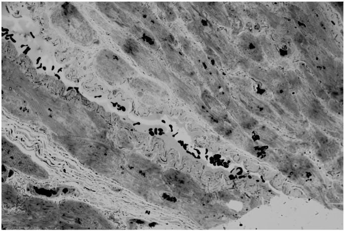
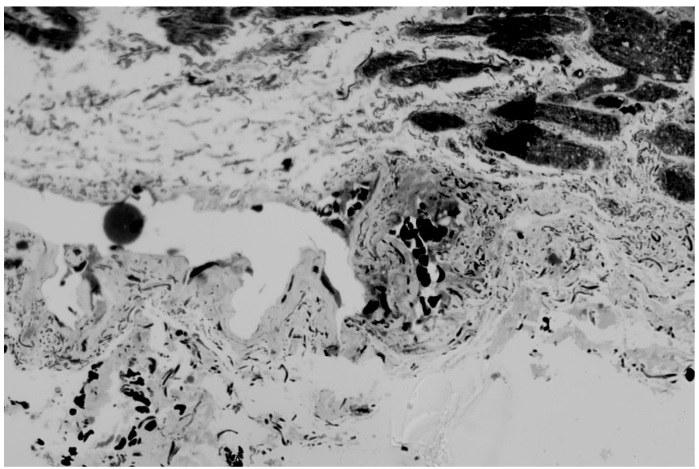
Cardiomyocytes are not wide in transverse diameter. Interstitial edema is expressed. Microcirculatory bed vessels vary in profile lumen width and wall thickness. The vessel wall is unevenly thickened, especially in large arterioles. Opposite walls merge and form bridges that unevenly block the profile lumen in several areas. Closure of opposite walls in a large arteriole leads to the formation of a new profile lumen in the vessel wall. And also closure of opposite walls of a large arteriole leads to the formation of a convolute consisting of forming chambers with the presence of erythrocytes in the lumen of the chambers. Such a convolute fences off part of the arteriole from the general lumen. On the pathological growth scale 3 points.
2. PATIENT IS A 66 YEAR-OLD MALE.
The closure of opposite walls in a large arteriole leads to the formation of a new lumen of the profile in the vessel wall. And also the closure of opposite walls of a large arteriole leads to the formation of a convolute consisting of forming chambers with the presence of erythrocytes in the lumen of the chambers. Such a convolute fences off part of the arteriole from the general lumen. On the scale of pathological growth 3 scopes.
3. PATIENT IS A 66 YEAR-OLD MALE.
Cardiomyocytes are different in width with prevalence of thicker ones. Walls of small arterioles are slightly thickened. Large arterioles with pronounced thickening of the walls and their numerous invaginations into the lumen of the vessel profile are presented. Takes place formation of bridges in such vessels. It is interesting to observe a very large arteriole of unusual configuration, having two separate large lumen profiles and invaginations of the walls into these lumens. And also taking place closure of opposite walls of the arteriole and the presence of an isolated lumen in the wall of the arteriole. This patient has a small arteriole that has formed a convolute of several chambers. And also, there are small arterioles De Novo located close to each other. On the scale of pathological growth from 3 to 4 points.
4. PATIENT IS A 54 YEAR-OLD MALE.
Cardiomyocytes vary in diameter, many are wider. Proliferation of connective tissue between cardiomyocytes. Interstitial edema. Arterioles vary in profile size from large to medium and small. Large arterioles with unevenly thickened walls as well proliferation of cellular elements of the vessel wall are observed. Large arterioles have a pronounced process of invagination of opposite walls into the profile lumen. Bridges are formed in the profile lumen of large arterioles, sometimes in large quantities. Small arterioles are also characterized by the presence of a bridge and the formation of two profile lumens. Several profile lumens are formed in a large arteriole in the vessel wall, which indicates the presence of a convolute. According to the pathological growth scale, 3 points.


Discussion
Ischemic heart disease is a rather insidious disease. A significant role here is played by atherosclerotic damage to both the coronary main vessels and the primary link of the microcirculatory system, the arterioles. The main resistance vessels of coronary arteries are arterioles (with a diameter of <200 mk) and play a key role in the physiological regulation of myocardial reperfusion. The regulatory mechanisms of arterioles can be further divided into large arterioles (100-200 mk that mediate dilation), medium arterioles (40-100 mk), and small arterioles (less than 40 mk). The studies we conducted on atherosclerosis of arterioles in men of different ages visualized more or less the same changes, while the assessment scores varied from 3 points in one patient to between 3 and 4 in 3 patients. The visualization of the biopsy from patient 1 indicates that this is a relatively young heart. The patient is 46 years old. There is no hypertension in the cardiomyocytes and no proliferation of myofibrils. Ischemic heart disease reveals atherosclerotic arterioles. There is pathological growth of the intussusception type. Characteristic invagination into the lumen of the arterioles and the formation of bridges take place. Unlike the first patient, who had a slight wall thickness, the three others showed changes in wall thickness of varying degrees of severity. This is also a negative factor in atherosclerosis of arterioles.
The increase in diameter and thickening of the walls of arterioles during atherosclerosis occurs due to the proliferation of cellular elements, where smooth muscle cells play a very important role. While under physiological conditions, during reperfusion, arterioles can change the sizes of their luminal diameters, in ischemic disease, the growth factor plays a significant role. The increase in the diameter of arterioles occurs up to a certain limit, after which further changes occur in the manner of intussusception. The number of arterioles involved in this process is significant, whether they are large with a diseased diameter or small. This determines what arterioles can subsequently form De Novo. At the same time, the formation of bridges is also a negative process, as it can obstruct normal blood flow. Four patients are characterized by the presence of formed or forming convolutis. This is also a negative point, as it leads to a violation of blood flow. The term convolutes means multi-chambered, similar to the capillaries of the brain in hypertension. We can assess the visualization of atherosclerotic arterioles on a 5-point scale of pathological growth in the manner of intussusception. The term of intussusception angiogenesis, arteriogenesis is indicate the ways of new capillaries arteries and venous formation from previous.
The scale of pathological growth of microvessels by intussusceptive type.
| Group | Investigation signs | Evaluation of vessels growth | Mark |
|---|---|---|---|
| I | The growth of vessels lumen. A bit expanding of vessels wall, with rarely presence of invagination into some vessels walls, and presence of thin wall in other vessels. | Insignificant changes | 1 |
| II | The lumen of vessels are different, often present expended one with involving of some vessels. Takes place decreasing process of uneven expanding, with often presence of invagination into wall, with connection of neighbour area. In such vessels wall forming of 5 new lumen. In other vessel observed thin, sometimes scrolled wall, as well as convergence of opposite sides of first vessels. | Slightly viewable changes | 2 |
| III | Extra growth of vessels profile in interstitial space between tissue cells. The lumen of vessel is thin – expanded. The wall of many vessels is uneven expended, scrolled, invaginated to the wall direction, forming new lumen in the vessel wall and thin scrolled wall presence in others. Visible enclosing of opposite walls of vessels. Often obtained formation of new bridges in the vessel lumen. Often observed presence of vessels with not completely divided wall close to each other. | Temperately viewable changes | 3 |
| IV | Extragrowth of vessels profile in interstitial space between tissue cells with termination of lasts. The lumen of vessels is thin-small. The vessel wall is quite uneven expanded and scrolled up to one profiles and thin, extremely scrolled up to others. The opposite walls of many of vessels reached close to each other creating bridges in vessels lumen. At the same time observed vessels profiles located close to each other, which have the one wall, and are not separated totally — multiple. | Viewable changes | 4 |
| V | Formation of de novo profiles from small to very small: multiple | Brightly viewable changes | 5 |
Sensitive measures of atherosclerosis may be able to quit the therapeutic benefit directed from more complete revascularization with coronary artery bypass graft. It will give additional information to cardiac surgeons and cardiologists which will be useful in their further work.
Conclusion
The conducted study allowed for the visualization and scoring of atherosclerotic arterioles in patients with ischemic heart disease who underwent surgical intervention. It confirmed previously obtained results and identified a more precise direction of the pathological process characteristic of these patients, which provides an opportunity for a more carefully tailored pharmacological therapy for such patients in the postoperative and long-term periods.
References:
- Burry P.H., Hlushchuk K.R, Djonov V Intussusceptiive angiogenesis, it’s emergent, it’s characteristics, and it significance. Dev Dyn ..2004 231:474 – 88. Doi:10 1002 dvdy 2018
- Gerd Heusch, “Myocardial ischemia reperfusion: Translational pathophysiology of ischemic heart disease” Med. volum 5, issue 1. 12 January, 2024, pages 10-31. https://doi.org/10.1016/medj.2023
- Gulevskia T.S., Enduring P.A.Ŕossia Modern Problems of Science and Education. 2017, 4(26); 86-9101. URL://www science – education,ru view?D=26720
- Kukurtchyan N.S., Karapetyan G.R., “European Visualisation and Evaluation Study of Some Patients Arteriosclerosis Microvessels with Coronary Artery Disease at Coronary Artery Bypass Grafting” Society of Medicine. Medical Research Archives 2024. 12(3). http://doi.org/10.18103/mra.v12
- Kukurtchyan N.S., Karapetyan G.R. “Heart microvessels and it morphological evaluation at open heart surgery RJBPT 2023, 10(8).
- Kukurtchyan N.S., Karapetyan G.R. “Heart microvessels research at ischemic heart disease at open heart surgery “ EJBPT, 2021, 8(8); 71-75
- Kukurtchyan N.S. Karapetyan G.R. Patent 2844 A Rep. Armenia Staining method of histological material
- K. Nishimya, J. Takahashi, K. Oyama, Y. Metsunoto, S. Vasuda, H. Shimokawa “Mechanisms of coronary artery spasm” Eur. Cardiol., 18(2023), p.39. [Scopus], [Google Scholar].
- K.Welen Schof, P. Tornvall, J.Alfredsson, E Hagstron, A. Ravin-Fisher, S. Soderberg, T. Yndigego, T. Jenborg.” Prevalence of angina pectoris and association with coronary atherosclerosis in general population” Heart. 109(2023), pp. 1450-1459. [Scopus] [Google Scholar].
- Klinbongart, G. Heusch “A fresh look at coronary microembolization” Nat. Rev. Cardiol. 19(2022) pp 265-580.
- Pries A.R Coronary microcirculation in ISCHEMIC Heart Disease. PubMed. Curry Pharm Des …2018
- Shimokawa H., 2014 Williams Harvey Lecture: importance coronary vasomotion abnormalities from bench to bedside”. Eur. Heart j. 2014, 35: 3180-93 [Doi], [Pub.Med], [Google Scholar].
- Sorrob O., Van Dr Wouw, Chadler S. Ohanyan V, Tuni J.D. Chilian W.M. Mercus D. Bender S.B. Duncker D.J. Experimental animal model of coronary microvascular dysfunction. Cardiovascular research 2020;116(4);756-787 Doi.org/10.1093/CV2/CV as 002
- Jivarny R. Taguety, Marcelo F. Dicarli “Coronary microvascular disease Pathogenic mechanisms and Therapeutic Options: j. Acc. Stable -of-the Art Review j. Am. Call Cardiol. 2018,v.27; 72(21); 2625-2641. Doi:10.1016/j.jacc.2018.09.042.
- W.W. Chilian Coronary microcirculation in health abdominal disease summary of NHL PI Workshop Circulation, 95(2), 1997, p. 552-528.