


Developmental Delay: 1q42 Duplication & 7p22 Deletion Case
A Case of Developmental Delay and Congenital Anomalies Associated with 1q42.12q44 Terminal Duplication and 7p22.3 Terminal Deletion
Roman Yusupov MD, 1 Eian D. Wallenhorst MS 2 Sajel Lala Kana MD 3 Haiying Ming, MD, PhD 2 Paul J. Benke MD, PhD 1,4*
- Joe DiMaggio Children’s Hospital, Hollywood, FL.
- Quest Diagnostics, Chantilly, VA.
- Nicklaus Children’s Hospital, Miami FL.
- Charles Schmidt School of Medicine, Boca Raton, FL.
Email: [email protected]
OPEN ACCESS
PUBLISHED 30 April 2026
CITATION Yusupov, R., Wallenhorst, ED., et al., 2026. A Case of Developmental Delay and Congenital Anomalies Associated with 1q42.12q44 Terminal Duplication and 7p22.3 Terminal Deletion. Medical Research Archives, [online] 14(4). https://doi.org/10.18103/mra.v14i4.7345
COPYRIGHT © 2026 European Society of Medicine. This is an open-access article distributed under the terms of the Creative Commons Attribution License, which permits unrestricted use, distribution, and reproduction in any medium, provided the original author and source are credited.
DOI https://doi.org/10.18103/mra.v14i4.7345
ISSN 2375-1924
ABSTRACT
We report a child with a 1q42.12q44 terminal duplication and a 7p22.3 terminal deletion detected by chromosomal microarray. He presented with a history of feeding problems, poor weight gain, short stature, mild facial dysmorphic features, pes planus, horseshoe kidney, and mild congenital heart disease. Parental karyotyping showed that the mother was a balanced translocation carrier between chromosomes 1q and 7p, (46,XX,t(1;7)(q42.1;p22). This combination of chromosomal abnormalities has not been previously reported. The case is notable for its clinical features as well as a potential underlying mechanism of lowered chromosomal breakage and translocation of chromosome arms secondary to excess chromosomal fragile sites on 1q42 and 7p22.3.
Keywords: Chromosome translocation, developmental delay, chromosome fragile site.
INTRODUCTION
Constitutional cytogenetic abnormalities include aneuploidy and structural aberrations such as partial chromosomal gains and losses, translocations, inversions, insertions, and marker chromosomes. Among them, unbalanced chromosomal abnormalities are often associated with a range of multiple congenital anomalies and developmental delays. The chromosome microarray analysis has been accepted as an appropriate first-tier test for the evaluation of imbalances associated with intellectual disability, autism, and multiple congenital anomalies by ACMG (American College of Medical Genetics). Large data curated at ClinGen and systemic documentation of phenotype/karyotype correlation for rare diseased initiated by multiple nonprofit organizations such as Orphanet and Unique have increased the accuracy of interpretation and symptom prediction for single copy gain or loss. However, interpretation and symptom prediction can be a puzzle when there is more than one abnormality in one patient, such as an unbalanced rearrangement resulting in a terminal duplication and a terminal deletion. Here we report a child with an approximately 22.6 Mb terminal gain at 1q42.12q44 and an approximately 2.8 Mb terminal loss at 7p22.3 identified via chromosomal microarray. His clinical presentation included hypotonia, feeding problems, poor weight gain, short stature, dysmorphic features and mild developmental delay. Parental karyotyping showed the mother to be carrier of a balanced form of the reciprocal translocation, (46, XX, -t(1;7)-(q42.1;p22). While individual cases of 1q42.12 duplication1-3 or 7p22.3 deletion4-5 have been reported, this combination of chromosome abnormalities is a new finding.
CASE REPORT
The mother was diagnosed with gestational hypothyroidism. A second trimester fetal ultrasound revealed a renal cyst and possible macrocephaly. The third trimester was complicated by abruptio placenta, and a cesarean section was performed at 35 weeks gestation. Birth weight was 2.155 kg, below the 1st percentile. Height was at the 3rd percentile for age, with a normal-sized head circumference.
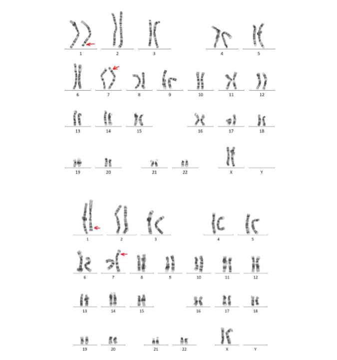
Problems were not apparent at birth and he was sent home after a few days in a newborn nursery. At home the parents noted excess drowsiness and poor feeding leading to a hospital admission at 1 week of age. He was also noted to have an inguinal hernia, undescended testicle, and a sacral dimple. Feeding difficulties persisted. Genetic testing in the form of a chromosomal microarray (Quest, Oligo-SNP) revealed a 1q42.12q44 terminal duplication and a 7p22.3 terminal deletion (Figure 1), possibly the product of an unbalanced translocation inherited from a parent. Parental karyotyping showed that the mother had a balanced form of the reciprocal translocation (Figure 2). A maternal aunt also carried the same balanced translocation.
![Chromosome microarray results; arr [hg19] 1q42.12q44(226,602,460-249,224,683 X3, 7p22.3 (42,360-2,799,184 X1 shown in Santa Clara Genome Browser. Top; all chromosome 1, showing chromosome 1q duplication. Middle; all chromosome 7 showing chromosome 7p deletion. Bottom; enlargement of 7p chromosome showing chromosome 7p deletion.](/pdf-to-wp-converter/uploads/images/developmental-delay-1q42-duplication-7p22-deletion-case-img-1.png)
Brain MRI revealed several brain cysts, diffuse hypomyelination, periventricular white matter volume loss, enlarged subarachnoid spaces with mild ventriculomegaly and thinning of the corpus callosum. Additionally, there was a small chronic subdural hygroma in the left frontal region, ependymitis granularis, pars intermedia cyst of the pituitary gland, and retro-cerebellar arachnoid cysts. An upper gastrointestinal x-ray showed minimal spontaneous reflux and a postnatal renal ultrasound revealed bilateral hydronephrosis and multiple right-sided renal cysts. Voiding cystourethrogram (VCUG) was normal. Spinal ultrasound indicated a possible low-lying conus medullaris with minimal movement of the cauda equina nerves.
An MRI of the abdomen revealed a right posterolateral extradural arachnoid cyst at L5-S1 level, measuring 2.3 x 1.4 x 0.9 cm, a horseshoe kidney associated with right sided multicystic dysplastic and/or markedly hydronephrotic kidney and a possible neurogenic bladder. An echocardiogram revealed an atrial septal defect (ASD). Hypotonia and feeding difficulties persisted, and the patient had a gastrojejunostomy (GJ) tube placed at 7 months of age. Attempts to discontinue the G-tube feeding were unsuccessful, and he remained below the 1st percentile for weight for more than three years and required a G-tube until age 3.5 years. He became ketotic on a mildly high-fat diet which was corrected by modifying the diet. Acylcarnitine profile revealed elevated C2 levels, despite continuous feeding without fasting. He had difficulty chewing and demonstrated poor oral intake, requiring continued supplemental feeding. The use of L-carnitine (250-500 mg/day) appeared to improve muscle strength but was limited due to side effects of loose stools. He had an episode of malignant hyperthermia (MH) during inguinal hernia repair and orchiopexy surgery at 2.5 years of age; halothane was used as an anesthetic. Genetic testing for known MH genes including CACNAIS, RYR1, and STAC3 was negative.
The patient was noted to have moderate developmental delays. He was crawling and babbling at age 1, but was unable to walk or speak beyond a few words. At age 3, he began to walk independently and say a few words. By age 5, he was able to use an augmentative communication (AAC) device with approximately 50 words. Psychological testing at age 5 revealed that the child’s cognitive function was largely at the level of a 3-year-old, with non-verbal abstract reasoning and verbal comprehension both below 3 years of age. He was described as “good natured” and loving personality, with a conceptual skill set in the extremely low range, his functioning in the low range, and “self-care” in the extremely low range.
DISCUSSION
Chromosome duplications and deletions have been associated with developmental delay and congenital anomalies from the early days of cytogenetics discovery, starting with the early discovery of translocation chromosome trisomy 21 and familial Down Syndrome6. Isolated duplications of the long arm of chromosome 1 are rare and present with variable degrees of developmental delay, minimally dysmorphic features, and occasional autism1-4. Case reports of 1q+ are variable because of small and large 1q involvement, or other chromosome changes that contribute to the clinical findings. However, there is a single presentation with duplication of the sub-bands 1q42.11 and 1q42.12 found in a boy and his mother so mild that microarray testing would not have been done except to examine for a potential cause for his short stature5. Distal 7p22.3 deletion is also rarely noted in the literature. Here, affected cases have some element of developmental delay, most have heart or kidney findings, and some have seizures4. With mild, not severe expected outcome of the small 1q duplication, it is likely that the 7p deletion is the major reason for the presentation of short stature, failure to thrive and developmental delay, and mild heart and kidney anomalies found. The loss of genes within the deleted interval are likely responsible. These genes, specifically the haploid status of PDGFA (growth factor subunit, PDGF receptor kinase), PRKAR1B (pathway of messenger cAMP ion transport/metabolism, and MAD1L1 (component of mitotic spindle assembly; cell cycle control), all within the segment Chr7:43360-2799185 (hg19/GRCh37)8, all potentially play a role in growth and development of the patient. The effects of PRKAR1B8 on metabolism and mitochondrial function may potentially play a role in the history of high fat sensitivity. A second mitochondrial gene in the interval, NUDT18 (MTH1), involved in nucleoside degradation, likely played little role in in the pathogenesis of the clinical findings. Learning, developmental, and cardiac issues in 7p deletion patients have been linked to the SNX8 gene found in the 7p22.3 segment9 and deleted here. A few cases of chromosome 7p22 deletions associated with other chromosome duplications and patients with developmental delay and congenital anomalies have been described10,11. However, the combination of 1q42.12 duplication and 7p22.3 deletion has not been previously reported. The unbalanced translocation was inherited from the mother, who was a carrier of the balanced translocation. This suggests the genetic rearrangement occurred through non-homologous end joining (NHEJ), which pairs the DNA end fragments in a multiprotein synaptic complex to promote their direct ligation. NHEJ is a highly versatile pathway that uses an array of DNA specific processing enzymes to repair damaged DNA ends and enable their ligation12. A possible mechanism limiting breaks in these particular chromosomal regions is described in a study from 1988 exploring the synergistic effect of DAPI and thymidylate stress conditions on the induction of common fragile sites. An early study notes that “when supplied to human leukocytes grown in complete medium (RPMI 1640), DAPI (4′,6-diamidino-2-phenylindole), a non-intercalating compound specific for the AT bases of DNA, induces the appearance of three common fragile sites (CFRA) mapped at 1q42, 2q31, and 7p22” 13. The results of that study agree with the hypothesis that DAPI-induced CFRA sites are DNA late-replicating chromosomal areas rich in AT bases suggesting that chromosomal regions 1q42 and 7p22 are less susceptible to breaks than other sections of human chromosomes, potentially the reason this combination is rare and has not been previously reported. Alternatively, these areas with a lower chance of breaking and recombination have a lesser chance of being “flanked by large (usually >10 kb), highly homologous low copy repeat (LCR) structures that can act as recombination substrates. Recombination between non-allelic LCR copies, also known as non-allelic homologous recombination, can result in deletion or duplication of the intervening segment”14. Whether the original genesis of the chromosome deletion/duplication originated in a grandparent by chance in spite of CFRA sites on 1q42 and 7p22 with decreased potential for breakage and rearrangement described above, or because of a low presence of LCR structures on these chromosomes would require additional study of translocations in areas of regions of high AT bases of DNA and areas of high common fragile sites (CFRA) leading to a low potential for chromosomal translocations.
CONCLUSION
This case represents a novel combination of a 1q42.12q44 terminal duplication and 7p22.3 terminal deletion, associated with a range of congenital anomalies and developmental delays. The findings underscore the importance of genetic testing, including microarray and whole exome sequencing (WES), now accomplished by whole genome sequencing (WGS) in patients with unexplained developmental delays and congenital abnormalities. Further studies are needed to explore the molecular mechanisms underlying the 1q42 and 7p22 combination of chromosomal breaks and recombination to confirm that these 2 areas are less susceptible to breaks than other sections of human chromosomes.
Conflict of Interest Statement: None.
Consent for un-blurred picture: Agree that un-blurred picture can be used.
Funding Statement: Gordon Foundation.
Acknowledgements: Supported by the Gordon Foundation. We thank the family for their cooperation, input, and consent for publication and Dr. Parul Jayakar for assistance.
REFERENCES
- Morris ML, Baroneza JE, Teixeira P, Medina CT, Cordoba MS, Versiani BR, et al; Partial 1q Duplications and Associated Phenotype. Mol Syndromol. 2016 Feb;6(6):297-303. doi: 10.1159/000443599. PMID: 27022331.
- Watanabe S, Shimizu K, Ohashi H, Kosaki R, Okamoto N, Shimojima K, et al; Detailed analysis of 26 cases of 1q partial duplication/triplication syndrome. Am J Med Genet A. 2016 Apr;170A(4):908-17. doi: 10.1002/ajmg.a.37496. PMID: 26782913.
- Balasubramanian M, Barber JC, Collinson MN, Huang S, Maloney VK, Bunyan D, Foulds N. Inverted duplication of 1q32.1 to 1q44 characterized by array CGH and review of distal 1q partial trisomy. Am J Med Genet A. 2009 Feb 15;149A(4):793-7. doi: 10.1002/ajmg.a.32463. PMID: 19248177.
- Skvortsova L, Perfilyeva A, Bespalova K, Kuzovleva Y, Kabysheva N, Khamdiyeva O. 7p22.3 microdeletion: a case study of a patient with congenital heart defect, neurodevelopmental delay and epilepsy. Orphanet J Rare Dis. 2024 Aug 16;19(1):301. doi: 10.1186/s13023-024-03321-8. PMID: 39152504.
- Bortotto L, Piovan E, Furlan R, Rivera H, Zuffardi O. Chromosome imbalance, normal phenotype, and imprinting. J Med Genet. 1990 Sep;27(9):582-7. doi: 10.1136/jmg.27.9.582. PMID: 2231652.
- Li R, Wang C, Zhang Z, Li D, Li L, Zhao D, Xu Z. Partial trisomy 9p and partial monosomy 7p of an infant inherited from maternal balanced translocation: a case report. BMC Pediatr. 2023 Apr 13;23(1):168. doi: 10.1186/s12887-023-03986-3. PMID: 37046298.
- Carter CO, Hamerton JL, Polani PE, Gunalp A, Weller SD. Chromosome translocation as a cause of familial mongolism. Lancet. 1960 Sep 24;2(7152):678-80. doi: 10.1016/s0140-6736(60)91749-9. PMID: 13691131.
- San Diego Genome Browser Chr7:43360-2799185 (GRCh 38/hg38).
- Mastromoro G, Capalbo A, Guido CA, Torres B, Fabbretti M, Traversa A, et al; Small 7p22.3 microdeletion: Case report of Snx8 haploinsufficiency and neurological findings. Eur J Med Genet. 2020 Apr;63(4):103772. doi: 10.1016/j.ejmg.2019.103772. PMID: 31568860.
- Kohannim O, Peredo J, Dipple KM, Quintero-Rivera F. Clinical findings associated with a de novo partial trisomy 10p11.22p15.3 and monosomy 7p22.3 detected by chromosomal microarray analysis. Case Rep Genet. 2011;2011:131768. doi: 10.1155/2011/131768. PMID: 23074670.
- Schmidt B, Udink ten Cate F, Weiss M, Koehler U. Cardiac malformation of partial trisomy 7p/monosomy 18p and partial trisomy 18p/monosomy 7p in siblings as a result of reciprocal unbalanced malsegregation–and review of the literature. Eur J Pediatr. 2012 Jul;171(7):1047-53. doi: 10.1007/s00431-012-1682-z. PMID: 22302461.
- Stinson BM, Loparo JJ. Repair of DNA Double-Strand Breaks by the Nonhomologous End Joining Pathway. Annu Rev Biochem. 2021 Jun 20;90:137-164. doi: 10.1146/annurev-biochem-080320-110356. PMID: 33556282.
- Rocchi A, Pelliccia F. Synergistic effect of DAPI and thymidylate stress conditions on the induction of common fragile sites. Cytogenet Cell Genet. 1988;48(1):51-4. doi: 10.1159/000132585. PMID: 3180848.
- Shaw CJ, Lupski JR. Implications of human genome architecture for rearrangement-based disorders: the genomic basis of disease. Hum Mol Genet. 2004 Apr 1;13 Spec No 1:R57-64. doi: 10.1093/hmg/ddh073. PMID: 14764619.