

Femoral Head Insufficiency Fracture: Case Study Insights
Femoral Head Insufficiency Fracture after Overuse Injury: Case Report of a Rare Ent
Mazin Usama MD¹, Ahmed Al Ghaithi MD¹, Humaid Al Farii MD FRCSC¹, Wafa Al Baluki MD FRCSC¹
- Department of Surgery, Orthopaedic division, Sultan Qaboos University Hospital. Muscat. Oman
OPEN ACCESS
PUBLISHED: 31 January 2024
CITATION: Al Farii, H., 2025. Femoral Head Insufficiency Fracture after Overuse Injury; Case Report of a Rare Entity and Literature Review. Medical Research Archives, [online] 13(1).
https://doi.org/10.18103/mra.v13i1.6148
COPYRIGHT: © 2025 European Society of Medicine. This is an open-access article distributed under the terms of the Creative Commons Attribution License, which permits unrestricted use, distribution, and reproduction in any medium, provided the original author and source are credited.
DOI https://doi.org/10.18103/mra.v13i1.6148
ISSN 2375-1924
ABSTRACT
Background: Overuse injuries develop gradually over time without a specific traumatic event. They are more commonly reported as the cause of lower limb injuries compared to acute incidents. The incidence of overuse injuries varies among different sport types but comparing them across sports or establishing specific links is challenging due to methodological heterogeneity. Youth sports’ growing popularity in the region has led to a rise in sports-related overuse injuries. Raising awareness is crucial for proper management and prevention of overuse injuries in this age groups.
Case Summary: Hence, we present a case report concerning a 10-year-old boy who experienced an overuse injury in a relatively uncommon area, specifically the femoral head.
Conclusion: Our intention is to contribute to the existing knowledge and comprehension of such injuries.
Keywords
- Femoral head insufficiency fracture
- Overuse injury
- Young athletes
- Growth plate
- Psychosocial implications
Introduction
Overuse injuries are a growing concern in youth sports, developing gradually over time without a specific traumatic event. The skeletal maturity of young athletes plays a crucial role in their susceptibility to overuse injuries¹,². The growth plate, a region of developing cartilage near the ends of long bones, is particularly vulnerable to stress during periods of rapid growth³,⁵. Arnold et al. highlighted that the growth plate undergoes structural changes during maturation, which may weaken its anchoring points and make it more susceptible to injury²,⁴,⁶. Additionally, the rate of bone mineralization may lag behind bone linear growth during pubescent growth spurts, further compromising the integrity of the growth plate⁴.
The location of overuse injuries can vary depending on the sport and repetitive movement patterns involved. Gymnasts commonly experience overuse injuries around the wrist¹,², while long-distance runners often sustain injuries in the distal femur⁷. Baseball pitchers frequently develop shoulder overuse injuries¹,²,⁴,⁸, emphasizing the importance of sport-specific training programs and conditioning to address the unique demands placed on different body regions⁹.
Among the various sites affected, the femoral head is a relatively rare location for overuse injuries. Understanding and managing overuse injuries in the femoral head is crucial for proper treatment and prevention of long-term complications. This case report presents a rare case of a 10-year-old boy with a femoral head overuse injury, aiming to enhance understanding and awareness of this condition. The report discusses the clinical presentation, diagnostic evaluation, treatment approach, and patient outcome, emphasizing the importance of early recognition and appropriate management of overuse injuries in the femoral head.
Case Presentation
A 10-year-old boy presented with right hip pain that had been present for approximately two weeks.
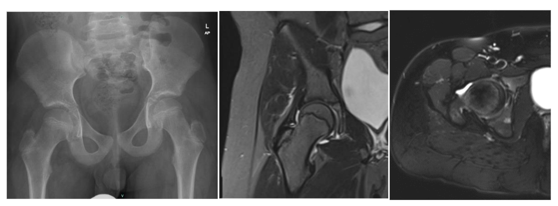
The pain started immediately after his karate training sessions. The child reported difficulty sitting with crossed legs and a subtle limp while walking. Initial clinical evaluation showed normal range of motion in both hips, with slight pain during rotational movements. Laboratory tests, including white blood cell count and C-reactive protein level, were within normal limits. Radiographs showed no fractures, and an ultrasound examination revealed no abnormalities. The case was initially categorized as an unspecified musculoskeletal injury, and observation was planned. However, due to persistent pain and limping, further investigations were conducted included MRI that was unremarkable (Fig. 1).
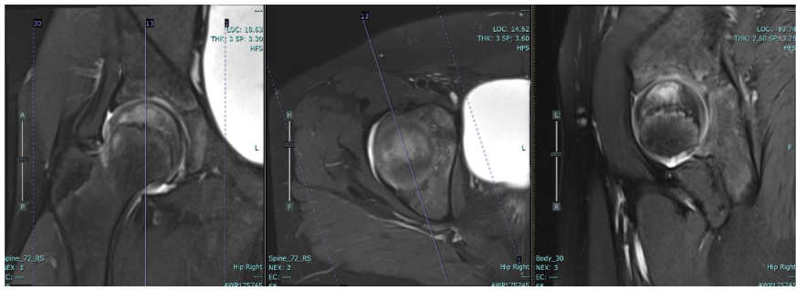
A follow-up visit, a few weeks later showed no improvement in pain and limping. Repeat MRI revealed bone marrow oedema and a subchondral hypointense linear signal abnormality along the weight-bearing surface of the femoral head, consistent with a subchondral insufficiency fracture. Necrotic areas suggestive of conditions like Perthes disease were not observed. (Fig. 2) Therefore, final diagnosis was an overuse injury of the right femoral head.
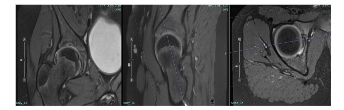
Conservative treatment was initiated, including toe-touch weight bearing with crutches and maintaining range of motion in the hip. Follow-up evaluations showed gradual resolution of symptoms, complete resolution of pain at the three-month mark, and radiographic improvement with no signs of avascular necrosis (Fig. 3). The patient was allowed to gradually resume weight-bearing and progressively return to normal activities.
Discussion
All authors declare that they have no conflicts of interest.
Funding/Sponsorship
No funding has been received to produce this work.
Patient/ Guardian Consent
A written consent obtained from the father to publish the case. No Clinical picture of the child is published.
References
- Guerra MVR, Estelles JRD, Aboudiya YA, Falcón JPR, Losa JPR. Epidemiology of the growth plate injury in young athletes at a training center. Acta Ortop Bras. 2016;24(4):204-207. doi:10.1590/1679-775720162404204.
- DiFiori JP, Puffer JC, Aish B, Borey F. Wrist pain, distal radial physical injury, and ulnar variance in young gymnasts: Does a relationship exist? Am J Sports Med. 2020;36(3):879-885.
- Arnold A, Thigpen C, Beattie PF, Kissenbh M, Shanley E. Overuse physical injuries in youth athletes: Risk factors, prevention, and treatment strategies. J Athl Train. 2019;54(1):S23-S30. doi:10.1177/1914731818668467.
- Stacioli C, Ascanio L, Levy Friedman H. et al. Pediatric sports injuries: A review of the literature. J Pediatr Orthop. 2015;35(6): 965-972. doi:10.1177/0034654315225211.
- Magri D, Nussbaum ED, Rizzone KH, Brown NJ. Femoral neck bone stress injuries in pediatrics and adolescence: Diagnosis, etiology, and treatment. J Bone Joint Surg Am. 2021;103(2). doi:10.55275/JPSNA-2021-0204.
- Bencardino JT, Stone TL, Roberts CC, et al. ACR Appropriateness Criteria® Stress (fatigue/ insufficiency) fracture: excluding other vertebrae. J Am Coll Radiol. 2017;14(5): S293-S306.
- Bernstein EM, Shay PH, Cotterak GK. The management of physical injury: Overuse. Sports Health. 2015;7(2): 142-153. doi:10.1177/1941738114567886.