


Fluorescent Assay Failure in Detecting M. avium in MS
Failure of a proprietary fluorescent in situ hybridization assay to detect M. avium subspecies paratuberculosis in archived frozen brain from patients with Multiple Sclerosis
Robert J. Greenstein MD FRCS (England) FACS 1,2; Peter S. Fam M.Sc. 3; Sheldon T. Brown MD 4,5
- Department of Surgery James J. Peters Veterans Affairs Medical Center Bronx NY USA.
- Laboratory of Molecular Surgical Research James J. Peters Veterans Affairs Medical Center Bronx NY USA.
- Alzheimer’s Research Institute James J. Peters, Veterans Affairs Medical Center Bronx NY USA.
- Infectious Disease Section. James J. Peters Veterans Affairs Medical Center Bronx NY USA.
- Department of Medicine, Icahn School of Medicine at Mt. Sinai. New York. New York. USA.
OPEN ACCESS
PUBLISHED: 30 November 2024
CITATION: Greenstein, R.J., Fam, P.S., et al., 2024. Failure of a proprietary fluorescent in situ hybridization assay to detect M. avium subspecies paratuberculosis in archived frozen brain from patients with Multiple Sclerosis. Medical Research Archives, [online] 12(11). https://doi.org/10.18103/mra.v12i11.5853
COPYRIGHT: © 2024 European Society of Medicine. This is an open- access article distributed under the terms of the Creative Commons Attribution License, which permits unrestricted use, distribution, and reproduction in any medium, provided the original author and source are credited.
DOI https://doi.org/10.18103/mra.v12i11.5853
ISSN 2375-1924
Abstract
Objectives: Multiple Sclerosis is a chronic, enigmatic, progressive central nervous system “inflammatory” disease, with no know etiology. As with other “inflammatory” diseases there is the possibility that a cryptic infectious trigger may play a role in initiating Multiple Sclerosis. M. avium subspecies paratuberculosis causes Johne’s disease in ruminants and may be an infectious trigger in Crohn’s disease. In this study, frozen archived brains from patients with Multiple Sclerosis, pure culture of multiple bacteria and circulating WBCs were assayed with proprietary (Affymetrix RNA view®) Tissue and Cell fluorescent in situ hybridization assay for MAP RNA. Results: Repetitively, false positive signal was observed in the “No-Probe” negative control. Despite advice from the technical staff at Affymetrix, multiple experimental modifications could not prevent positive signal in the “No-Probe” negative control. When studying human white blood cells under specific storage conditions, we observe positive signal with human house-keeping genes, when no signal is seen in the No-Probe controls. Conclusions: We conclude, that when performed according to manufactures instructions and with multiple variations on the manufactures recommended suggestions to correct for false positive signal, that the Affymetrix RNA view® TISSUE assay cannot be used to detect M. avium subspecies paratuberculosis in pre-frozen brains of humans with Multiple Sclerosis. In contrast, using the Affymetrix RNA view Cell fluorescent in situ hybridization system, evaluating human white blood cells, we reliably identify human house-keeping genes. This indicates that the Cell fluorescent in situ hybridization assay may be useful when evaluating circulating cells for specific pathogens.
Keywords
In situ hybridization; multiple sclerosis; Mycobacterium avium subspecies paratuberculosis; Mycobacteria; Crohn disease; Johne disease
Introduction
Multiple Sclerosis (MS) was first definitively described by Charcot in the French medical literature in 1868 and subsequently in the English medical literature in 1873. MS remains a chronic, enigmatic, progressive neurological disease of the central nervous system, that has no known etiology (see 3 for review.) The possibility that the etiology of MS may be consequent to an infectious cause is the cause of innumerable studies and speculations (see 4-6 for reviews.) None of which have been proven or are accepted. Intriguingly, the relevance of vitamin D in the prevalence, incidence and clinical course of MS has revealed noteworthy correlates. Circumstantial data show the relevance of vitamin D include migration, its time of life and destination influence the incidence of MS. Observational studies show a positive clinical course when vitamin D levels are elevated and an exacerbation when vitamin D levels are low. Randomized control trials of vitamin D supplementation in MS did NOT meet their primary endpoints (which was no evidence of disease activity) but did show improvements in secondary endpoints (annualized relapse rates and new or hypointesnse T1 Lesions on MRI. Habitually, the role of vitamin D is assigned to its well documented effect on the immune response of the afflicted individual. We have shown that vitamin D directly inhibits mycobacteria in culture, suggesting a possible correlate between an infectious etiology of MS and the role of vitamin D. Johne’s disease is a chronic wasting intestinal infection in animals, universally acknowledged to be caused by M. avium subspecies paratuberculosis (MAP.) There is increasing concern that MAP may be zoonotic. Crohn’s disease is an affliction evocative of Johne’s disease. MAP is frequently implicated as a possible infectious trigger in the “inflammatory” bowel condition best known as Crohn’s disease. Evidence of polymicrobial (fungal as well as bacterial) have been documented in MS. In 1986, an unexplained association of MS with inflammatory bowel disease was observed. Subsequently, in a series based primarily on immunological studies, a possible MAP/MS etiological connection has been investigated. It must be emphasized, that these studies did not identify MAP in MS lesions. Accordingly, it would be of noteworthy interest if viable MAP, could be identified in MS CNS lesions. The presence of MAP RNA or culture of MAP would be an acceptable indication of viability. A proprietary (Affymetrix RNA view®) fluorescent in situ hybridization (FISH) assay identifies RNA. We have shown that this FISH assay cannot be used to reliably identify MAP RNA in frozen archived intestine from ruminants with Johne disease. Likewise we show that this FISH assay cannot reliably identify MAP RNA in frozen archived resected intestine from patients with Crohn’s disease. The purpose of this present study was to evaluate whether the MAP Affymetrix RNA view® FISH assay could reliably detect MAP RNA from frozen brain autopsy specimens of humans who had demonstrable multiple sclerosis as well as normal brain controls.
Methods
This study was approved by the Research & Development Committee at the VAMC Bronx NY (0720-06-038.) The methods and results with bovine ileal intestinal tissue, with and without Johne’s disease, and with human tissue in Crohn disease have been published. Non-identifiable brain tissue from individuals with and without multiple sclerosis were obtained from Wallace W. Tourtellotte, M.D., Ph.D. Human Brain and Spinal Fluid Resource Center. Neurology Research [email protected]. Specimens were shipped on dry ice and stored at -80°C until processed as below. The tissue and assay were handled in an identical manner to that of the published bovine study, with one exception. Previously, at our request Affymetrix had generated probes that were species specific from published gene sequences. In this (and the parallel Crohn disease study, the housekeeping gene was Human Specific β-actin (Affymetrix Catalog # VA6-10506-1 Probe type 6) As in the previous study for MAP, an Affymetrix generated probe designed using the published sequence. (Affymetrix name: M. tuberculosis Is900: Cat # VF1 19496: Lot # 195634523: Probe type 1.) Previously, the house keeping gene for ruminants was bovine β-actin (Bos Taurus actb: NCBI Reference Sequence: NM_173979.3 (Affymetrix name: Bos Taurus Actb: Cat# VF6 20062: Lot # 200642784: Probe Type 6.) All these probes are proprietary to Affymetrix. Excepting for using a Human Specific β-actin probe and non-identifiable human brain tissue (instead of bovine and or human intestine), the assay was carried out identically as published.
| Figure #’s | Tissue/Cell With Probes | Figure #’s No Probe Control | Indicate reliable FISH data |
|---|---|---|---|
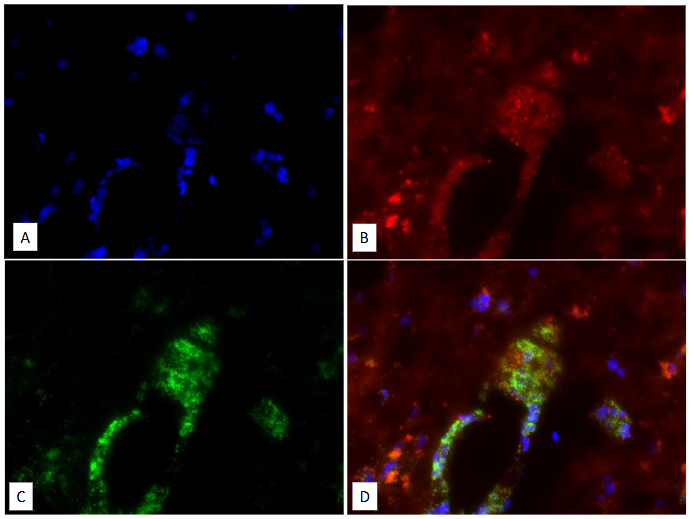
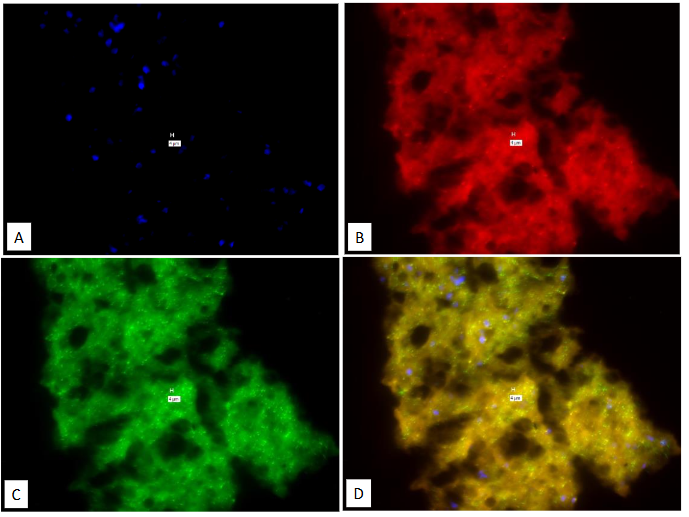
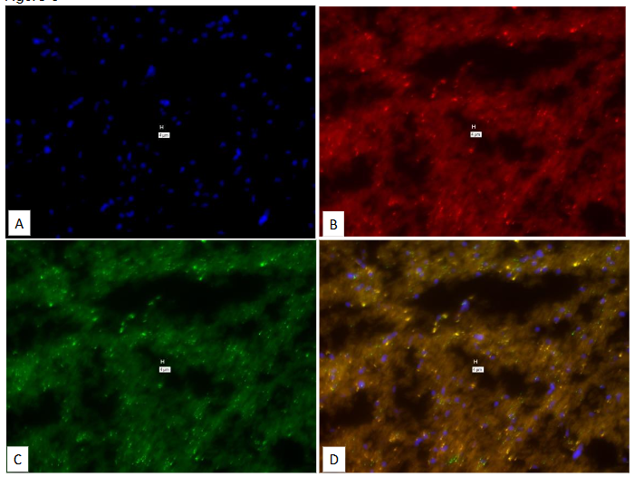
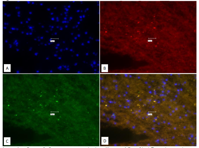
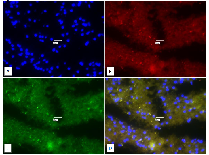
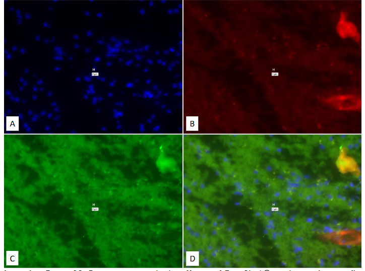
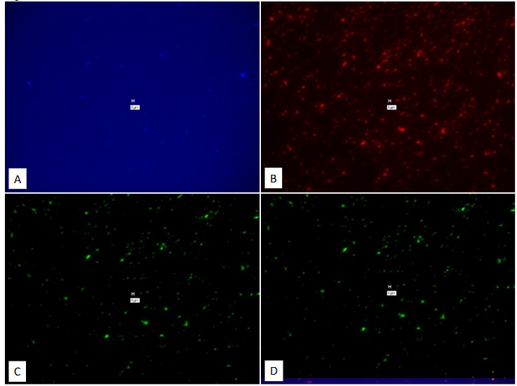
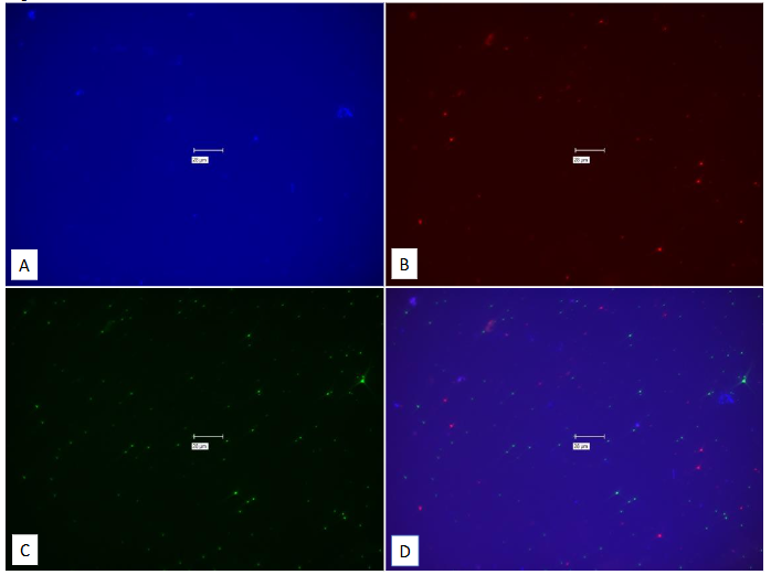
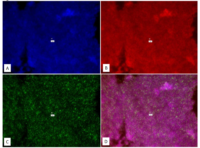
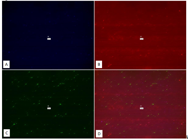
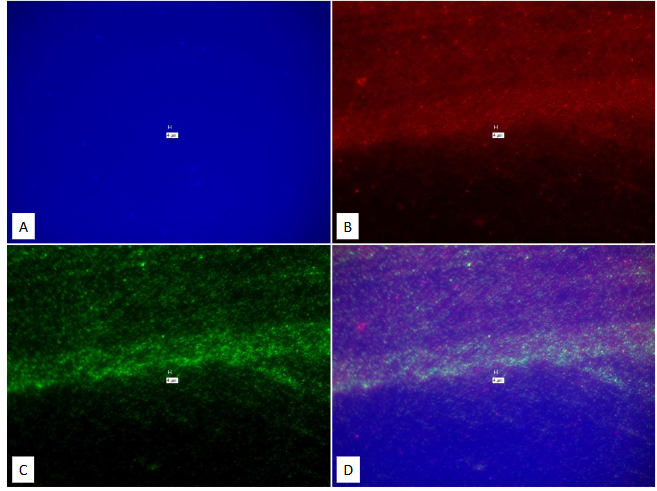
| 1-8 | Human Brain MS | 1,3,6 | No |
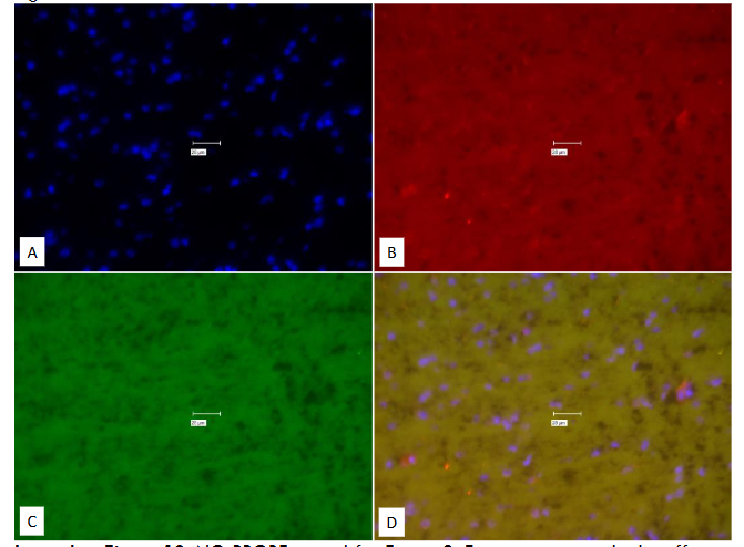
| 9-12 | Human Brain Normal | 9 | No |
| 13-15 | BCG | 13 | No |
| 16-18 | MAP Dominic | 16 | No |
| 19-21 | E. Coli | 19 | No |
| 22-24 | M. tb. | 22 | No |
| 25-28 | M. avium avium | 25,26 | No |
| 25-31 | MAP Dominic | 29 | No |
| 32-33 | Murine Cells | 32 | No |
| 34-43 | Human WBC’s | 34,36,38,40,42 | Possibly |
Results
In Table 2 we show the overview of all the experiments and their results. Note that it is only in Figures 35-43 that we obtain reliable results, specifically with WBCs.
| Figure # | Probes / Stains used | Technique Modification | Filters used | FISH “No-Probe” Control | Positive/Negative Signal | |
|---|---|---|---|---|---|---|
| 1. (Brain) (IS900) Human B-Actin | None | Texas Red | Cy-5 | Yes | “Positive” | |
| 2. (Brain) | None | None | Texas Red | Cy-5 | Yes (for #1) | “Positive” |
| 3. (Brain) | None | None | Texas Red | Cy-5 | Yes (for #1) | “Positive” |
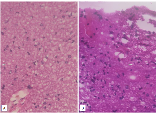
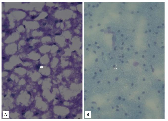
| 4. (Brain) | H&E | NA | ||||
| 5. (Brain) | Luxol Blue | NA | ||||
| 6. (Brain) IS900/ B-Actin | None | Texas Red | Cy-5 | Yes | “Positive” | |
| 7. (Brain) | None | None | Texas Red | Cy-5 | Yes (for #6) | “Positive” |
| 8. (Brain) | None | None | Texas Red | Cy-5 | Yes (for #6) | “Positive” |
| 9. (Brain) IS900/ B-Actin | True Black® | Texas Red | Cy-5 | Yes | “Positive” | |
| 10. (Brain) | None | True Black® | Texas Red | Cy-5 | Yes (for #9) | “Positive” |
| 11. (Brain) | None | None | Texas Red | Cy-5 | Yes (for #9) | “Positive” |
| 12. (Brain) | None | True Black® | Texas Red | Cy-5 | Yes (for #9) | “Positive” |
| 13. (BCG) 16S (Red) 16S(Green) | RNA View® Tissue | Texas Red | Cy-5 | Yes | “Positive” | |
| 14. (BCG) | None | RNA View® Tissue | Texas Red | Cy-5 | Yes (for #13) | “Positive” |
| 15. (BCG) | None | RNA View® Tissue | Texas Red | Cy-5 | Yes (for #13) | “Positive” |
| 16. (Dominic) 16S (Red) 16S(Green) | RNA View® Tissue. Trit C Hope | Yes | “Positive” | |||
| 17. (Dominic) | None | RNA View® Tissue. Trit C Hope | Yes (for #16) | “Positive” | ||
| 18. (Dominic) | None | RNA View® Cell | Texas Red | Cy-5 | Yes (for #16) | “Positive” |
| 19. (E. Coli) 16S (Red) 16S(Green) | RNA View® Tissue. | Texas Red | Cy-5 | Yes | “Positive” | |
| 20. (E. Coli) | None | RNA View® Tissue. | Texas Red | Cy-5 | Yes (for #19) | “Positive” |
| 21. (E. Coli) | None | RNA View® Cell | Texas Red | Cy-5 | Yes (for #19) | “Positive” |
| 22. (M. tb) IS 6110 (Red) | RNA View® Tissue | Texas Red | Yes | “Positive” |
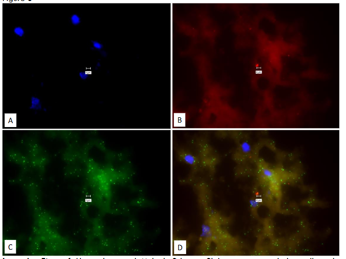
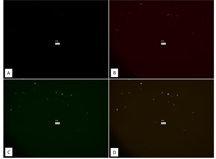

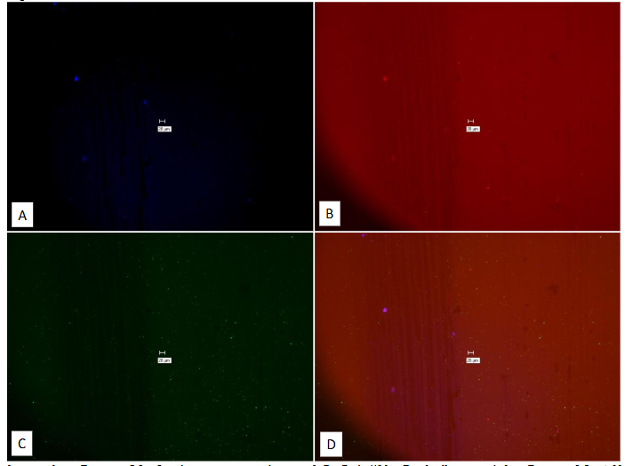
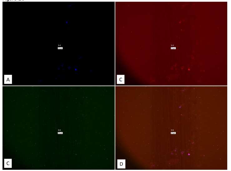
Legend to Figure 24. Study on pure culture of M.tb. This is the “No-Probe” control for Figures 22 & 23, using Affymetrix RNA View® ISH CELL Assay Kit: Thermo Fisher Catalog #: QVT 0001. Panel A = DAPI Panel B = Texas Red Panel C = Cy-5. Panel D = composite of A, B & C. Note “positive” signal in Panels B, C & D. Marker bars in µm indicates magnification of × 60.
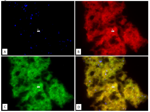
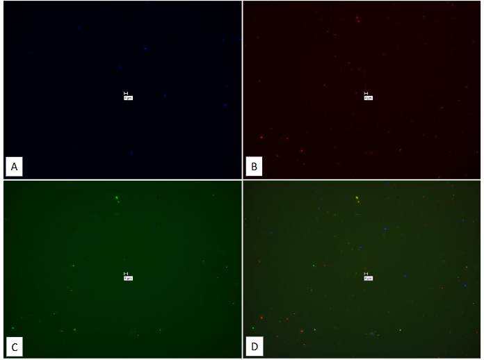
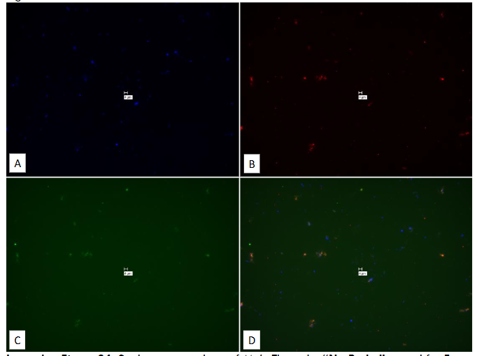
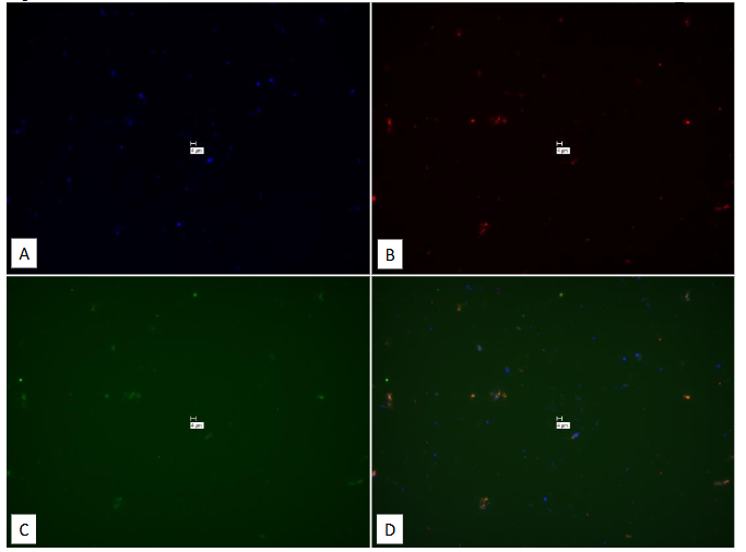
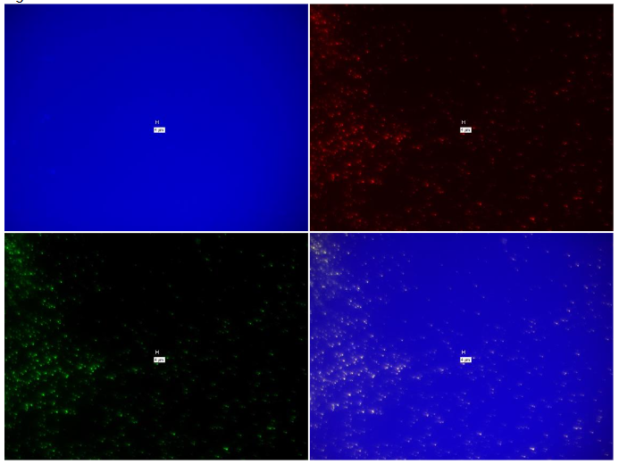
Cultured M. avium subspecies avium gives positive signal (Figures 25 & 26) with different “With-Probes.” Note that spurious positivity in Figure 26 Panel B which should only be positive for MAP is here “positive” for M. avium subspecies avium. The “No-Probe” control for both the TISSUE (Figure 27) and CELL assay (Figure 28) both show false positive signal.
Figure 25
Legend to Figure 25. Study on pure culture of M. avium subspecies avium. “With-Probe” using Affymetrix RNA View® ISH TISSUE Assay Kit Catalog # QVT 0013. Panel A = DAPI Panel B = (Mycobacterium 16S: Type 1 = Red.) Panel C = All bacteria 16S (Type 6 = Green, see Main Text.) Panel D = composite of A, B & C. Note “positive” signal in Panels B, C & D. Marker bars in µm indicates magnification of × 40.
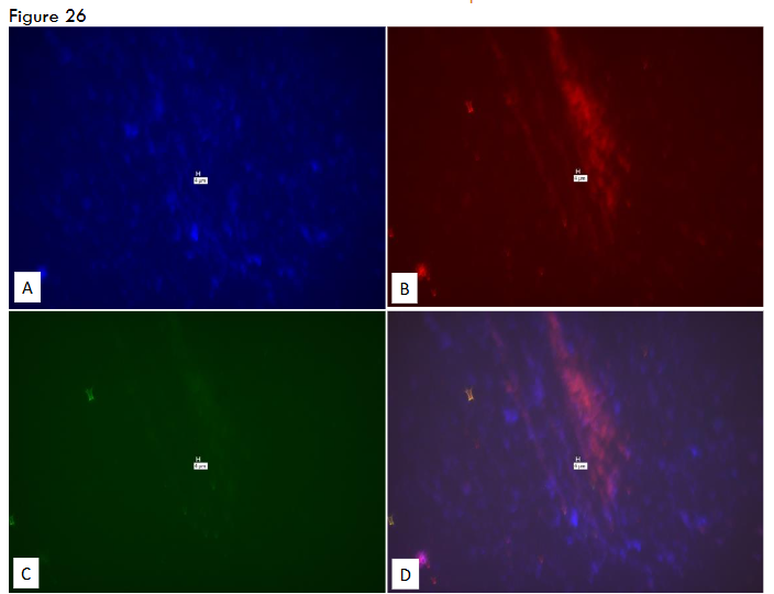
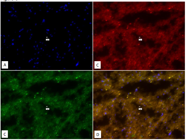
Legend to Figure 26. Study on pure culture of M. avium subspecies avium. Different probes than those used in Figure 25. “With-Probe” using Affymetrix RNA View® ISH TISSUE Assay Kit Catalog # QVT 0013. Panel A = DAPI Panel B = (MAP IS900: Type 1 = Red.) Panel C = All-bacteria 16S (Type 6 = Green, see Main Text.) Panel D = composite of A, B & C. Note “positive” signal in Panels B, C & D. Marker bars in µm indicates magnification of × 40.
Note the “positive” signal in Panel B, which should be specific to MAP, is here positive for M. avium subspecies avium.
Figure 27
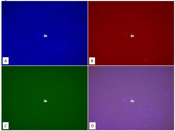
Legend to Figure 27. Study on pure culture of M. avium subspecies avium. “No-Probe” control for Figures 25 & 26 using Affymetrix RNA View® TISSUE Assay Kit Catalog # QVT 0013. Panel A = DAPI Panel B = Texas Red Panel C = Cy-5. Panel D = composite of A, B & C. Note “positive” signal in Panels B, C & D. Marker bars in µm indicates magnification of × 40.
Failure of a proprietary fluorescent in situ hybridization assay to detect M. avium subspecies paratuberculosis in archived frozen brain from patients with MS
Figure 28
Legend to Figure 28. Study on pure culture of M. avium subspecies avium. “No-Probe” control for Figures 25 & 26, using Affymetrix RNA View® ISH CELL Assay Kit: Thermo Fisher Catalog #: QVT 0001. Panel A = DAPI Panel B = Texas Red Panel C = Cy-5. Panel D = composite of A, B & C. Note “positive” signal in Panels B, C & D. Marker bars in µm indicates magnification of × 60.
We next studied a pure culture of MAP Dominic with probes that should only have identified M. tb specific IS 6110.
Figure 29
Legend to Figure 29. Study on pure culture MAP Dominic. “With-Probe” using Affymetrix RNA View® ISH TISSUE Assay Kit Catalog # QVT 0013. Panel A = DAPI Panel B = IS6110 (Tb specific: Type 1 = Red.) Panel C = IS900 (Type 6 = Green, see Main Text.) Panel D = composite of A, B & C. Note “positive” signal in Panels B, C & D. In particular, Panel B should be negative as IS6110 is M. tb specific. Marker bars in µm indicates magnification of × 40.
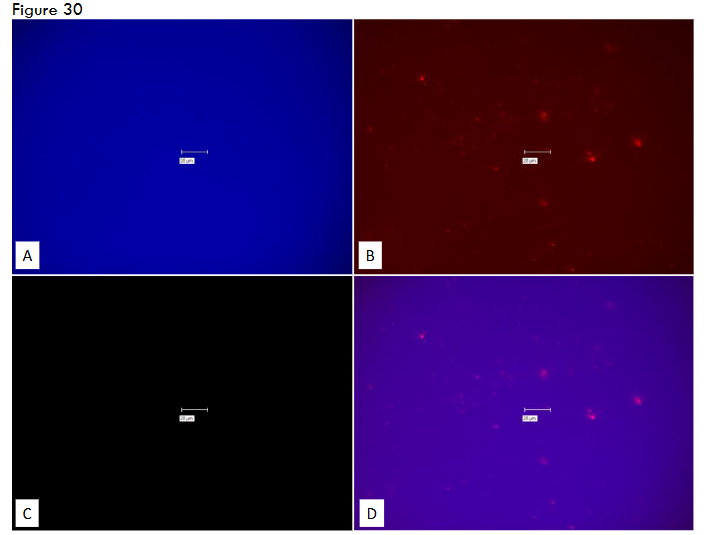
Legend to Figure 30. Study on pure culture of MAP Dominic. “No-Probe” control for Figure 29. Affymetrix RNA View® ISH TISSUE Assay Kit Catalog # QVT 0013. Panel A = DAPI Panel B = Texas Red Panel C = Cy-5. Panel D = composite of A, B & C. Note “positive” signal in Panels B, C & D. Marker bars in µm indicates magnification of × 40.
Figure 31
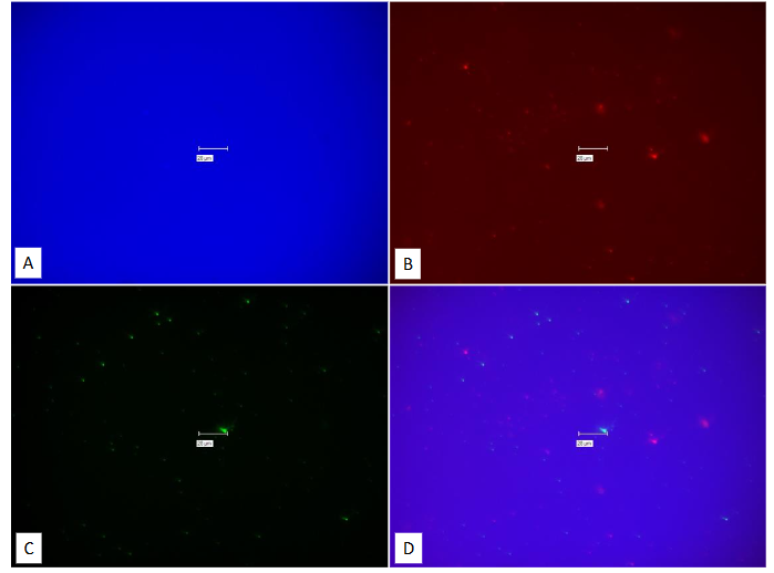
Legend to Figure 31. Study on pure culture of MAP Dominic. “No-Probe” comparison of different filters from Figure 26. Affymetrix RNA View® ISH TISSUE Assay Kit Catalog # QVT 0013. Panel A = DAPI Panel B = TrIt.C. Panel C = “Hope” (see Main Text.) Panel D = composite of A, B & C. Note “positive” signal in Panels B, C & D. Marker bars in µm indicates magnification of × 40.
In contrast to the prediction of the Affymetrix Technical staff (see Main Text), Cy-5 gives less spurious signal than “Hope” in the identical view. Compare Panel C from Figure 30 with Panel C in Figure 31.
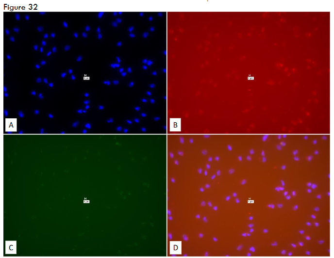
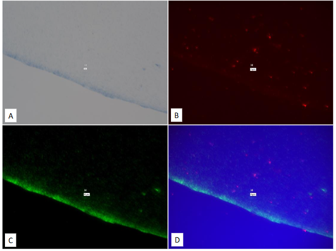
Legend to Figure 32. Pure culture of eukaryotic cells (Mouse RAW264 NIH 93 cells.) “With-Probe” using Affymetrix RNA View® ISH TISSUE Assay Kit Catalog # QVT 0013. Panel A = DAPI Panel B = (MAP IS900: Type 1 = Red.) Panel C = All-bacteria 16S (Type 6 = Green, see Main Text.) Panel D = composite of A, B & C. Note “positive” signal in Panels B & D. Marker bars in µm indicates magnification of × 40
In this pure culture of an uncontaminated mouse cell line, panels B (MAP IS900) & D should be negative.
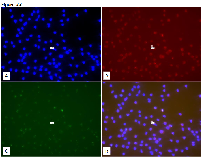
Legend to Figure 33. “No-Probe” control for Figure 32. Pure culture of eukaryotic cells (Mouse RAW264 NIH 93 cells.) Affymetrix RNA View® ISH TISSUE Assay Kit Catalog # QVT 0013. Panel A = DAPI Panel B = Texas Red. Panel C = Cy-5. Panel D = composite of A, B & C. Note “positive” signal in Panels B, C & D. Marker bars in µm indicates magnification of × 40
Figure 34
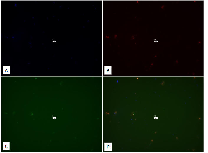
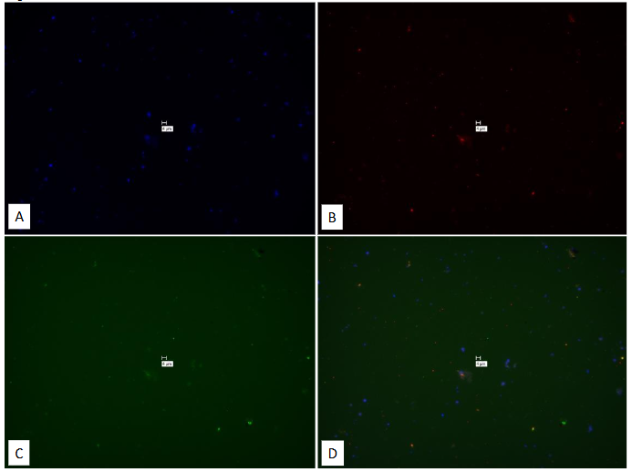
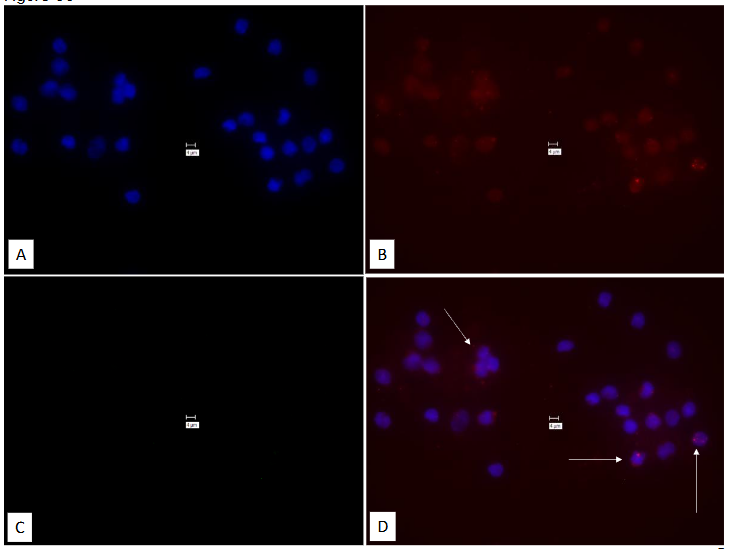
Legend to Figure 34. Studies on Ficoll gradient buffy coat human WBC’s. Affymetrix RNA View® ISH CELL Assay Kit: Thermo Fisher Catalog #: QVT 0001. Blood left at room temperature for one hour. Probes were two human housekeeping genes: Human β-actin (Type 1 = Red) and Human GAPD (glyceraldehyde-3-phosphate dehydrogenase) (Type 6 = Green.) Panel A = DAPI; Panel B = Texas Red; Panel C = Cy-5. Panel D = composite of A, B & C. Marker bars in µm indicates magnification of × 100.
Note positive signal, samples indicated with white arrows, always associated with DAPI positive signal, in Panel D.
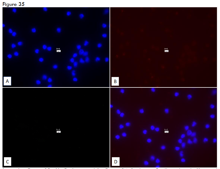
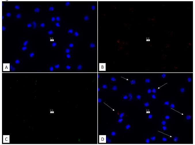
Legend to Figure 35. No-Probe control for Figure 34. Studies on Ficoll gradient buffy coat human WBC’s. Affymetrix RNA View® ISH CELL Assay Kit: Thermo Fisher Catalog #: QVT 0001. Blood left at room temperature for one hour. Panel A = DAPI; Panel B = Texas Red; Panel C = Cy-5. Panel D = composite of A, B & C. Marker bars in µm indicates magnification of × 100.
Note, in this Cell assay Thermo Fisher Catalog #: QVT 000, there is virtually no spurious signal in Panel B & D and zero spurious signal in Panel B. Indicating that the signal in Figure 34 may be reliable.
Figure 36
Legend to Figure 36. Studies on Ficoll gradient buffy coat human WBC’s. Affymetrix RNA View® ISH CELL Assay Kit: Thermo Fisher Catalog #: QVT 0001. Blood stored at 4°C for 24 hours prior to centrifuging for Ficoll gradient. Probes were two human house-keeping genes: Human β-actin (Type 1 = Red) and Human GAPD (glyceraldehyde-3-phosphate dehydrogenase) (Type 6 = Green.) Panel A = DAPI; Panel B = Texas Red; Panel C = Cy-5. Panel D = composite of A, B & C. Marker bars in µm indicates magnification of × 100.
Note positive signal, samples indicated with white arrows, always associated with DAPI positive signal, in Panel D.
In contrast to the one-hour period of RT storage, this signal is predominantly Human β-actin (Type 1 = Red) and should show no signal. “No-Probe” control for Figure 27 TISSUE assay, Filters are Texas Red (Panel B) and Cy-5 (Panel D). The false positive signal is additionally seen when TrItC and “Hope” are the filters used in the identical image (Figure 31.) We conclude that using different filters, as recommended by the Affymetrix technical staff, does not eliminate false positivity. And that Cy-5 (Figure 30; Panel C) gives clearer background that “Hope”, that was predicted by Affymetrix technical staff.
Next the TISSUE assay was evaluated with cultured eukaryotic cells (Mouse RAW264 NIH 93 cells.) “With-Probe” TISSUE Assay; both MAP and bacteria 16S are positive. Similarly, the “No-Probe” control is positive (Figure 33), indicating that the TISSUE assay cannot be used to study cultured eukaryotic cells.
A series of studies were then performed on buffy coat human WBC’s using the CELL (Catalog #: QVT 0001) assay. The initial study evaluated two, human specific, house-keeping genes, (human β-actin (Type 1 = Red) and Human GAPD (glyceraldehyde-3-phosphate dehydrogenase; Type 6; Green). Positive signal was seen, always associate with DAPI positive regions: indicating association with cells (Figure 34). In marked contrast, in the “No-Probe” control, limited signal is seen in panel B and zero spurious signal in panel C. (Figure 35.) We consider that this may indicate that on circulating WBC’s the CELL assay may give reliable signal. Accordingly, the conditions under which this signal could be obtained was studied.
Blood was stored at 4°C for 24 hours prior to being processed With-Probe (Figure 36) and “No-Probe” (Figure 36.) There is zero spurious signal in the “No-Probe” control. Human β-actin may be more stable and an appropriate house-keeping gene than GAPD. Blood was then stored at room temperature (RT) for 24 hours prior to processing.
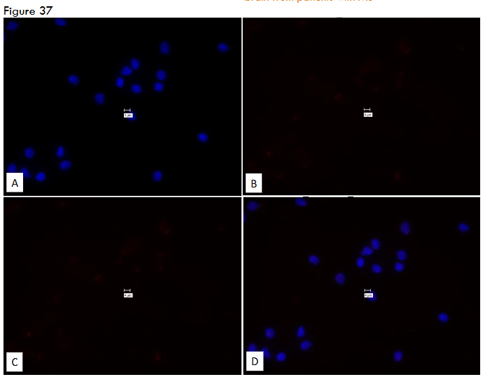
Legend to Figure 37. No-Probe control for Figure 36. Studies on Ficoll gradient buffy coat human WBC’s. Affymetrix RNA View® ISH CELL Assay Kit: Thermo Fisher Catalog #: QVT 0001. Blood stored at 4°C for 24 hours prior to centrifuging for Ficoll gradient. Panel A = DAPI; Panel B = Texas Red; Panel C = Cy-5. Panel D = composite of A, B & C. Marker bars in µm indicates magnification of × 100.
Note, in this Cell assay Thermo Fisher Catalog #: QVT 000, there is zero spurious signal in Panels B, C & D. Possibly indicating that the signal in Figure 36 may be reliable. It may also indicate that Human β-actin may be more stable and an appropriate house-keeping gene than Human GAPD.
Figure 38
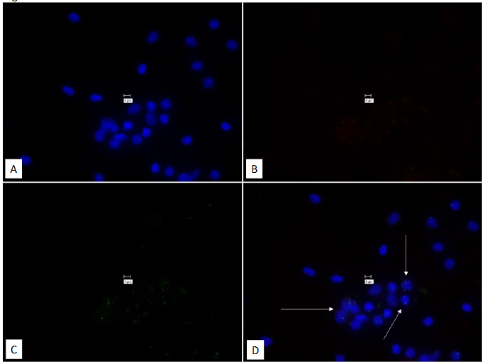
Legend to Figure 38. Studies on Ficoll gradient buffy coat human WBC’s. Affymetrix RNA View® ISH CELL Assay Kit: Thermo Fisher Catalog #: QVT 0001. Blood stored at Room Temperature for 24 hours prior to centrifuging for Ficoll gradient.
gradient. Probes were two human house-keeping genes: Human β-actin (Type 1 = Red) and Human GAPD (glyceraldehyde-3-phosphate dehydrogenase) (Type 6 = Green.) Panel A = DAPI; Panel B = Texas Red; Panel C = Cy-5. Panel D = composite of A, B & C. Marker bars in µm indicates magnification of × 100.
Note positive signal, samples indicated with white arrows, always associated with DAPI positive signal, in Panel D
Figure 39
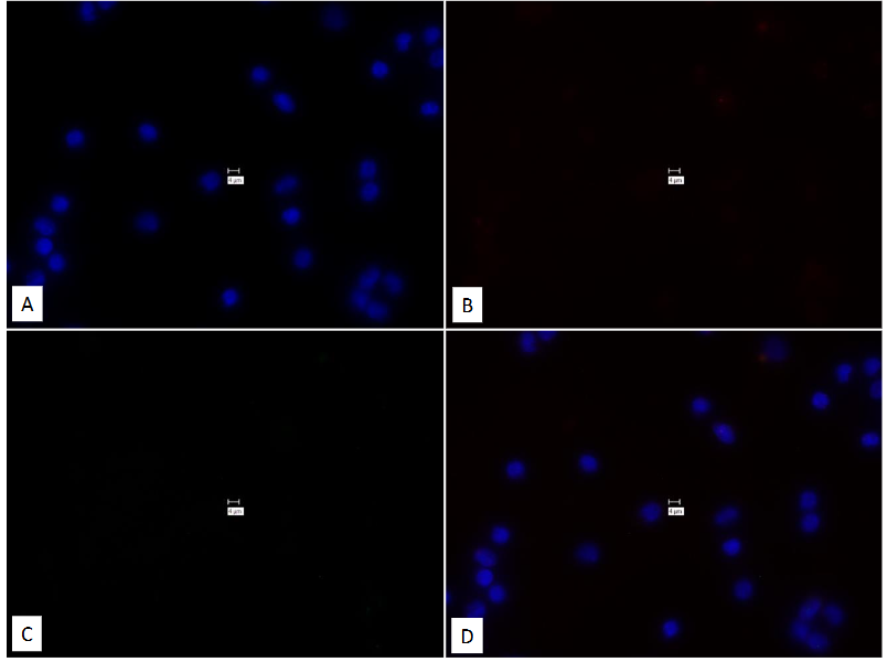
Legend to Figure 39. “No-Probe” control for Figure 38. Studies on Ficoll gradient buffy coat human WBC’s. Affymetrix RNA View® ISH CELL Assay Kit: Thermo Fisher Catalog #: QVT 0001. Blood stored at Room Temperature for 24 hours prior to centrifuging for Ficoll gradient. Panel A = DAPI; Panel B = Texas Red; Panel C = Cy-5. Panel D = composite of A, B & C. Marker bars in µm indicates magnification of × 100.
Note, in this Cell assay Thermo Fisher Catalog #: QVT 000, there is zero spurious signal in Panels B, C & D. Possibly indicating that the signal in Figure 38 may be reliable. Intriguingly, human GAPD is also present (see Figure 38, Panel D, white arrows, green signal.) It may be possible that transportation of blood samples may be possible at ambient temperature for 24 hours.
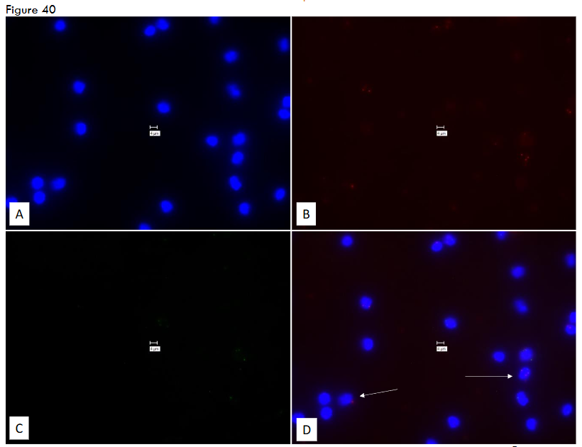
Legend to Figure 40. Studies on Ficoll gradient buffy coat human WBC’s. Affymetrix RNA View® ISH CELL Assay Kit: Thermo Fisher Catalog #: QVT 0001. Blood stored at 4°C for 74 hours prior to centrifuging for Ficoll gradient. Probes were two human house-keeping genes: Human β-actin (Type 1 = Red) and Human GAPD (glyceraldehyde-3-phosphate dehydrogenase) (Type 6 = Green.) Panel A = DAPI; Panel B = Texas Red; Panel C = Cy-5. Panel D = composite of A, B & C. Marker bars in µm indicates magnification of × 100.
Note the limited positive signal, (Human β-actin (Type 1 = Red) samples indicated with white arrows, always associated with DAPI positive signal, in Panel D.
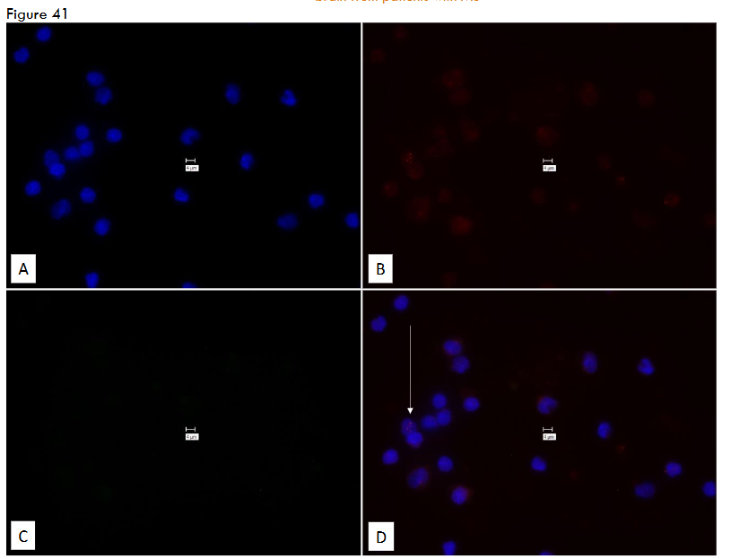
Legend to Figure 41. “No-Probe” control for Figure 40. Studies on Ficoll gradient buffy coat human WBC’s. Affymetrix RNA View® ISH CELL Assay Kit: Thermo Fisher Catalog #: QVT 0001. Blood stored at 4°C for 74 hours prior to centrifuging for Ficoll gradient. Temperature for 74 hours prior to centrifuging for Ficoll gradient. Panel A = DAPI; Panel B = Texas Red; Panel C = Cy-5. Panel D = composite of A, B & C. Marker bars in µm indicates magnification of × 100. Note, in this Cell assay Thermo Fisher Catalog #: QVT 000, there is minimal signal in Panels B & D (see Figure 41, Panel D, white arrows, Red signal.) This may indicate that storage at 4°C for 74 hours prior to centrifuging for Ficoll gradient may not result in reliable signal.
Next blood was stored at 4°C for 74 hours: the “With-Probe” (Figure 40) whereas there is zero spurious signal in the “No-Probe” control (Figure 41.) There is minimal Texas Red signal in the “No-Probe” control (see Figure 41, Panel D white arrow.)
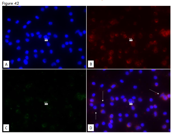
Legend to Figure 42. Studies on Ficoll gradient buffy coat human WBC’s. Affymetrix RNA View® ISH CELL Assay Kit: Thermo Fisher Catalog #: QVT 0001. Blood stored at RT for 74 hours prior to centrifuging for Ficoll gradient. Probes were two human house-keeping genes: Human β-actin (Type 1 = Red) and Human GAPD (glyceraldehyde-3-phosphate dehydrogenase) (Type 6 = Green.) Panel A = DAPI; Panel B = Texas Red; Panel C = Cy-5. Panel D = composite of A, B & C. Marker bars in µm indicates magnification of × 100.
Note positive signal for both Human β-actin (Type 1 = Red) and Human GAPD (Type 6 = Green.) See Figure 42 Panel D white arrows.
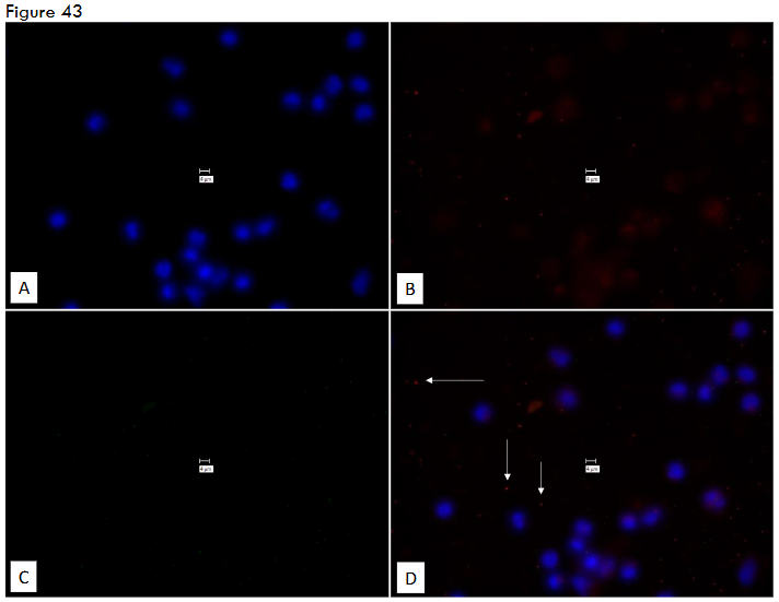
Legend to Figure 43. “No-Probe” control for Figure 42. Studies on Ficoll gradient buffy coat human WBC’s. Affymetrix RNA View® ISH CELL Assay Kit: Thermo Fisher Catalog #: QVT 0001. Blood stored at Room Temperature for 74 hours prior to centrifuging for Ficoll gradient. Panel A = DAPI; Panel B = Texas Red; Panel C = Cy-5. Panel D = composite of A, B & C. Marker bars in µm indicates magnification of × 100.
Note, in this CELL assay Thermo Fisher Catalog #: QVT 000, although some Texas Red signal is seen (Figure 43: Panel D white arrows), it is not associated with DAPI positive regions. This indicates that not associated with WBC’s. It may be possible that transportation of blood samples may be possible at ambient temperature for 74 hours, prior to processing for Buffy coat.
Finally, blood was stored at RT for 74 hours prior to processing. Signal is seen for both Human β-actin and GAPD (Figure 42 see Panel D white arrows.) In contrast, in the “No-Probe” control any spurious signal is not DAPI associated (Figure 43 see Panel D white arrows.) It is therefore possible, that reliable signal might be obtained following transportation of blood samples at ambient temperature for 74 hours, prior to processing for Buffy coat.
As previously stated, ⁴² we repetitively contacted the Technical staff at ThermoFisher Affymetrix in an attempt to prevent false positive signal in the No-probe controls. We have shown that this false positive “No-probe” signal cannot be ascribed to contamination of the negative control slide by probes during the post hybridization wash, by increasing the stringency or duration of the HCL pretreatment, nor using different filters (TrItC for Texas-Red, and for Cy-5 a custom recommended filter set “Hope.” ⁴² Several of these modifications were recommended by the technical staff at Affymetrix.
A proprietary FISH assay has been performed according to the recommended conditions of the vendor. Purportedly positive signal was detected for both MAP (IS900) and our eukaryotic housekeeping gene, human β-actin. However, repetitively the “No-Probe” negative control for a given experiment showed obviously false “positive” signal.
Discussion
It is concluded that when the assay is performed according to the Affymetrix ViewRNA ISH Tissue 2-Plex Assay recommended instructions, it cannot be used for FISH studies to identify the RNA of MAP on previously frozen human brain from individuals with and without Multiple Sclerosis or on pure bacterial cultures. In contrast, when studying human WBC’s, we observed reliable signal with no spurious background. These studies (see Figures 34-43) were performed using probes that identified human house-keeping genes, not mycobacterial pathogens. They were used as controls and were not the purpose of the study, which was to evaluate whether we could detect FISH signal of mycobacterial pathogens in human brain from subjects with MS. These arguably reliable experiments were achieved using the Affymetrix ViewRNA ISH “CELL” Assay Kit; Invitrogen by Thermo-Fisher Scientific: Catalog Number: QVC0001) and not the Affymetrix ViewRNA ISH “TISSUE” Assay Kit. These appropriate WBC data may indicate the possibility, in future studies, of identifying pathogens in circulating macrophages that have ingested infecting disease-causing organisms. Accordingly, we studied WBCs under a variety of conditions. We find that storage of WBCs at room temperature (but not 4°C) for up to 72 hours permitted reliable data to be achieved. These observations are the same as the timespan that we use with a phage assay that identified MAP on circulating WBCs in cattle with Johne disease.
Limitations:
In this study we asked a binary question. Is MAP RNA present or absent in brains of humans with and without Multiple Sclerosis? Especially when the target is expected to be in low abundance, any background may result in a false positive interpretation and is unacceptable. In contrast, when a change in gene expression is being quantified, for example comparing normal with inflamed tissue, a low FISH signal to noise background may be acceptable. Accordingly, our conclusions apply only to frozen human brain from individuals with and without Multiple Sclerosis. We cannot comment on other scientific investigations that employ the Affymetrix ViewRNA ISH Tissue 2-Plex Assay on human brain. Likewise, our conclusions only apply to frozen, not fresh human brain.
Declarations:
- Ethics approval: This study was approved by the Research & Development Committee at the VAMC Bronx NY (0720-06-038.)
- Consent to participate: Tissue is non-identifiable. No consent is necessary.
- Consent for publication: Not Applicable
- Availability of data and material: Competing interests
Dr. Greenstein has three MAP related patents based on his published work in this field.
Funding:
The project was funded intramurally by the Bronx Veterans Research Fund Inc. C. Heffner provided an unrestricted grant which was used to purchase the Keyence microscope. Neither the Bronx Veterans Research Fund Inc., nor C. Heffner had any role in the study design, data collection and analysis, decision to publish, or preparation of the manuscript.
Authors’ Contributions:
- Conceived and designed the study: RJG.
- Performed the study: PSF, RJG.
- Reviewed the data: RJG, PSF, STB.
- Wrote the manuscript. RJG.
- Read and approved the final version of the manuscript: RJG, PSF, STB.
- Each author agrees to be accountable for all aspects of this work in ensuring that questions related to the accuracy or integrity of any part of the work are appropriately investigated and resolved. RJG, PSF, STB.
Acknowledgements:
The Bronx Veterans Affairs Research Foundation provided the facility in which this study was performed (see Funding above.) We thank L. Su for her technical assistance.
References:
- Charcot M. Histologie de le sclerose en plaque. Gaz Hop Paris 1868;141:554-8.
- Anonymous. Guy’s Hospital: Case of Insular Sclerosis of the Brain and Spinal Cord (under the care of Dr. Moxon). Lancet 1873;1:236.
- Thompson AJ, Baranzini SE, Geurts J, Hemmer B, Ciccarelli O. Multiple sclerosis. Lancet 2018;391(10130):1622-1636. Doi: 10.1016/S0140-6736(18)30481-1.
- Libbey JE, Cusick MF, Fujinami RS. Role of pathogens in multiple sclerosis. Int Rev Immunol 2014;33(4):266-83. (In eng). Doi: 10.3109/08830185.2013.823422.
- Libbey JE, Fujinami RS. Potential triggers of MS. Results Probl Cell Differ 2010;51:21-42. Doi: 10.1007/400_2008_12.
- Marrodan M, Alessandro L, Farez MF, Correale J. The role of infections in multiple sclerosis. Mult Scler 2019;25(7):891-901. Doi: 10.1177/1352458518823940.
- Alelyani M, Gameraddin M, Alshahrani R, et al. Assessment of vitamin D status and associated risk factors in high-altitude populations affected by multiple sclerosis: A case-control study. Medicine 2024;103(22):e38369. (In eng). Doi: 10.1097/md.0000000000038369.
- Gombash SE, Lee PW, Sawdai E, Lovett-Racke AE. Vitamin D as a Risk Factor for Multiple Sclerosis: Immunoregulatory or Neuroprotective? Front Neurol 2022;13:796933. Doi: 10.3389/fneur.2022.796933.
- Rotstein DL, Marrie RA, Maxwell C, et al. MS risk in immigrants in the McDonald era: A population-based study in Ontario, Canada. Neurology 2019;93(24):e2203-e2215. Doi: 10.1212/WNL.0000000000008611.
- Sabel CE, Pearson JF, Mason DF, Willoughby E, Abernethy DA, Taylor BV. The latitude gradient for multiple sclerosis prevalence is established in the early life course. Brain : a journal of neurology 2021;144(7):2038-2046. Doi: 10.1093/brain/awab104.
- Alter M, Leibowitz U, Speer J. Risk of multiple sclerosis related to age at immigration to Israel. Archives of neurology 1966;15(3):234-7. (In eng). Doi: 10.1001/archneur.1966.00470150012002.
- Dean G, Kurtzke JF. On the risk of multiple sclerosis according to age at immigration to South Africa. Br Med J 1971;3(5777):725-9. (In eng). Doi: 10.1136/bmj.3.5777.725.
- Munger KL, Zhang SM, O’Reilly E, et al. Vitamin D intake and incidence of multiple sclerosis. Neurology 2004;62(1):60-5. (In eng). Doi: 10.1212/01.wnl.0000101723.79681.38.
- Mowry EM. Vitamin D: evidence for its role as a prognostic factor in multiple sclerosis. Journal of the neurological sciences 2011;311(1-2):19-22. (In eng). Doi: 10.1016/j.jns.2011.06.035.
- Pierrot-Deseilligny C, Souberbielle JC. Is hypovitaminosis D one of the environmental risk factors for multiple sclerosis? Brain : a journal of neurology 2010;133(Pt 7):1869-88. (In eng). Doi: 10.1093/brain/awq147.
- Smolders J, Torkildsen Ø, Camu W, Holmøy T. An Update on Vitamin D and Disease Activity in Multiple Sclerosis. CNS Drugs 2019;33(12):1187-1199. (In eng). Doi: 10.1007/s40263-019-00674-8.
- Bruce D, Ooi JH, Yu S, Cantorna MT. Vitamin D and host resistance to infection? Putting the cart in front of the horse. Exp Biol Med (Maywood) 2010;235(8):921-7.
- Hewison M. Vitamin D and the intracrinology of innate immunity. Mol Cell Endocrinol 2010;321(2):103-11.
- Hewison M. Vitamin D and immune function: an overview. The Proceedings of the Nutrition Society 2012;71(1):50-61. (In eng). Doi: 10.1017/S0029665111001650.
- Wang TT, Dabbas B, Laperriere D, et al. Direct and indirect induction by 1,25-dihydroxyvitamin D3 of the NOD2/CARD15-defensin beta2 innate immune pathway defective in Crohn disease. J Biol Chem 2010;285(4):2227-31.
- White JH. Vitamin D deficiency and the pathogenesis of Crohn’s disease. The Journal of steroid biochemistry and molecular biology 2018;175:23-28. Doi: 10.1016/j.jsbmb.2016.12.015.
- Talafha MM, Qasem A, Naser SA. Mycobacterium avium paratuberculosis Infection Suppresses Vitamin D Activation and Cathelicidin Production in Macrophages through Modulation of the TLR2-Dependent p38/MAPK-CYP27B1-VDR-CAMP Axis. Nutrients 2024;16(9) (In eng). Doi: 10.3390/nu16091358.
- Greenstein RJ, Su L, Brown ST. Vitamins A & D inhibit the growth of mycobacteria in radiometric culture. PLoS ONE 2012;7(1):e29631. (In eng). Doi: 10.1371/journal.pone.0029631.
- Greenstein R. Human genetic defects and misinterpreted pharmacological data indicate that Crohn disease is consequent to a mycobacterial infection. Medical Research Archives 2024;12(7). Doi: 10.18103/mra.v12i7.5541.
- Johne HA, Frothingham L. Ein eigenthumlicher fall von tuberculose beim rind (A particular case of tuberculosis in a cow). Dtsch Zeitschr Tiermed, Vergl Pathol 1895;21:438-454.
- Schurr E, Gros P. A common genetic fingerprint in leprosy and Crohn’s disease? N Engl J Med 2009;361(27):2666-8.
- Greenstein RJ, Collins MT. Emerging pathogens: is Mycobacterium avium subspecies paratuberculosis zoonotic? Lancet 2004;364(9432):396-7. Doi: 10.1016/S0140-6736(04)16781-0.
- Dalziel TK. Chronic intestinal enteritis. British Medical Journal 1913;ii:1068-1070.
- Crohn BB, Ginzberg L, Oppenheimer GD. Regional Ileitis. J Amer Med Assoc 1932;99:1323-1328.
- Mishina D, Katsel P, Brown ST, Gilberts EC, Greenstein RJ. On the etiology of Crohn disease. Proceedings of the National Academy of Sciences of the United States of America 1996;93(18):9816-9820.
- Greenstein RJ. Is Crohn’s disease caused by a mycobacterium? Comparisons with leprosy, tuberculosis, and Johne’s disease. The Lancet infectious diseases 2003;3(8):507-14.
- Alonso R, Fernandez-Fernandez AM, Pisa D, Carrasco L. Multiple sclerosis and mixed microbial infections. Direct identification of fungi and bacteria in nervous tissue. Neurobiol Dis 2018;117:42-61. Doi: 10.1016/j.nbd.2018.05.022.
- Minuk GY, Lewkonia RM. Possible familial association of multiple sclerosis and inflammatory bowel disease. N Engl J Med 1986;314(9):586. Doi: 10.1056/NEJM198602273140921.
- Cossu D, Cocco E, Paccagnini D, et al. Association of Mycobacterium avium subsp. paratuberculosis with multiple sclerosis in Sardinian patients. PLoS One 2011;6(4):e18482. Doi: 10.1371/journal.pone.0018482.
- Cossu D, Masala S, Cocco E, et al. Association of Mycobacterium avium subsp. paratuberculosis and SLC11A1 polymorphisms in Sardinian multiple sclerosis patients. Journal of infection in developing countries 2013;7(3):203-7. (In eng). Doi: 10.3855/jidc.2737.
- Cossu D, Masala S, Frau J, Cocco E, Marrosu MG, Sechi LA. Anti Mycobacterium avium subsp. paratuberculosis heat shock protein 70 antibodies in the sera of Sardinian patients with multiple sclerosis. Journal of the neurological sciences 2013;335(1-2):131-3. Doi: 10.1016/j.jns.2013.09.011.
- Cossu D, Masala S, Frau J, et al. Antigenic epitopes of MAP2694 homologous to T-cell receptor gamma-chain are highly recognized in multiple sclerosis Sardinian patients. Molecular immunology 2014;57(2):138-40. Doi:10.1016/j.molimm.2013.09.001.
- Frau J, Cossu D, Coghe G, et al. Role of interferon-beta in Mycobacterium avium subspecies paratuberculosis antibody response in Sardinian MS patients. Journal of the neurological sciences 2015;349(1-2):249-50. Doi: 10.1016/j.jns.2015.01.004.
- Masala S, Cossu D, Palermo M, Sechi LA. Recognition of zinc transporter 8 and MAP3865c homologous epitopes by Hashimoto’s thyroiditis subjects from Sardinia: a common target with type 1 diabetes? PLoS One 2014;9(5):e97621. Doi: 10.1371/journal.pone.0097621.
- Mameli G, Cossu D, Cocco E, et al. Epstein-Barr virus and Mycobacterium avium subsp. paratuberculosis peptides are cross recognized by anti-myelin basic protein antibodies in multiple sclerosis patients. J Neuroimmunol 2014;270(1-2):51-5. Doi: 10.1016/j.jneuroim.2014.02.013.
- Chiodini RJ. Mycobacterium paratuberculosis. JClinMicrobiol 1987;25:796-801.
- Greenstein RJ, Su L, Fam PS, Stabel JR, Brown ST. Failure to detect M. avium subspecies paratuberculosis in Johne’s disease using a proprietary fluorescent in situ hybridization assay. BMC Res Notes 2018;11(1):498. Doi: 10.1186/s13104-018-3601-5.
- Greenstein RJ, Su L, Fam PS, Gurland B, Endres P, Brown ST. Crohn’s disease: failure of a proprietary fluorescent in situ hybridization assay to detect M. avium subspecies paratuberculosis in archived frozen intestine from patients with Crohn’s disease. BMC Res Notes 2020;13(1):96. Doi: 10.1186/s13104-020-04947-0.
- Green EP, Tizard ML, Moss MT, et al. Sequence and characteristics of IS900, an insertion element identified in a human Crohn’s disease isolate of Mycobacterium paratuberculosis. Nucleic acids research 1989;17(22):9063-73.
- Greenstein RJ, Su L, Grant IR, et al. Comparison of a mycobacterial phage assay to detect viable Mycobacterium avium subspecies paratuberculosis with standard diagnostic modalities in cattle with naturally infected Johne disease. Gut pathogens 2021;13(1):30. Doi: 10.1186/s13099-021-00425-5.