


Free Radicals and Antioxidants: A New Paradigm in Medicine
Free Radicals and Antioxidants: A new Paradigm has emerged
Volkmar Weissig¹*
- ¹Midwestern University, College of Pharmacy, Department of Pharmaceutical Sciences, 19555 59th Avenue, Glendale, AZ, USA
OPEN ACCESS
PUBLISHED: 31 July 2024
CITATION: Weissig V, 2024. Free Radicals and Antioxidants: A new Paradigm has emerged. Medical Research Archives, [online] 12(7).
https://doi.org/10.18103/mra.v12i7.5456
COPYRIGHT: © 2024 European Society of Medicine. This is an open-access article distributed under the terms of the Creative Commons Attribution License, which permits unrestricted use, distribution, and reproduction in any medium, provided the original author and source are credited.
DOI https://doi.org/10.18103/mra.v12i7.5456
ISSN 2375-1924
ABSTRACT
Despite the tremendous progress Redox Biology has made since the turn of the century, several misconceptions about free radicals with Reactive Oxygen Species most prominently among them and about their neutralization by antioxidants still seem to permeate the literature and common thinking as well: First, all Reactive Oxygen Species are toxic byproducts of mitochondrial respiration and as such cause oxidative damage to biological macromolecules. Second, fundamental differences in terms of reactivity and stability between Reactive Oxygen Species are generally neglected and they are widely still being treated as one chemical entity. Third, mitochondria are the major source of toxic Reactive Oxygen Species and fourth, low-molecular weight natural or synthetic antioxidants scavenge free radicals and thereby diminish oxidative damage. This review will debunk these four faulty assumptions, critically address the failure of large-scale clinical trials involving low-molecular weight antioxidants and subsequently propose a new mechanism of action underlying the undisputable health benefits of nutritional antioxidants in fruits and vegetables.
In summary, it will be shown that data published during the last two decades by leading investigators in the field of Redox Biology provide the ground for a paradigm change regarding free radicals and antioxidants in human health and disease.
Introduction: The Antioxidant Paradox
The concept of free radicals, commonly also known as “Reactive Oxygen Species” (ROS) being involved in the etiology and/or pathogenesis of human diseases such as cancer, cardiovascular diseases, atherosclerosis, asthma, diabetes, inflammatory joint diseases as well as numerous neurodegenerative diseases appears as generally accepted in the scientific community¹⁻³ and has found its way into biomedical textbooks⁴˒⁵. By oxidizing biological macromolecules like proteins, lipids and nucleic acids ROS can cause damage to cellular components and ultimately may cause cellular injury and cell death. Likewise, it has been textbook knowledge for decades⁶˒⁷, that there are a large variety of synthetic and natural low-molecular compounds universally known as “antioxidants” which are able to scavenge free radicals in vitro and deactivate ROS thereby preventing oxidative damage to other molecules. In conclusion it seems very reasonable to assume that by giving antioxidants to patients one should be able to prevent, treat or cure all human diseases which are associated with oxidative stress caused by ROS. However, the results of many if not all clinical trials using antioxidants as an intervention have been very disappointing and no FDA-approved antioxidant-based therapy has reached the clinic so far⁸.
In an article with the title “Why Have Antioxidants Failed in Clinical Trials?” Steven Steinhubl (Scripps Research, La Jolla) writes: *“Antioxidant therapies have been evaluated in placebo-controlled trials involving tens of thousands of patients. These clinical trial results have been, to date, mostly negative…”*⁹. Likewise, Anthony William Linnane (1930–2017), in summarizing all failed attempts at developing clinically relevant protocols for antioxidant therapies writes: *“No compelling evidence exists to support the claims that the ingestion of small-molecule antioxidants such as vitamins C, E, b-carotene and others prevent/ameliorate the development of age-associated human diseases.”*¹⁰. Moreover, in several clinical trials the administration of antioxidants has worsened the outcome in many patients, reviewed in⁸. Just two examples shall be given. A multicenter, randomized, double-blind, placebo-controlled prevention trial involving the administration of a mixture of retinol and of b-carotene with over 18,000 enrolled smokers had to be terminated prematurely due the increasing overall mortality in the treatment group¹¹. Further, a trial aimed at reducing the risk of myocardial infarction and cardiovascular death in patients with ischemic heart diseases using alpha-tocopherol treatments resulted in an increase of cardiovascular deaths in the treatment group¹². Even more surprising if not alarming are Martin Bergo’s (Karolinska Institute) discoveries that dietary antioxidants accelerate lung cancer growth and metastasis in mice. Bergo’s group has shown that supplementing the diet with the antioxidants vitamin E and N-acetylcysteine (NAC) significantly increases the progression of the tumor and subsequently reduces survival rate in a mouse model¹³. The same authors also demonstrated that the administration of N-acetylcysteine (NAC) increases lymph node metastases in a melanoma mouse model but has no impact on the number and size of primary tumors¹⁴.
The widespread failure of antioxidants to prevent or treat human diseases in clinical trials, despite the assumed promise they seem to show, is considered the “antioxidant paradox”¹⁵˒¹⁷.
Rather surprisingly yet, antioxidants are increasingly added to food and beverages based on the health benefits they presumably provide¹⁶˒¹⁸ and the belief that “antioxidant is good, more antioxidant is better” appears not only to be embedded in the public mind¹⁶ but also in the mind of a large fraction of the scientific community. As of February 8ᵗʰ, 2023, the Clinical Trial Database at the U.S. National Library of Medicine¹⁹ lists 5301 clinical trials using “antioxidants” as an intervention, among them are 548 trials which are currently recruiting and 188 ongoing trials which do not recruit anymore.
Several factors contributing to the failure of supplementary antioxidants to have an impact on the prevention or therapy of human diseases are being discussed (reviewed in⁸). First, the unknown baseline oxidative stress of the enrolled patients, second, the choice of the specific antioxidants, third, the proper timing of the antioxidant treatment as well as the dose and fourth the bioavailability of the chosen antioxidant at different physiological sites. To overcome the bioavailability problem significant efforts are being made in the drug delivery community to exploit pharmaceutical nanocarriers²⁰˒²¹ for the targeted delivery of a sufficient amount of antioxidant to specific tissues and cells suffering from oxidative stress. Examples are “Antioxidant Liposomes”²², “Nanoantioxidants”²³, “Superoxide Dismutase Enzymosomes”²⁴ and many more (reviewed in⁸). The believe that pharmaceutical nanocarriers might eventually be able to properly address the antioxidant paradox is alive, for example the development of “novel nanotechnology-based systems … for therapeutic delivery of natural antioxidants with improved bioavailability” has recently been reviewed in²⁵.
It is the purpose of this review to demonstrate that the significant progress made in the field of Redox Biology during the last two decades has laid the foundation for a dramatic paradigm shift in what is commonly known or assumed about free radicals and antioxidants in human health and disease.
Four myths surrounding free radicals and Reactive Oxygen Species in Biology
Free radicals are molecules with at least one unpaired valence electron. They are, with a few exceptions, very unstable and highly reactive. Most common examples (more details below) are the superoxide radical anion made by electron transfer (reduction) to molecular oxygen, hydrogen peroxide generated from superoxide radicals by enzymatic dismutation and hydroxyl radicals formed from hydrogen peroxide in the presence of heavy metals like iron cations. Though hydrogen peroxide, in contrast to superoxide radicals and hydroxyl radicals does not possess unpaired electrons, all three oxygen derived species are commonly grouped together (among others) under the name “Reactive Oxygen Species (ROS)”, a fallacy to be discussed below.
Two free radicals made a significant impact on the progress of science, one over a hundred years ago, the other one more recently. Around 1900 Moses Gomberg (1866–1947) at the University of Michigan (USA) tried to synthesize hexaphenylethane from triphenylmethyl chloride in the presence of zinc. To his surprise he isolated a species with an unexpectedly high reactivity, which eventually was identified as the triphenylmethyl radical, the very first isolated organic molecule with an unpaired electron²⁶˒²⁷. Although his discovery was not immediately accepted by many of his fellow scientists, Moses Gomberg is nowadays considered the founder of free radical organic chemistry. The second free radical of historic interest is nitric oxide, which was named in 1992 by Science magazine “Molecule of the Year” and which six years later earned the Nobel Prize in Physiology and Medicine for Robert F. Furchgott (1916–2009), Louis J. Ignarro and Ferid Murad. The Nobel Assembly at the Karolinska Institute in Stockholm has awarded the Nobel Prize to these three US pharmacologists *“for their discovery that the unstable gas, nitric oxide (NO), is an essential regulator of vasodilation”*²⁸. The revelation that NO, a small redox-active molecule with an unpaired electron, which has for decades been considered biologically insignificant, plays a significant role in the human body for inter-cellular messaging has worldwide triggered an enormous interest in Redox Signaling research. Prior to 1998, free radicals (or ROS) were generally regarded as dangerous cellular garbage produced in mitochondria as a byproduct of aerobic ATP generation. Rather surprising to the author, the National Cancer Institute at the NIH (USA) still described as of March 2023 a “free radical” as an “…unstable molecule that is made during normal cell metabolism… Free radicals can build up in cells and cause damage to other molecules, such as DNA, lipids, and proteins.”
This damage may increase the risk of cancer and other diseases”*²⁹.
Despite the tremendous progress Redox Biology has made since the turn of the century there are still four widely held beliefs, the author would like to call “myths”:
- All Reactive Oxygen Species (ROS) are “the same”, they can be treated as one chemical entity.
- All ROS are toxic byproducts of mitochondrial respiration and cause oxidative damage.
- Mitochondria are the major source of ROS.
- All antioxidants “scavenge” free radicals and therefore will diminish oxidative damage.
In the following, these four myths will be discussed based on original data and reviews published during the last two decades by leading investigators in the field of Redox Biology.
1st Myth: All Reactive Oxygen Species are “the same.”
Although it has become evident over the last two decades that the use of “ROS” as an “umbrella term” for oxygen-based free radicals and related reactive molecules is inappropriate, the use of the indiscriminate term “ROS” in the literature appears to be on the rise. While in the year 2000 about 200 publications referenced on Medline had the term “ROS” in their title, this number went up to about 1,400 twenty years later.
All ROS have markedly different reactivities, different species are formed at different concentrations under different conditions at different locations and over different time courses³⁰˒³². For that reason, the leading researchers in this field have in 2022 issued *“Guidelines for measuring reactive oxygen species and oxidative damage in cells in vitro and in vivo”*³¹. In their consensus statement they highlight problems that can arise when utilizing commonly used methods for measurements of ROS and cellular oxidative damage. The authors go even so far to state that *“the application and interpretation of such measurements are fraught with challenges and limitations. This can lead to misleading claims entering the literature and impeding progress…”*³¹.
As Table 1 (adapted from³³) shows, the half-life of different ROS spans over 10 orders of magnitude.
Table 1: Half-life of selected ROS
| ROS species | Symbol | Half-life [seconds] |
|---|---|---|
| Superoxide radical anion | O₂•⁻ | 10⁻⁶ |
| Hydroxyl radical | OH• | 10⁻¹⁰ |
| Alkoxyl radical | RO• | 10⁻⁶ |
| Peroxyl radical | ROO• | 17 |
| Hydrogen peroxide | H₂O₂ | Stable |
| Singlet oxygen | ¹O₂ | 10⁻⁶ |
| Organic peroxide | ROOH | Stable |
While the hydroxyl radical has with 10⁻¹⁰ seconds the shortest half-life, hydrogen peroxide and organic peroxides are considered stable, provided the absence of peroxide reducing agents. Noteworthy, hydrogen peroxide was already shown in 1970 to be a normal feature of aerobic life by Helmut Sies³⁴, who is widely recognized as “Redox Pioneer”³⁵. Sies also developed the model of oxidative stress³⁵, a fundamental concept which has been guiding all research into oxidants and antioxidants ever since.
There are also marked differences in the reactivity of different ROS towards different biological macromolecules, as summarized in³¹. A few examples: The highly reactive superoxide radical anion does not attack most biological molecules; it can quickly react with nitric oxide to form peroxynitrite and it is rapidly converted by superoxide dismutase into hydrogen peroxide. Hydrogen peroxide in turn can oxidize specific protein cysteine residues but does not react with most biomolecules. In the presence of transition metal ions hydrogen peroxide can be converted via the Fenton reaction to hydroxyl radicals, which are indiscriminately highly reactive. As will be discussed in detail below, the generation of superoxide radical anions and its subsequent rapid conversion into hydrogen peroxide play an essential role in redox signaling.
2ⁿᵈ Myth: All Reactive Oxygen Species are toxic byproducts of mitochondrial respiration and cause oxidative damage
The assumption that ROS are toxic byproducts of mitochondrial respiration and subsequently cause oxidative damage is perhaps one of the most prevailing myths. Despite the tremendous new insights into the nature and function of ROS gained since the turn of the century which in summary clearly debunk this myth, a comment published in Science in 2015 still states *“Ever since living systems began to use molecular oxygen… they have had to deal with its detrimental by-products… ROS, which are mainly formed in the mitochondria, cause oxidative damage to cellular components…”*³⁶.
The presumption all ROS being a harmful and unavoidable consequence of an aerobic lifestyle has historic roots³⁷. At the beginning of the 1950s, using a rabbit model, Rebecca Gerschman (1903–1986) studied the protective action against oxygen poisoning by a variety of compounds also known to increase resistance to irradiation and she concluded that just like x-radiation also oxygen poisoning involves the formation of damaging free radicals. In a landmark paper published in 1954 she proposed that free radicals play a major role in oxygen poisoning and contribute to cell aging and death³⁸. Obviously influenced by Rebecca Gerschman’s work³⁹, Denham Harman (1916–2014) proposed in 1956 that *“… aging and the degenerative diseases associated with it are attributed to the attacks of free radicals on cell constituents”*⁴⁰. During the following decade Harman transformed his “Free Radical Theory” into the “Mitochondrial Free Radical Theory of Aging (MFRTA)”⁴¹. In brief, the MFRTA proposes that mitochondrial free radicals, produced as toxic by-products of mitochondrial respiration cause oxidative damage to lipids, proteins and nucleic acids, which in turn is a major driving force in the aging process⁴². Under physiological conditions mitochondria, namely complex I and ubiquinone radicals generated from complex III of the ETC, are indeed the source of superoxide radicals⁴³˒⁴⁴. But the crucial question in the context of the “2ⁿᵈ myth” is whether there are other intracellular sources of ROS, or whether mitochondria are truly the only source of free radicals. In 1974 Bernard Babior (1935–2004) made the seminal discovery that phagocytotically active leukocytes produce superoxide radicals through cytosolic NADPH oxidase⁴⁵˒⁴⁶. This enzyme, also referred to as “NOX” remained until 1999 the only example of a purposeful generation of oxygen free radicals. Its purpose is to generate superoxide radicals as a bactericidal agent during the so-called “respiratory burst”³⁷ which is an event describing the rapid release of reactive oxygen species for the oxidative destroying of pathogens⁴⁷.
In 1999 Young-Ah Suh et al⁴⁸ described the cloning of a gene (called “Mox1” at that time), which encodes a homologue of the catalytic subunit of the superoxide-generating NADPH oxidase of phagocytes. Messenger RNA transcribed from mox1 was found in colon, prostate, uterus and vascular smooth muscle. From their data Suh’s group concluded that there seems to be a link between ROS production by Mox1 to growth control in non-phagocytic cells. During the following two years, i.e. between 1999 and 2001,
five more homologues of the superoxide-generating subunit of the NADPH oxidase were identified⁴⁹–⁵³. They all are transmembrane proteins residing in the cytoplasm membrane of non-phagocytotic cells in many different tissues³⁷. By 2020 the number of identified extra-mitochondrial hydrogen peroxide and superoxide generating human enzymes has grown up to over 40 which obviously presents evidence that the deliberate generation of ROS seems to be a general trait of many if not all cells³². This raises the question why would the generation of ROS be a general feature of perhaps all cells? As mentioned above, hydrogen peroxide does not react with most biomolecules, but it is able to oxidize cysteine residues in proteins, yet the reaction rate is in general very low. For specific proteins however it has been found that the reactivity of hydrogen peroxide towards cysteine residues can increase about 100-fold to 10⁷ M⁻¹ sec⁻¹, depending on the specific protein environment of a particular cysteine residue. Overall, the pKₐ of cysteines in proteins lies between 8 and 9, i.e. the thiol group is not dissociated. Yet neighboring amino acids like serine, tyrosine, threonine, histidine, lysine and arginine are able to stabilize negatively charged thiolate groups, the dissociation products of thiols, via hydrogen bonds, thereby significantly reducing the pKₐ of cysteine residues. The thiolate form in turn is highly sensitive to oxidative modification⁵⁴. Further, the sulfenate, the first oxidation product of cysteines can readily be reduced either via reduction of the disulfide with thioredoxin or via reduction of glutathionylated cysteines with glutaredoxin³².
In conclusion, hydrogen peroxide can selectively and reversibly modify certain cysteine residues in specific target proteins. The obvious question now is what for? What exactly is the purpose of redox modifications of specific cysteine residues of certain target proteins? The answer to this question encompasses perhaps one of the most important discoveries Biochemists and Cell Biologists made during the last three decades. In the middle of the 1990s a new term entered the scientific literature, which is **“redox signaling”**⁴⁶. In brief, this term describes the orchestration and control of most important intracellular processes like proliferation, differentiation, migration, and angiogenesis via redox modification of a large variety of proteins. For example, the reversible cysteine oxidation of target proteins with hydrogen peroxide alters the protein activity and hence is responsible for either launching or terminating intracellular pathways.
The enzyme responsible for turning superoxide radicals into hydrogen peroxide via dismutation, named “superoxide dismutase (SOD)”, was discovered by Joe M. McCord and Irwin Fridovich (1929–2019) at the end of the 1960s⁵⁵˒⁵⁶. In retrospective these researchers wrote in 2014 *“This was an enzyme with the sole apparent function of getting rid of a little-known oxygen-derived free radical called superoxide. To some, it may have seemed to be a solution in search of a problem, as it was neither appreciated that superoxide was produced in significant amounts in biological systems, nor that it was harmful.”*⁴⁶.
Humans possess three types of superoxide dismutase distinguished from each other by their metal cofactor as well as by their protein structure and intracellular location⁵⁷˒⁵⁹. The cytosolic form of Cu/Zn-SOD is named SOD1 or, when localized in the extracellular matrix, SOD3. Mn-SOD is restricted to mitochondria and is referred to as SOD2. Historically, superoxide dismutases have been considered exclusively as antioxidant enzymes, as the exterminators of free superoxide radicals. However, in view of what is known today about redox signaling it appears justified stating that the generation of hydrogen peroxide as a signaling molecule is another, if not the major function of superoxide dismutases. As an antioxidant enzyme or as a generator of a redox-active messenger molecule, superoxide dismutases regulate the steady-state concentrations of superoxide radicals and hydrogen peroxide. Obviously, any interference with this physiological steady-state concentration, for example by ingesting mega doses of antioxidant molecules might hypothetically explain the antioxidant paradox. Anthony Linnane wrote: *“Any random scavenging or deactivation of superoxide anions, hydrogen peroxide, nitrogen oxide or peroxynitrite presumably has the potential to catastrophically derange their second messenger function, which is essential for the regulation of the metabolome’s activities”*¹⁰.
Figure 1 (reprinted with permission from⁶⁰) shows the range of possible fluctuations of intracellular hydrogen peroxide concentrations.
Figure 1: Estimated ranges of hydrogen peroxide concentration in oxidative stress with regard to cellular responses. The intracellular physiological range likely spans between 1 and 10 up to approx. 100 nM H₂O₂, the arrow indicates data from normally metabolizing liver. Stress and adaptive stress responses occur at higher concentrations. Even higher exposure leads to inflammatory response, growth arrest and cell death by various mechanisms. Green and red coloring denotes predominantly beneficial or deleterious responses, respectively. An estimated 100-fold concentration gradient from extracellular to intracellular is given for rough orientation (reprinted with permission from⁶⁰).
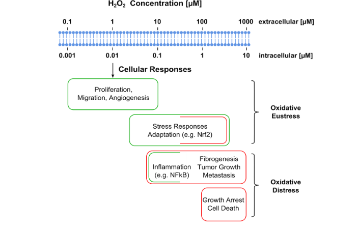
Under physiological conditions in the absence of any type of cellular stress the intracellular concentration of hydrogen peroxide lies between 1 and 100 nM. At this low concentration hydrogen peroxide plays a major signaling function for cell proliferation, differentiation, migration, and angiogenesis. A variety of stress factors including hypoxia and inflammation as well as metabolic keys lead to elevated intracellular hydrogen peroxide concentrations of up to about 1 µM which in turn is responsible for cellular adaptations to stress. A major cellular stress response is based on the activation of the Nrf2 pathway, which will be discussed in more detail below. This elevated level
of hydrogen peroxide is referred to as “oxidative eustress”. Only above about 1 µM hydrogen peroxide levels become toxic to the cell, eventually resulting in cell death. Such conditions are referred to as “oxidative distress”. In summary, there are three intracellular levels of RONS. At so-called physiological concentrations they act as a messenger and are responsible for the metabolome regulation. At elevated levels protective pathways are being activated to enable the cell to adapt to stress and only at a pathological level caused by an unregulated over production of RONS the cell suffers oxidative damage eventually resulting in cellular death. Coming back to the “antioxidant paradox” it becomes obvious that a major problem for any antioxidant-based prevention or therapy of human diseases is the inability of the antioxidant molecule to distinguish between these three levels. But this is not the only hurdle ingested antioxidant molecules would have to overcome to become effective in reducing oxidative stress. Another stumbling block involves the theoretically needed intracellular concentration of administered antioxidant molecules, which will be discussed in detail further below.
3rd Myth: Mitochondria are the major source of Reactive Oxygen Species
The above discussion of the 2ⁿᵈ myth has clearly established that mitochondria are not the only source of RO(N)S. But are mitochondria still the major source, or in other words, the main contributor to the intracellular ROS pool? Due to the lack of appropriate quantitative assays, establishing the relative contributions of mitochondrial and cytosolic sources of superoxide radicals and hydrogen peroxide has proven elusive. A first step towards solving this problem has recently been taken by Martin D. Brand and his group from the Buck Institute for Research of Aging in Novato, CA. Using different inhibitors which specifically affect only one and not any other pathway of cellular hydrogen peroxide production the authors measured the rate of appearance of hydrogen peroxide in the extracellular medium of resting myoblasts in vitro⁶¹. They found that approximately 40% of the total cellular amount of hydrogen peroxide was produced by NADPH oxidases outside mitochondria and around 45% was generated by two mitochondrial sites; 30% by complex III and 15% by complex I. The remaining 15% were attributed to other enzymatic sources. One can of course argue now that mitochondria are the major contributor to the hydrogen peroxide pool in resting myoblasts as the authors did⁶¹, but equally it is justified to say that the major amount of cellular hydrogen peroxide is generated by cytosolic sources. Nevertheless, it is unquestionable that the exact contribution of each source to the intracellular pool of ROS depends on a large variety of factors including the cell cycle, the metabolic state and finally pathologic events.
4th Myth: All antioxidants “scavenge” free radicals and therefore will diminish oxidative damage to biological macromolecules
How did the dogma of antioxidants scavenging free radicals arise? Since the 19ᵗʰ century the chemical and the food industry have been using additives for preventing “fatigue” in rubber and rancidity in food. Although not known yet at that time, all these additives either degrade peroxides or indeed scavenge free radicals by transferring (“donating”) a hydrogen atom, i.e., a single electron and a proton, and turning into a relatively stable radical themselves. The transfer of an electron from an antioxidant molecule onto a reactive free radical, i.e. the scavenging of that free radical by an antioxidant, has been experimentally verified in countless test tubes (in vitro) and has become standard knowledge described in all relevant Chemistry textbooks.
Following Gomberg’s first description of an organic molecule with an unpaired electron around 1900 (see above), studies on the chemistry of oxidation of organic molecules during the first half of the 20ᵗʰ century brought Leonor Michaelis (1875–1949) (better known for his seminal work on enzyme kinetics) to the general conclusion that all biological oxidations involved free radicals⁶².
Apparently inspired by the increasing interest in the chemistry of free radicals, Albert Szent-Györgyi (1893–1986) began to emphasize in the early 1930s their biological significance. Szent-Györgyi, who was awarded the Nobel Prize in 1937 for the discovery of Krebs cycle intermediates and Vitamin C, developed the general concept that *“incorrect free radical formation or elimination is the ultimate cause of cancer”*⁶³. Following Michaelis’ and Szent-Györgyi’s postulates, although never experimentally verified during their lifetime⁶⁴ the idea that free radicals are involved in biological pathways gained progressively hold. Ultimately it was believed that free radicals cause cellular damage and that subsequently eliminating free radicals must be health protective⁶⁴. Gerschman’s work about radicals playing a major role in oxygen poisoning and Harman’s “Free Radical Theory of Aging” (discussed above) further enforced what has now become a dogma.
The historical development as briefly outlined above *“led to the substantiation of a syllogism”*⁶⁴. Because free radicals produce damage and antioxidants scavenge free radicals, antioxidants must provide health benefits. *“The more antioxidants one could pack into cells, the greater would be the resistance to pathology caused by free radicals”*⁶⁴. During the last 30 years, in particular following the passing of the “Dietary Supplement Health and Education Act” by US Congress on May 11, 1994, a law which was called “Snake Oil Protection Act” by the New York Times on October 5, 1993, *“a whole industry has risen based on the proposition that the administration of small antioxidant molecules can be used as a strategy to treat or prevent a large variety of diseases…”*⁸.
So, what exactly is an antioxidant? As of May 2024, the National Cancer Institute at the NIH (USA) defines antioxidants on its dictionary webpage as *“…a substance that protects cells from the damage caused by free radicals (unstable molecules made by the process of oxidation during normal metabolism). Free radicals may play a part in cancer, heart disease, stroke, and other diseases of aging. Antioxidants include beta-carotene, lycopene, vitamins A, C, and E, and other natural and manufactured substances.”*⁶⁵. Noteworthy to the author, this definition seems to ignore the progress redox biology made over the last three decades, there is no mentioning at all of the critical role free radicals play as signaling molecules. There is of course no doubt that free radicals can be neutralized, or “scavenged”, by oxidizable molecules which in turn are much less reactive in their reduced form. Vitamin C may serve as a textbook example. By donating electrons to reactive free radicals, i.e. scavenging them, ascorbic acid is reduced to stable dehydroascorbic acid. Likewise, Vitamin E, tocopherol, embedded in biological membranes reduces free radicals while being oxidized to tocopherol quinone. The crucial role of tocopherol for preventing the oxidation of phospholipid membranes by lipid peroxyradicals remains undisputed (for reasons to be discussed later), so does the regeneration of tocopherol quinone with ascorbic acid. The question to be raised now is whether other low-molecular weight antioxidant molecules, so-called natural or synthetic ones, can scavenge free radicals under physiological conditions thereby protecting proteins, lipids, nucleic acids and carbohydrates from oxidative damage. Already 10 years ago, Henry Jay Forman from the University of Southern California undertook some kinetic considerations and came to a conclusion which has significant implications regarding the efficiency of low-molecular weight antioxidants in vivo⁶⁴. The reaction rate constant for the reaction of for example hydroxyl radicals (HO•) with organic molecules is larger than 10⁹ M⁻¹ s⁻¹ thus approaching limitations of diffusion. In other words, no compound has more HO• scavenging activity than the thousands of molecules present inside the cell. Subsequently, to be 50% effective, any antioxidant would have to be present at equal or greater concentration than all those macromolecules already present inside the cell⁶⁴. Interestingly, as one of the reasons for the failure of numerous clinical trials with antioxidants low intracellular concentrations of the exogenous antioxidant have been discussed and many attempts have been and still are being made to increase the intracellular concentration, i.e. the bioavailability of antioxidants via administering them using different types of pharmaceutical nanocarriers, as already mentioned above and reviewed in⁸. It stands to reason to assume that any exogenous low-molecular weight compound present inside a cell at a concentration equal or larger than that of all endogenous macromolecules already present might have a detrimental effect on cellular homeostasis and metabolism.
Nevertheless, the literature is filled with reports about free radical scavengers, aiming at the identification of potentially useful antioxidants for clinical use. A Medline search in May 2024 using as search term “evaluation of free radical scavenging activity of plant extracts” yielded 3,171 hits. In all studies this author read, all employed different radical scavenging assays were conducted in vitro, i.e. in an environment in which the antioxidant does not have to compete with any other biological macromolecule, or in reverse, in such assays the free radical has nothing to react with but the free radical scavenger.
Free radical scavenging by a non-enzymatic antioxidant mechanism seems only possible for much slower reactions. For example, the rate constants for the reactions of lipid peroxyl radicals with lipids lie in the range between 2–10 × 10¹ M⁻¹ s⁻¹, while their reaction rate with tocopherol is much faster having rate constants between 10³–10⁶ M⁻¹ s⁻¹. Therefore, as already indicated above, tocopherol (Vitamin E) is being considered the only non-enzymatic free radical scavenger in vivo³⁰˒³²˒⁶⁴. In summary, Henry Jay Forman concludes that *“…a free radical scavenger mechanism for antioxidants cannot be substantiated on a kinetic basis in vivo…”*⁶⁴.
Nutritional antioxidants
Does Forman’s conclusion about the inability of antioxidants to scavenge free radicals in vivo render fruits and vegetables as a widely and commonly acknowledged source of antioxidants useless? A recent analysis even highlighted the pharmacological relevance of fruits and plants as natural sources of antioxidants and their beneficial effect on human health⁶⁶. The authors of that analysis concluded that *“adopting functional foods with high antioxidant potential will improve the effective and affordable management of free radical diseases while avoiding the toxicities and unwanted side effects caused by conventional medication”*⁶⁶. How can this apparent contradiction be explained? According to Meccariello and D’Angelo, the most abundant antioxidants in our diet are plant polyphenols⁶⁷. According to these authors, the intake of polyphenols ameliorates age-related phenotypes, including oxidative stress, inflammation, and cellular senescence, both in vitro and in vivo⁶⁷. But are these polyphenols really acting as “radical scavenging antioxidants”? The last 20 years provided new and quite surprising insight into plant polyphenols⁶⁴˒⁶⁷˒⁷². To summarize, it has been demonstrated that although flavonoids have powerful antioxidant activities in vitro (they are able to scavenge a wide range of reactive oxygen, nitrogen and chlorine species), in vivo no antioxidant activities have been established yet. Moreover, it has been found that polyphenols oxidize readily in beverages, in cell culture, in the mouth and in the GI tract. Henry Jay Forman and co-authors concluded that *“it now appears that the chemically important properties of antioxidants in vivo are actually pro-oxidant, i.e. electrophilic”*⁶⁴.
Do nutritional antioxidants paradoxically provide their health benefits by oxidation, and if so, what is their nucleophilic target? This will be discussed in the following and last section of this review.
The Nuclear factor erythroid 2-related factor 2/Antioxidant Response Element The Nuclear factor erythroid 2-related factor 2 /Antioxidant Response Element
The Nuclear factor erythroid 2-related factor 2 /Antioxidant Response Element (abbreviated from here on as Nrf2) was discovered in 1994⁷³. It is a basic leucine zipper transcription factor responsible for the transcriptional regulation of about 250 genes all of which have in common an enhancer sequence in their promoter region called “antioxidant response element”⁷⁴. Its activation results in the expression of numerous enzymes involved in xenobiotic biotransformation, antioxidant metabolism and other cytoprotective pathways. Among the enzymes Nrf2 is coordinating in response to cellular stress are for example NAD(P)H quinone oxidoreductase 1⁷⁵, Sulfiredoxin 1 and Thioredoxin reductase 1⁷⁶, Heme oxygenase-1⁷⁷ and the Glutathione S-transferase family⁷⁸. In summary, Nrf2 activation launches a multitude of cellular defense mechanisms against a variety of pathologies with oxidative stress in the context of this review being the most important one.
Under normal, i.e. stress-free conditions, the Nrf2 protein remains localized in the cytosol and has a half-life of about 20 minutes only⁷⁹. It is associated with another protein, called Kelch-like ECH-associated protein 1 (KEAP1), an E3 ubiquitin ligase substrate adaptor that prepares Nrf2 for fast degradation via the proteasomal pathway⁸⁰˒⁸¹. In other words, KEAP1, under stress-free conditions, controls the short half-life of Nrf2 and renders it a protein of very low abundance. Once the association between Nrf2 and KEAP1 is interrupted, Nrf2 escapes its rapid proteasomal degradation, accumulates in the nucleus and subsequently triggers the expression of cytoprotective proteins with antioxidant enzymes from the perspective of this review the most relevant ones.
Above the question was raised whether nutritional antioxidants like polyphenols, following their quick oxidation before entering tissues and cells, paradoxically provide their health benefits by oxidation. This indeed appears to be the case. The KEAP1 protein (just like other redox sensitive proteins) contains several cysteine residues which are very sensitive to oxidation. As already discussed above, such cysteines are localized in a microenvironment in which neighboring amino acids stabilize their thiolate form via hydrogen bonds thus rendering them sensitive to oxidative post-translational modifications⁵⁴. In addition, there are also dietary antioxidants which do not need to be oxidized to exert their beneficial effects. For example, curcumin, the “Indian gold”, contains electrophilic α,β-unsaturated carbonyl groups, which can easily react in a “Michael addition” with cysteine thiolates. In other compounds, like carnosic acid, known for its antioxidant, anti-inflammatory, and anticarcinogenic activities⁸², an α,β-unsaturated carbonyl group can be formed through oxidation⁶⁴.
Conclusion
Tremendous progress over the last two decades in redox biology and kinetic considerations about the reactivity of free radicals seem to cast doubt about the benefits of natural or synthetic low-molecular weight antioxidants. However, the health benefits of dietary antioxidants in fruits, seed, vegetables, and plants remains unchallenged, though new insights into their stability and chemistry appear to ask for a revision of their supposed mechanism of action. Instead of acting in human cells as an antioxidant, or as a free radical scavenger, such dietary plant-based antioxidants appear to exert their health benefit paradoxically via intracellular oxidation, namely by oxidizing cysteine residues in the Nrf2/Keap1 complex which triggers the launch or activation of cytoprotective pathways.
Conflict of Interest Statement:
The author reports no conflict of interest pertaining to this work.
Funding Statement:
For writing this review the author did not receive any specific grant from funding agencies in the public, commercial or not-for-profit sectors.
Acknowledgement Statement:
None
Major segments of this review were part of a “Theodor Billroth Lecture” hosted by the International Consortium of Research Excellence of the Theodor Billroth-Academy®, given by the author on February 8th, 2023, under the title “Antioxidants: Promise or Delusion?”
References
1. Florence TM. The Role of Free-Radicals in Disease. Australian and New Zealand Journal of Ophthalmology. 1995;23(1):3-7.
2. Lobo V, Patil, A., Phatak, A., Chandra, N. Free radicals, antioxidants and functional foods: Impact on human health. Pharmacognosy Reviews. 2010;4(8):118-126.
3. Sharifi-Rad M, Kumar NVA, Zucca P, et al. Lifestyle, Oxidative Stress, and Antioxidants: Back and Forth in the Pathophysiology of Chronic Diseases. Frontiers in Physiology. 2020;11.
4. McPhee SJ, Ganong, W. F. Pathophyiology of Disease: An Introduction to Clinical Medicine. 5th Edition ed. New York, Chicago, San Francisco, Lisbon, London, Madrid, Mexico City, Milan, New Dehli, San Juan, Seoul, Singapore, Sydney, Toronto: McGraw-Hill, Medical Publishing Division; 2006.
5. Kumar V, Abbas, A. K., Aster, J. C. Robbins Basic Pathology. Philadelphia: Elsevier; 2018.
6. Denisov ET, Afanasef, I. B. Oxidation and Antioxidants in Organic Chemistry and Biology. Boca Raton, FL: CRC Press; 2005.
7. Graham Solomons TW, Fryhle, C. B., Snyder, S. A. Organic Chemistry. 12th ed: Wiley; 2016.
8. Weissig V, Guzman-Villanueva D. Nanocarrier-based antioxidant therapy: promise or delusion? Expert Opin Drug Deliv. 2015;12(11):1783-1790.
9. Steinhubl SR. Why have antioxidants failed in clinical trials? American Journal of Cardiology. 2008;101(10a):14d-19d.
10. Linnane AW, Kios M, Vitetta L. Healthy aging: regulation of the metabolome by cellular redox modulation and prooxidant signaling systems: The essential roles of superoxide anion and hydrogen peroxide. Biogerontology. 2007;8(5):44 5-467.
11. Omenn GS, Goodman GE, Thornquist MD, et al. Effects of a combination of beta carotene and vitamin A on lung cancer and cardiovascular disease. N Engl J Med. 1996;334(18):1150-1155.
12. Stephens NG, Parsons A, Schofield PM, Kelly F, Cheeseman K, Mitchinson MJ. Randomised controlled trial of vitamin E in patients with coronary disease: Cambridge Heart Antioxidant Study (CHAOS). Lancet. 1996;347(9004):781-786.
13. Sayin VI, Ibrahim MX, Larsson E, Nilsson JA, Lindahl P, Bergo MO. Antioxidants accelerate lung cancer progression in mice. Sci Transl Med. 2014;6 (221):221ra215.
14. Le Gal K, Ibrahim MX, Wiel C, et al. Antioxidants can increase melanoma metastasis in mice. Sci Transl Med. 2015;7(308):308re308.
15. Halliwell B. The antioxidant paradox. Lancet. 2000;355(9210):1179-1180.
16. Halliwell B. The antioxidant paradox: less paradoxical now? British Journal of Clinical Pharmacology. 2013;75(3):637-644.
17. Bonner MY, Arbiser JL. The antioxidant paradox: what are antioxidants and how should they be used in a therapeutic context for cancer. Future Medicinal Chemistry. 2014;6(12):1413-1422.
18. Finley JW, Kong AN, Hintze KJ, Jeffery EH, Ji LL, Lei XG. Antioxidants in Foods: State of the Science Important to the Food Industry. Journal of Agricultural and Food Chemistry. 2011;59(13):683 7-6846.
19. Medicine UNLo. Clinical Trials Database. In. https://clinicaltrials.gov/ct2/home2023.
20. Weissig V, Pettinger TK, Murdock N. Nanopharmaceuticals (part 1): products on the market. Int J Nanomedicine. 2014;9:4357-4373.
21. Weissig V, Guzman-Villanueva D. Nanopharmaceuticals (part 2): products in the pipeline. Int J Nanomedicine. 2015;10:1245-1257.
22. Stone WL, Smith M. Therapeutic uses of antioxidant liposomes. Mol Biotechnol. 2004;27(3): 217-230.
23. Du L, Li J, Chen C, Liu Y. Nanocarrier: a potential tool for future antioxidant therapy. Free Radic Res. 2014;48(9):1061-1069.
24. Corvo ML, Marinho HS, Marcelino P, et al. Superoxide dismutase enzymosomes: carrier capacity optimization, in vivo behaviour and therapeutic activity. Pharm Res. 2015;32(1):91-102.
25. Vaiserman A, Koliada A, Zayachkivska A, Lushchak O. Nanodelivery of Natural Antioxidants: An Anti-aging Perspective. Front Bioeng Biotechnol. 2019;7:447.
26. Gomberg M. An instance of trivalent carbon: triphenylmethyl. Journal of the American Chemical Society. 1900;22(11):757-771.
27. Gomberg M. On trivalent carbon. Journal of the American Chemical Society. 1902;24(7):597-628.
28. Smith O. Nobel Prize for NO research. Nat Med. 1998;4(11):1215.
29. (NIH) NCI. Free radical https://wwwcancergov/publications/dictionaries/cancer-terms/def/free-radical. 2023.
30. Forman HJ, Zhang H. Targeting oxidative stress in disease: promise and limitations of antioxidant therapy. Nat Rev Drug Discov. 2021;20 (9):689-709.
31. Murphy MP, Bayir H, Belousov V, et al. Guidelines for measuring reactive oxygen species and oxidative damage in cells and in vivo. Nat Metab. 2022;4(6):651-662.
32. Sies H, Jones DP. Reactive oxygen species (ROS) as pleiotropic physiological signalling agents. Nat Rev Mol Cell Biol. 2020;21(7):363-383.
33. Phaniendra A, Jestadi DB, Periyasamy L. Free radicals: properties, sources, targets, and their implication in various diseases. Indian J Clin Biochem. 2015;30(1):11-26.
34. Sies H, Chance B. The steady state level of catalase compound I in isolated hemoglobin-free perfused rat liver. FEBS Lett. 1970;11(3):172-176.
35. Jones DP, Radi R. Redox pioneer: professor Helmut Sies. Antioxid Redox Signal. 2014;21(18): 2459-2468.
36. Krengel U, Tornroth-Horsefield S. Biochemistry. Coping with oxidative stress. Science. 2015;347(6218):125-126.
37. Lambeth JD. NOX enzymes and the biology of reactive oxygen. Nat Rev Immunol. 2004;4(3):181-189.
38. Gerschman R, Gilbert DL, Nye SW, Dwyer P, Fenn WO. Oxygen poisoning and x-irradiation: a mechanism in common. Science. 1954;119 (3097):623-626.
39. Pomatto LCD, Davies KJA. Adaptive homeostasis and the free radical theory of ageing. Free Radic Biol Med. 2018;124:420-430.
40. Harman D. Aging: a theory based on free radical and radiation chemistry. J Gerontol. 1956; 11(3):298-300.
41. Harman D. The biologic clock: the mitochondria? J Am Geriatr Soc. 1972;20(4):145-147.
42. Sanz A, Stefanatos RK. The mitochondrial free radical theory of aging: a critical view. Curr Aging Sci. 2008;1(1):10-21.
43. Grivennikova VG, Vinogradov AD. Generation of superoxide by the mitochondrial Complex I. Biochim Biophys Acta. 2006;1757(5-6):553-561.
44. Winterbourn CC. Biological Chemistry of superoxide radicals. ChemTexts. 2020;6:1-13.
45. Babior BM, Kipnes RS, Curnutte JT. Biological defense mechanisms. The production by leukocytes of superoxide, a potential bactericidal agent. J Clin Invest. 1973;52(3):741-744.
46. McCord JM, Fridovich I. Superoxide dismutases: you’ve come a long way, baby. Antioxid Redox Signal. 2014;20(10):1548-1549.
47. Hampton LMT, Jeffries MKS, Venables BJ. A practical guide for assessing respiratory burst and phagocytic cell activity in the fathead minnow, an emerging model for immunotoxicity. MethodsX. 2020;7:100992.
48. Suh YA, Arnold RS, Lassegue B, et al. Cell transformation by the superoxide-generating oxidase Mox1. Nature. 1999;401(6748):79-82.
49. Cheng G, Cao Z, Xu X, van Meir EG, Lambeth JD. Homologs of gp91phox: cloning and tissue expression of Nox3, Nox4, and Nox5. Gene. 2001;269(1-2):131-140.
50. Geiszt M, Kopp JB, Varnai P, Leto TL. Identification of renox, an NAD(P)H oxidase in kidney. Proc Natl Acad Sci U S A. 2000;97(14): 8010-8014.
51. Banfi B, Molnar G, Maturana A, et al. A Ca(2+)-activated NADPH oxidase in testis, spleen, and lymph nodes. J Biol Chem. 2001;276(40):375 94-37601.
52. Edens WA, Sharling L, Cheng G, et al. Tyrosine cross-linking of extracellular matrix is catalyzed by Duox, a multidomain oxidase/ peroxidase with homology to the phagocyte oxidase subunit gp91phox. J Cell Biol. 2001;154(4) :879-891.
53. De Deken X, Wang D, Many MC, et al. Cloning of two human thyroid cDNAs encoding new members of the NADPH oxidase family. J Biol Chem. 2000;275(30):23227-23233.
54. Butturini E, Butera G, Pacchiana R, Carcereri de Prati A, Mariotto S, Donadelli M. Redox Sensitive Cysteine Residues as Crucial Regulators of Wild-Type and Mutant p53 Isoforms. Cells. 2021;10(11).
55. McCord JM, Fridovich I. Superoxide dismutase. An enzymic function for erythrocuprein (hemocuprein). J Biol Chem. 1969;244(22):6049-6055.
56. McCord JM, Fridovich I. The utility of superoxide dismutase in studying free radical reactions. I. Radicals generated by the interaction of sulfite, dimethyl sulfoxide, and oxygen. J Biol Chem. 1969;244(22):6056-6063.
57. Antonyuk SV, Strange RW, Marklund SL, Hasnain SS. The structure of human extracellular copper-zinc superoxide dismutase at 1.7 A resolution: insights into heparin and collagen binding. J Mol Biol. 2009;388(2):310-326.
58. Borgstahl GE, Parge HE, Hickey MJ, et al. Human mitochondrial manganese superoxide dismutase polymorphic variant Ile58Thr reduces activity by destabilizing the tetrameric interface. Biochemistry. 1996;35(14):4287-4297.
59. Cao X, Antonyuk SV, Seetharaman SV, et al. Structures of the G85R variant of SOD1 in familial amyotrophic lateral sclerosis. J Biol Chem. 2008;283(23):16169-16177.
60. Sies H. Hydrogen peroxide as a central redox signaling molecule in physiological oxidative stress: Oxidative eustress. Redox Biol. 2017;11: 613-619.
61. Wong HS, Benoit B, Brand MD. Mitochondrial and cytosolic sources of hydrogen peroxide in resting C2C12 myoblasts. Free Radic Biol Med. 2019;130:140-150.
62. Michaelis L. Free radicals as intermediate steps of oxidation-reductions. Cold Spring Harb Symp Quant Biol. 1939;7:33-49.
63. Moss RW. Free Radical: Albert Szent-Gyorgyi and the Battle over Vitamin C. New York: Paragon House Publishers; 1988.
64. Forman HJ, Davies KJ, Ursini F. How do nutritional antioxidants really work: nucleophilic tone and para-hormesis versus free radical scavenging in vivo. Free Radic Biol Med. 2014;66:24-35.
65. Institute NC. Dictionary. https://wwwcancergov/publications/dictionaries/cancer-terms/def/antioxidant. 2024.
66. Rahaman MM, Hossain R, Herrera-Bravo J, et al. Natural antioxidants from some fruits, seeds, foods, natural products, and associated health benefits: An update. Food Sci Nutr. 2023;11(4): 1657-1670.
67. Meccariello R, D’Angelo S. Impact of Polyphenolic-Food on Longevity: An Elixir of Life. An Overview. Antioxidants (Basel). 2021;10(4).
68. Jhoo JW, Lo CY, Li S, et al. Stability of black tea polyphenol, theaflavin, and identification of theanaphthoquinone as its major radical reaction product. J Agric Food Chem. 2005;53(15):6146-6150.
69. Sang S, Hou Z, Lambert JD, Yang CS. Redox properties of tea polyphenols and related biological activities. Antioxid Redox Signal. 2005;7(11-12):1704-1714.
70. Sang S, Lee MJ, Hou Z, Ho CT, Yang CS. Stability of tea polyphenol (-)-epigallocatechin-3-gallate and formation of dimers and epimers under common experimental conditions. J Agric Food Chem. 2005;53(24):9478-9484.
71. Aoshima H, Hirase T, Tada T, et al. Safety evaluation of a heavy oil-degrading bacterium, Rhodococcus erythropolis C2. J Toxicol Sci. 2007;32(1):69-78.
72. Lambert JD, Kwon SJ, Hong J, Yang CS. Salivary hydrogen peroxide produced by holding or chewing green tea in the oral cavity. Free Radic Res. 2007;41(7):850-853.
73. Moi P, Chan K, Asunis I, Cao A, Kan YW. Isolation of NF-E2-related factor 2 (Nrf2), a NF-E2-like basic leucine zipper transcriptional activator that binds to the tandem NF-E2/AP1 repeat of the beta-globin locus control region. Proc Natl Acad Sci U S A. 1994;91(21):9926-9930.
74. Cuadrado A, Rojo AI, Wells G, et al. Therapeutic targeting of the NRF2 and KEAP1 partnership in chronic diseases. Nat Rev Drug Discov. 2019;18(4):295-317.
75. Venugopal R, Jaiswal AK. Nrf1 and Nrf2 positively and c-Fos and Fra1 negatively regulate the human antioxidant response element-mediated expression of NAD(P)H:quinone oxidoreductase1 gene. Proc Natl Acad Sci U S A. 1996;93(25):14960-14965.
76. Neumann CA, Cao J, Manevich Y. Peroxiredoxin 1 and its role in cell signaling. Cell Cycle. 2009;8(24):4072-4078.
77. Jarmi T, Agarwal A. Heme oxygenase and renal disease. Curr Hypertens Rep. 2009;11(1):56-62.
78. Hayes JD, Chanas SA, Henderson CJ, et al. The Nrf2 transcription factor contributes both to the basal expression of glutathione S-transferases in mouse liver and to their induction by the chemopreventive synthetic antioxidants, butylated hydroxyanisole and ethoxyquin. Biochem Soc Trans. 2000;28(2):33-41.
79. Kobayashi A, Kang MI, Okawa H, et al. Oxidative stress sensor Keap1 functions as an adaptor for Cul3-based E3 ligase to regulate proteasomal degradation of Nrf2. Mol Cell Biol. 2004;24(16):7130-7139.
80. Zhang DD, Lo SC, Cross JV, Templeton DJ, Hannink M. Keap1 is a redox-regulated substrate adaptor protein for a Cul3-dependent ubiquitin ligase complex. Mol Cell Biol. 2004;24(24):10941-10953.
81. Cullinan SB, Gordan JD, Jin J, Harper JW, Diehl JA. The Keap1-BTB protein is an adaptor that bridges Nrf2 to a Cul3-based E3 ligase: oxidative stress sensing by a Cul3-Keap1 ligase. Mol Cell Biol. 2004;24(19):8477-8486.
82. Mirza FJ, Zahid S, Holsinger RMD. Neuroprotective Effects of Carnosic Acid: Insight into Its Mechanisms of Action. Molecules. 2023;28(5).