


Herbal Nephropathy: The Risks of Aristolochic Acid
Herbal Nephropathy and Balkan Endemic Nephropathy: The Pathogenic Role of Aristolochic Acid
Koulouridis Ioannis MD, MSc, *¹˒², Koulouridis Efstathios MD, PhD², Maha Qari BDS, MS, DSc³
- King Faisal Specialist Hospital and Research Center Division. Department of Cardiovascular Medicine, Department of Electrophysiology. Jeddah, Saudi Arabia.
- Private Nephrology Clinic. Corfu, Greece.
- bn Sina National College. Assistant Professor, Department of Periodontology. Jeddah, Saudi Arabia.
*Corresponding author: [email protected]
ABSTRACT
Aristolochic acid nephropathy is a worldwide iatrogenic disease affecting individuals consuming herbal remedies derived from Aristolochia and Asarum species. The disease is characterized by prominent renal atrophy and extensive interstitial fibrosis. A considerable proportion of patients, about 30-45 % develop also transitional cell carcinoma mainly of the upper urinary tract. A special attention to the disease was paid in early 90’s because of an epidemic of rapidly progressive interstitial nephropathy affecting young women consuming slimming pills, containing Chinese herbs, supplied from the same clinic in Brussels, Belgium. Detailed investigation of slimming pills showed that they inadvertently contained Aristolochic acid considered as the causative factor of the disease. Soon thereafter it was showed that another devastating chronic interstitial nephropathy accompanied also with increased incidence of upper urothelial cancer, known as Balkan Endemic Nephropathy, is the result of chronic intoxication of affected individuals with low doses of Aristolochic acid derived from the soil contamination from the plant Aristolochia clematitis. Although Aristolochic acid has already characterized as class I carcinogen for humans, Aristolochia species continues to be used as herbal remedies especially in Asia countries and China as well as in westernized communities. It is noteworthy that although Aristolochia species are used as herbal remedies since antiquity there are no references for nephrotoxicity and carcinogenicity until the Belgium epidemic. In this review article we attempt to elucidate the historical evolution of our knowledge upon the etiology of Aristolochic acid nephropathy as well as the underlying mechanisms of Aristolochic acid cytotoxicity.
OPEN ACCESS
PUBLISHED: June 30, 2024
CITATION: Ioannis K, Efstathios K, et al., 2024. Herbal Nephropathy and Balkan Endemic Nephropathy: The Pathogenic Role of Aristolochic Acid, Medical Research Archives, [online] 12(6).https://doi.org/10.18103/mra.v12i6.5565
COPYRIGHT: © 2024 European Society of Medicine. This is an open-access article distributed under the terms of the Creative Commons Attribution License, which permits unrestricted use, distribution, and reproduction in any medium, provided the original author and source are credited.
DOI https://doi.org/10.18103/mra.v12i6.5565
ISSN 2375-1924
Keywords:
Herbal nephropathy, Balkan Endemic Nephropathy, Aristolochic acid toxicity.
Introduction
There is sufficient evidence that humans have been using herbs for relief of pain and other diseases since about 60,000 years ago. The first written document for the preparation of herbal remedies was found in a Sumerian clay slab dated 5,000 years BC. The use of Aristolochia species for preparation of herbal remedies is known since Hippocrates and detailed description of the plant as well as its medicinal use is referred to Theophrastus in 4th century BC.
Nevertheless, recognition of Aristolochia as a poison for humans with nephrotoxic and carcinogenic properties was delayed until early 90’s when an epidemic of rapidly progressing interstitial nephritis, affecting young women consuming slimming pills prepared from Chinese herbs, emerged in Belgium. Detailed investigation of the epidemic showed that aristolochic acid, derived from the plant Aristolochia fangchi, was the culprit of the disease which firstly named as “Chinese Herbs Nephropathy” (CHN). Soon thereafter it became evident that the new disease shares common clinical picture and histological findings with another chronic interstitial nephritis, confined in certain regions of Balkan Peninsula around Danube River, and is known as Balkan Endemic Nephropathy (BEN). Eventually it was showed that the causative factor of BEN was the chronic poisoning of the regional population with small amounts of aristolochic acid derived from the decoy of Aristolochia Clematitis plants growing in the cultivated fields of the endemic territory.
A few years later the first cases of upper urinary tract transitional cell carcinoma emerged among patients suffered from CHN and epidemiological studies among patients suffered from BEN showed also an increased incidence of upper urothelial transitional cell and urinary bladder carcinoma. Detailed histological and molecular studies revealed the presence of aristolochic acid-DNA adducts as well as mutations of p53 protein gene in the lesions derived from dissected kidneys and tumors from affected individuals either from CHN or BEN. These findings established unequivocally the nephrotoxicity and carcinogenicity of aristolochic acid.
Accumulated evidence suggests that the nephrotoxicity and carcinogenicity of aristolochic acid is due to certain metabolites, namely aristolactams, produced by the nitroreduction of the molecule, but until now the metabolic pathways of aristolochic acid are not fully elucidated, mainly because of difficulties in the determination of intermediate metabolic products.
After these discoveries aristolochic acid has been characterized by the International Agency for Research on Cancer (IARC) as a Group I carcinogen for humans and many countries around the World have prohibited the marketing of Aristolochia herbs in their territory because they possess a harm for public health. Unfortunately, they continue to be used especially in Asia countries and China as constituents of herbal medicines as well as in westernized communities supplied by marketing via the internet.
In this review article, we shall try to give a brief historical review of humankind’s attempts to discover plant and chemical remedies from the prehistoric era until nowadays. After that we focus upon the Belgian epidemic and the following investigation to elucidate the etiology of Balkan Endemic Nephropathy. At the end we deal with the molecular mechanisms of aristolochic acid leading to nephrotoxicity and carcinogenicity as well as with ongoing attempts to discover any effective therapy for aristolochic acid toxicity.
Aims and scope
Thirty-two years after the first description of Chinese herbs nephropathy in Belgium it became evident that the irrational use of herbs as medicines from the modern humans for relief of various diseases or pathologic conditions like obesity are related to harmful complications such as nephrotoxicity and carcinogenicity. Aristolochia and Asarum species are characteristic patterns of such complications. Although many Public Authorities over the World have emphasized the harms for public health arising from the unauthorized use of medicinal plants many people continue to use these products because they believe that ‘everything which is coming from nature is effective and safe”. On the other hand, there is no unique legislation system among westernized nations concerning the regulation of marketing and surveillance of safety and efficacy of herbal medicines and among undeveloped and or developing countries especially in Asia and Africa there is a traditional use of these remedies centuries ago which continue to be supported by the present health authorities for many reasons. The present paper aims to add a little evidence upon the hazards of public health coming from the irrational use of herbal medicines such as Aristolochia and Asarum species.
Methodology
We have searched the web via PubMed, Google Scholar and ChatGPT-4. We used search keys as follows: Herbal medicine history, herbs nephropathy, Balkan Endemic Nephropathy, aristolochic acid metabolism, aristolochic acid toxicity, aristolochic acid carcinogenicity. We found seventy (70) papers relevant to the scope of the present review. After thorough inspection of the available literature, we chose review articles, historical articles and research articles mainly published in the last decade. We included also papers referring to original discoveries or papers containing very early information upon aristolochic acid toxicity, which was ignored, and they were published even in the 19th century. At the end we used thirty-nine (39) papers for our references.
Brief historical overview
The use of medicinal plants for the treatment of pain and various diseases by prehistoric man is believed to have begun around 60,000 years ago. However, the oldest documented reference to the use of medicinal plants is found in a Sumerian clay inscription from 5000 B.C. which describes twelve recipes for therapeutic use and refers to more than 250 types of plants. In the broad sense of the term “book,” the first documentary evidence referring to medicinal plants is a collection of texts from ancient Egypt known as the “Ebers Papyrus,” written in 1500 B.C.
The use of germinated seeds for therapeutic purposes is attributed to the mythical emperor of China, Shen Nong, in 2737 B.C., and he is believed to have written the book Shen Nong Ben Cao Jing (The Divine Farmer’s Materia Medica). Historically, the first pharmacognosy book in China was written between 206 B.C. and 220 A.D. titled “Pen Cao Jing,” which, however, included references from previous written texts. It mentions 365 preparations with medicinal properties that came from plants, animals, or minerals.
The foundations of botany in ancient Greece were laid by Θεόφραστος ο Ερέσιος (Theophrastus of Eresos, 371-287 B.C.). In his book titled «Περί φυτών ιστορίαι» (Historia Plantarum), 500 medicinal plants are described, and it became the basis of Botany in the European continent, for which he is considered the “father” of European Botany.
The most important work on pharmacognosy in Europe appeared in the 1st century A.D. and was written by Διοσκουρίδης ο Πεδάνιος (Pedanius Dioscorides), titled «Περί ύλης Ιατρικής» (De Materia Medica). It contained detailed descriptions of more than 1,000 drugs, two-thirds of which were of plant origin, as well as the names of the plants they were derived from. The book became the basis of pharmacognosy in Europe until the Renaissance, for more than 1,500 years.
In the late 15th and early 16th century, Paracelsus laid the foundations for the creation of chemical drugs, and between the 16th and 18th centuries, there was an imperative need to produce synthetic drugs that included medicinal plants and extracts of animal and plant origin. In the early 19th century, alkaloids and glycosides were isolated in pure form from medicinal plants, laying the foundation of scientific pharmacy. The use of pure pharmaceutical substances gradually displaced the use of medicinal plants due to their rapid and potent action. At the beginning of the 20th century, it was found that the action of pure pharmaceutical substances lasted longer than that of plant preparations and had multiple effects and side effects. This led to the first research efforts for the cultivation and production of fresh medicinal plants.
The recognition of side effects and contraindications of synthetic pharmaceutical substances, as well as the public perception that everything derived from nature is healthy and safe, contributed to the renaissance of botanical pharmacy based on the latest scientific knowledge of the cause of diseases, as well as the action and effectiveness of products from medicinal plants. Despite this, in many regions of the world, due to cultural traditions, economic hardship, and difficulty in obtaining chemical drugs, the use of traditional medicines based on herbs continues, especially in China, India, Brazil, and many other Asian countries. It is estimated that even today, 70-80% of the global population, particularly in developing countries, relies on the primary therapeutic use of botanical pharmaceutical preparations. Since the end of the last century, there has been a rapid increase in the use of plant-based preparations even among citizens of developed countries. In the USA, it is estimated that 17.7% of the adult population uses medicinal preparations of plant origin (data from 2007). This fact has led many modern pharmaceutical companies to include herbal medicines in their product range. The World Health Organization estimates that the annual turnover of the global trade in herbal medicines amounts to approximately 80 billion dollars, a figure that seems to be underestimated.
It is well known that botanical pharmaceutical preparations are available on the market from many and varied suppliers, who are difficult to regulate due to the differing policies governing the legislation of individual states. There is also no internationally recognized body for the control and certification of these products, resulting in the recording of dangerous side effects arising from their non-therapeutic actions, contamination by mycotoxins, their interaction with conventional drugs already being taken by consumers, and from the potential presence of other toxic substances. For these reasons, the World Health Organization and the European Union have addressed the problem and have drafted guidelines that describe in detail the various stages of cultivation, harvesting, processing, and storage of medicinal plants intended for use by pharmaceutical industries.
The Belgian Epidemic
In 1993, Vanherweghem and colleagues described in Belgium nine cases of rapidly progressive nephropathy in young women under the age of 50, who were using herbal slimming preparations in pill form, supplied by a specific clinic in Brussels. The authors initially treated 2 cases at their own center and subsequently conducted an epidemiological study at other nephrology centers in Brussels during 1991 and 1992, identifying an additional seven cases of young women with the same characteristics of rapidly progressive nephropathy, who had used the same slimming preparations from the same Brussels clinic. The search for other cases revealed that since 1990, more than 105 women had been affected by the same disease in Belgium. In 8 of the 9 cases, kidney biopsies were performed, showing severe cortical atrophy and extensive interstitial fibrosis with ischemic necrosis of the renal glomeruli, mainly of the cortex. The lesions showed a gradual reduction from the cortex to the medulla and only the renal pyramids remained almost intact. The clinical picture was characterized by the rapid progression of kidney disease, disproportionate anemia relative to the level of kidney function, aseptic pyuria, glucosuria without hyperglycemia, mild proteinuria mainly of tubular origin (β2-microglobulin), and normal blood pressure. The progression of the disease was fast (doubling of serum creatinine within 3 months). 70% of the patients reached end-stage renal failure in less than 2 years.
A year later, Cosyns et al. studied four specimens of kidneys and ureters that were removed from 3 women involved in the Belgian cases who underwent ureteronephrectomy as part of the preparation for kidney transplantation. The thorough study of the specimens showed the presence of atrophic kidneys with smooth outlines and intense interstitial fibrosis, without cellular infiltration, affecting the cortical glomeruli and the renal tubules with a progressive reduction from the cortex to the medullary portion. Sclerosis also included the Bertini columns. Mucoid thickening of the inner layer, sclerosis, and hyalinization of the wall of the interlobar arteries were present in all specimens. Mild to moderate cellular atypia and hyperplasia of the upper urothelium were observed in all specimens. In some of them, the development of connective tissue was found encasing the ureter with moderate dilation of the pelvis.
Beyond the cellular atypia of the upper urothelium, which was observed from the beginning of the disease diagnosis, sporadic cases of urothelial cancer began to appear in patients with Belgian nephropathy who were already undergoing dialysis. This led Nortier et al. to recommend that patients being treated at their center undergo prophylactic total ureteronephrectomy. The results were disappointing. In a total of 39 patients, the presence of urothelial cancer was detected in 18 (46%), while the remaining 19 presented mild to moderate urothelial cellular atypia. The cancers were almost exclusively located in the upper excretory part of the kidney (pelvis – ureters with the same frequency), and only one male patient presented with cancer in the bladder.
The new pathological entity was named Chinese Herbs Nephropathy. However, it seems that the first reference to the disease was made in 1964 by the Chinese doctor Songhan Wu, who described two patients with acute renal failure after consuming a large amount of the plant Aristolochia manshuriensis. The publication was in Chinese and thus is not widely known. Subsequently, other cases of herb-induced nephropathy were reported in the international literature, particularly in Asian countries where there is extensive use of herbal medicines by the population, which are manufactured from herbs and their use is permitted by the Health Services. It is believed that the true incidence of the disease in the general population of Asian countries is underestimated due to its slow progression and the nonspecific symptoms.
Balkan Endemic Nephropathy
It was quickly realized that the new nephropathy presented a similar clinical picture and the same histological findings as the Balkan Endemic Nephropathy (BEN), which is endemic in specific Danubian regions of the Balkan Peninsula. The first description of the disease was made in 1953 in Vratza, Bulgaria, by Tanchev et al., who initially used the term “Endemic Nephropathy of Vratza”. After the first presentation of the disease, further cases were reported in neighboring Yugoslavia and then in Romania. Further investigation of the cases clearly showed that it was a form of interstitial nephropathy with characteristic atrophy of the kidneys and intense interstitial fibrosis, with insidious onset and slow progression, which had a specific geographical distribution and familial but not hereditary character, as it affected individuals living under the same roof with common dietary habits but not necessarily relatives. The geographic distribution of the disease was and remains localized to small villages along the Danube River and its tributaries in the rural areas of Bosnia, Croatia, Serbia, Romania, and Bulgaria. It was also observed that individuals from non-endemic areas who migrate to endemic regions after a stay of 15-20 years become affected, indicating an association with environmental factors, likely dietary habits. The disease has a long-term progression and manifests after the 4th – 5th decade of life. The clinical picture includes the early appearance of anemia, often before the rise of urea and creatinine, mild proteinuria of tubular origin (β2-microglobulin), and normal blood pressure.
The examination of kidneys from autopsy material of patients who died from BEN showed small, shrunken kidneys with smooth outlines, with the kidney weight ranging between 20-60 grams. Microscopic examination of biopsy material in early stages shows atrophy in the proximal convoluted tubules, accompanied by interstitial edema and fibrosis. In advanced stages, the disease is characterized by extensive interstitial fibrosis and tubular atrophy, primarily located in the cortical portion of the kidney. Sclerosis and hyalinization of renal glomeruli are mainly observed in the subcapsular region of the kidneys where the alterations are more pronounced. Apart from the extensive interstitial fibrosis, a characteristic finding of the disease is the absence of interstitial inflammatory reaction.
The frequency of cancer of the excretory part of the urinary system in patients with BEN also increased, ranging between 33-50%. Epidemiological studies have shown that it is 57 times higher in residents of endemic areas who suffer from BEN, while bladder cancer is 12 times more frequent. Urothelial cancer in patients with BEN presents some peculiarities compared to the same cancer in the general population. Specifically, it is more common in the upper excretory part of the kidney and is more frequent in women than in men. The tumors have multiple locations and are more malignant. Their composition is solid, whereas in the rest of the population, they mainly have a papillary structure.
The etiology of Balkan Endemic Nephropathy (BEN) has long been a subject of research and debate, showing significant epidemiological differences compared to herb-induced nephropathy, particularly in terms of the progression of the two diseases and gender affection. The later progresses rapidly and mainly affects women, while the former has a long-term progression, familial distribution, and affects both genders equally, with cohabitation and common dietary habits as common criteria. From the beginning, research focused on environmental factors such as polycyclic aromatic hydrocarbons, heavy metals, hydrocarbons from lignite sediments in endemic areas, and mycotoxins, with ochratoxin A (OTA) being a principal suspect. However, as early as 1969, Ivic, noting previous publications of nephropathy cases in horses fed on hay containing Aristolochia seeds, conducted field research in villages of endemic areas and found that farmers did not remove Aristolochia seeds from the wheat used for bread-making. Subsequent experiments on rabbits fed flour made from Aristolochia seeds developed nephropathy with histological findings similar to those of endemic nephropathy. Additionally, subcutaneous injections of Aristolochia extracts in rodents led to the development of sarcomas at the injection sites. Following these findings, Ivic proposed the theory of a causal association between endemic nephropathy and the accompanying cancers with chronic poisoning of the population in endemic areas by small quantities of Aristolochia seeds. These announcements were made in Croatian and remained obscure for many years.
For historical accuracy, it should be mentioned that according to Grollman, the first to isolate a toxic substance from extracts of the plants Aristolochia Clematitis, Aristolochia Rotunda, and Aristolochia Longa was the Austro-German pharmacologist Julius Pohl in 1891. He named it aristolochin and proposed the molecular formula: C32 H22 N2 O13 (notably, the contemporary molecular formula for aristolochic acid is C33 H20 N2 O13). Pohl subsequently administered aristolochin to rabbits and demonstrated its nephrotoxic action.
The incrimination of Aristolochic Acid
Following the Belgian epidemic, research quickly began to identify a common causative factor for the two diseases and to find possible carcinogenic activity. Tracing the origin and content of the slimming pills used by the Belgian nephropathy cases revealed that one of the herbs supposed to be included in the pills had been mistakenly replaced. Specifically, the ingredient from the plant Stephania tetrandra was replaced by that of the plant Aristolochia fangchi because these two plants have similar pronunciations in the Pin Yin system of Chinese language (Han Fang Ji for the first and Guang Fang Ji for the second). Following this, suspicions regarding the causal association of the disease turned towards Aristolochic Acid, which was eventually isolated by two independent groups, one in Hong Kong and one in Belgium, from a sample of the supposed pure “Stephania” powder procured by Belgium.
Laboratory research on nephroureterectomies from 5 Belgian cases conducted by Schmeiser et al. showed for the first time the presence of Aristolochic Acid-DNA adducts in the lesions from the kidneys and the excretory system of all the cases examined. The results of the study directly linked the causal relationship of Aristolochic Acid with the nephrotoxicity of the Chinese herbs and the cellular atypia of the upper urothelium and the bladder presented by these patients, making it responsible for carcinogenesis as well.
Regarding Balkan Endemic Nephropathy (BEN), Grollman et al. demonstrated the presence of DNA adducts and aristolactams (dA-Aristolactam and dG-Aristolactam) in histological preparations of kidneys and upper urinary tract cancers from affected cases in endemic areas. Additionally, they searched for the presence of mutations in the tumor suppressor protein p53. The most frequent mutations they identified were in the Adenine: Thymine (A: T) pair, accounting for 89%, specifically the transversion A:T → T:A in 78% of cases, whereas this particular mutation does not exceed a frequency of 5% in transitional epithelium tumors from non-endemic areas. The authors also demonstrated the presence of DNA-AL adducts in biopsy material from patients 3 years after cessation of their exposure to aristolochic acid.
Despite existing data directly linking Balkan Endemic Nephropathy (BEN) and the accompanying upper urothelial carcinogenesis to the toxicity of aristolochic acid, the disease’s precise geographic localization to specific Danubian regions of the Balkans remains enigmatic. A recent study by Ivan Brzic et al. showed that the primary ecological distribution of Aristolochia Clematitis spans a wide area covering Southern Europe, Anatolia, and the Caucasus, and then, possibly during the Middle Ages, spread anthropogenically to Eastern and Central Europe. The peculiarity of the endemic areas lies in the fact that Aristolochia plantations are particularly thriving in cultivated fields where they can mix their fruit with wheat or corn harvests. However, similar conditions were also found in Northern Italy and Southern Ukraine, where, to date, no cases of endemic nephropathy nor an increased frequency of urothelial cancers have been reported.
The initial explanation for the mixing of Aristolochia seeds with wheat or corn seeds and the entry of aristolochic acid into the food chain of the inhabitants of endemic areas has been questioned for the following reasons: 1. The ripening of the cereals does not coincide in time with the ripening of the Aristolochia fruits (there is a time difference of at least one month), and 2. The size of the Aristolochia seeds is multiple times larger than that of wheat and corn seeds, making them easily separable during the threshing process. The puzzle of how aristolochic acid entered the food chain of residents in endemic areas was ultimately solved by the studies of Chan, Li, and Pavlovic, who demonstrated the presence of aristolochic acid in the soil of regions affected by Balkan Endemic Nephropathy. They proceeded with experimental cultivations of corn, cucumbers, and onions in the same fields using the same irrigation water and found the presence of aristolochic acid in the edible parts of the plants in quantities sufficient to cause toxic effects. It is now believed that plants and seeds of Aristolochia decomposing in the soil of cultivated fields imbue the soil with aristolochic acid, which dissolves in water and is absorbed by the roots of plants grown in the area. It is then transferred to the edible parts and fruits of the plants, thus entering the food chain of the inhabitants, exerting its nephrotoxic and carcinogenic effects.
The Aristolochia genus includes more than 500 species of plants that thrive in many parts of the world such as Europe, Southeast Asia, Malaysia, China, tropical Africa, and South America. In Greece, 12 species of Aristolochia thrive (there is no mention of Aristolochia clematitis), while in the Danubian regions of the Balkans, the species Aristolochia clematitis grows as a wild weed in cultivated fields. These are perennial evergreen plants, most of which are herbaceous, but some are climbing. The name of the plant was given by the ancient Greek philosopher Θεόφραστος (Theophrastus) in the 4th century B.C., derived from the Greek words «άριστος» (best) and «λοχεία» (childbirth), because it was believed that its use facilitated the delivery of the placenta. A description of the plant and its therapeutic use is mentioned in Theophrastus’ treatise “Historia Plantarum” IX.8.3., which formed the basis of Botany from antiquity to the Middle Ages for the European continent, and thus the name has been preserved to this day.
Aristolochic acid is found abundantly in two plant genera, Aristolochia and Asarum (wild ginger), which are widely distributed around the world. To date, more than 178 plant-derived analogs of aristolochic acid are known. Chemically, the molecule of aristolochic acid is a polycyclic aromatic hydrocarbon of the nitrophenanthrene-carboxylic acid type. Plant extracts contain a plethora of similarly structured molecules, from which two are found in greater proportions: Aristolochic Acid-I (AA-I) and Aristolochic Acid-II (AA-II). Studies in experimental animals showed that the two derivatives of aristolochic acid (AA-I and AA-II) do not possess the same nephrotoxic and carcinogenic activity. It appears that only AA-I has nephrotoxic activity, while both have carcinogenic activity since both were found in tissue lesions of tumors from the urothelium of the experimental animals. After administering aristolochic acid to experimental animals, it was proven that it binds to a plethora of organs such as the stomach, intestine, bladder, liver, spleen, and kidney. In plasma, aristolochic acid is bound to albumin, which reduces its elimination through glomerular filtration and requires active excretion through organic transporters from the epithelium of the renal tubules.
The cumulative dose of aristolochic acid required for the manifestation of nephrotoxicity has been estimated at 0.5 grams, while it is known that not all users of Aristolochia preparations exhibit nephrotoxicity but about 24% do. Users of slimming pills in Belgium developed nephropathy at a rate of 15-20%, while residents of endemic areas in the Balkans present with BEN at a rate of 5-10%. Regarding the carcinogenesis of Aristolochia preparations, it has been estimated that the consumption of at least 200 grams of Aristolochia Fangchi is required for the appearance of urothelial cancer.
The diagnostic criteria for aristolochic acid poisoning are as follows:
- Presence of interstitial fibrosis with progressive reduction from the cortex to the medulla in kidney biopsy.
- Positive history of consuming herbs that contain aristolochic acid.
- The presence of aristolochic acid-DNA adducts or the identification of the transversion A: T → T: A in the p53 gene in samples of kidney tissue or urothelial tumors.
The presence of 2 of the above criteria securely establishes the diagnosis of aristolochic acid poisoning. The presence of one criterion is considered a strong indication and requires further investigation.
In 2002 and then again in 2012, the International Agency for Research on Cancer (IARC) classified aristolochic acid as a Group 1 carcinogen. After recognizing aristolochic acid as a nephrotoxic and carcinogenic agent, many countries around the world have banned the import and distribution of products containing aristolochic acid. Nonetheless, the supply and use of these products continue in Europe and America via the internet, while in China and other Asian countries, many herbs used to this day in traditional medicine contain aristolochic acid.
Metabolism of Aristolochic Acid and Cytotoxicity
Studies in animals and humans have shown that the main metabolites of aristolochic acid are the formation of aristolactams (AL-I and AL-II), which are created by the reduction of the nitro group at the C6 position of the molecule. To a lesser extent, the oxidation of the O-methyl group at the C8 position of the molecule has been demonstrated, leading to the creation of 8-(OH) aristolochic acid (AA-Ia). The nephrotoxic and carcinogenic action of AA is due to the formation of aristolactams, while the oxidation of the O-methyl group leads to the detoxification of the organism.
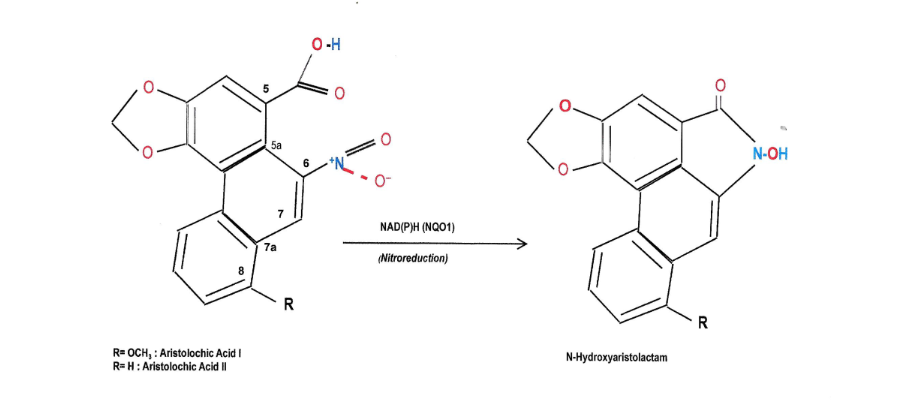
Subsequently, aristolactams are converted into nitrenium-aristolactams, which are potent electrophilic molecules (nitrenium ions). Nitrenium-aristolactams form complexes with the purine bases (adenine-guanine) of DNA at the C7 position of their molecule. The 7-deoxy-adenine-aristolactam-I (7-dA-AAI) and 7-deoxy-adenine-aristolactam-II (7-dA-AAII) are found in the tissue lesions caused by aristolochic acid for many years after an individual’s exposure. Of these, dA-AAI is found more frequently and for a longer duration in complexes with DNA. The reason why DNA-Aristolactam complexes remain for a long time in tissue lesions is because these complexes are primarily formed in areas of DNA that are not transcribed, and therefore, the repair of the damage is delayed.
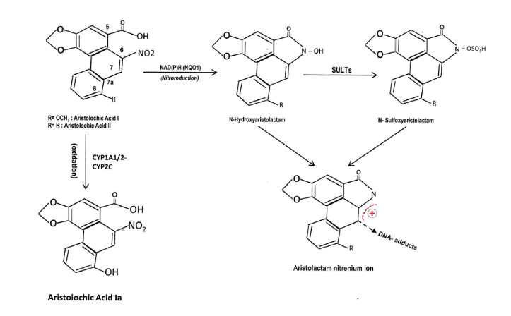
The formation of DNA complexes with nitrenium-aristolactams leads to DNA mutations, consisting of a transversion from adenine to thymine (A: T→T: A), which are responsible for the nephrotoxicity and carcinogenesis of aristolochic acid. The A: T→T: A transversion results in mutations of the tumor suppressor protein p53 molecule (TP53 mutations) and is found at a high frequency in urothelial tumors developed by patients exposed to derivatives of aristolochic acid. The presence of AA-DNA complexes and TP53 mutations in tissue lesions are reliable biomarkers of exposure to aristolochic acid.
The selective targeting of the proximal convoluted tubule by aristolochic acid implies the involvement of Organic Anion Transporters (OATs) in the entry of the acid into the epithelial cells of the renal tubule. It has been demonstrated in experimental animals that the entry of albumin-bound aristolochic acid into the epithelial cells of the kidney is achieved through OAT1, 2, and 3. The administration of probenecid prevents the accumulation of aristolochic acid in cell cultures. The excretion of aristolochic acid into the tubular lumen remains largely unclear. It is speculated that the transporters Multidrug Resistance-Associated protein 1,2 (MRP1,2), which are expressed on the brush border of the proximal convoluted tubule, play a significant role.
Experimental studies based on specific artificial devices known as “Micro Physiological Systems” showed that the nephrotoxic action of aristolochic acid is intensified during its passage through the liver. There are strong indications that the liver, instead of detoxifying the body from aristolochic acid, increases its nephrotoxic effect by approximately 5 times.
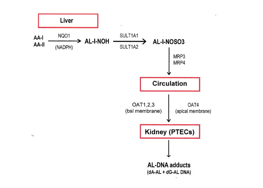
Today, it is believed that aristolochic acid, during its passage through the liver, undergoes reduction of its nitro group via Nicotinamide Adenine Dinucleotide Phosphate (NADPH) and is converted into N-hydroxyaristolactams (AL-I-NOH and AL-II-NOH). This process involves a six-electron reduction and leads to the creation of the electrically charged nitrenium/carbenium species, which can form complexes with the purine bases of DNA. Subsequently, aristolactams bond with a sulfhydryl root, through liver sulfotransferases (SULTs), leading to the creation of sulfonyl complexes of the type of AL-I-NOSO3 and AL-II-NOSO3. These complexes exit the liver cell through organic transporters anchored in the basolateral membrane, known as Multidrug-Resistance Proteins (MRPs), and are released into the bloodstream. The proteins involved in this transfer appear to be MRP3 and MRP4.
Following this pathway, the complexes AL-I-NOSO3 and AL-II-NOSO3 are transported to the kidney through the bloodstream, where they exert their nephrotoxic and carcinogenic action. The entry of the complexes into the renal cells of the proximal convoluted tubule occurs in two ways. One way is intracellular transport from the basolateral side of the cell membrane through Organic Anion Transporters OAT1,2, and 3, where they are abundantly expressed, and the other is from the luminal surface of the cell where the transporter OAT4 is expressed. The first way is based on the bloodstream flow of the complexes, while the second on glomerular filtration.
In vitro and in vivo studies on experimental animals have shown that the derivatives of aristolochic acid (AA-I and AA-II), in addition to the reduction of the nitro group at position 6 of their molecule and the creation of aristolactams, also undergo a smaller scale oxidation of the methoxy group of their molecule, resulting in the creation of 8-(OH) aristolochic acid (AA-Ia), which is less toxic than the aristolactams and is removed from the organism, thus achieving detoxification of the body from aristolochic acid. The problem is that such oxidation occurs almost exclusively in the molecule of AA-I, while the molecule of AA-II is oxidized very little or almost not at all. The oxidation of the methoxy group of aristolochic acid is achieved through the P450 enzyme system, specifically the CYP1A1 and CYP1A2 molecules. Experimental data show that the carbon atom of the methoxy group, at position 8 of the AA-I molecule, is more vulnerable to oxygen than the C8 carbon atom of the aromatic ring in the AA-II molecule, resulting in the creation of the α-C-hydroxylated derivative, which is unstable and easily converted to formaldehyde and AA-Ia (replacement of methoxy group with hydroxyl group). This specific development is thermodynamically favored because it requires less energy, resulting in the oxidative detoxification being almost exclusively limited to AA-I, while AA-II undergoes almost exclusively nitro-reduction to aristolactams and subsequently the formation of DNA complexes.
Therapeutic Efforts
Despite ongoing intensive research, the mechanisms of damage involved in the nephrotoxicity of aristolochic acid have not been fully clarified. Experimental data from numerous studies support that AA’s cellular-level toxicity manifests through increased cellular oxidative stress, apoptosis activation, inflammation induction, and ultimately, the development of interstitial fibrosis. On an experimental level, a plethora of substances that interfere in the evolution of these cellular mechanisms has been tested, showing potential in slowing down or reversing kidney damage progression. These substances, derived from either chemical or plant sources, are applied in cell culture environments or administered in vivo to experimental animals prior to or simultaneously with the administration of aristolochic acid. A few of these substances and their outcomes will be mentioned indicatively.
- Administration of probenecid inhibits the uptake of AA by epithelial cells of the proximal convoluted tubule and prevents renal damage when administered to experimental animals either orally or intraperitoneally.
- Induction of the CYP1A enzymatic system (A1/A2) with substances such as Sudan I, 3-methylcholanthrene, β-Naphthoflavone, Omeprazole, Baicalin, and Tansinone I. These substances have been used only in experimental animals.
- Inhibition of the NQO1 enzymatic system expression with the use of dicoumarol has been used in experimental animals.
- Inhibition of reactive oxygen species (ROS) production with the use of antioxidant vitamins such as vitamin C and E has been used in experimental animals.
- Inhibition of reactive nitrogen species production with the administration of L-arginine in experimental animals.
- Suppression of cell apoptosis with the use of estrogen derivatives and erythropoietin (17-β Estradiol and Erythropoietin) has been used in experimental animals.
- Reduction of the inflammation reaction with the use of corticosteroids (prednisolone) has been used in humans.
- Reduction of the fibrosis process with the use of Bortezomid, Hepatocyte Growth Factor (HGF), and Anti-TGF-β1 antibodies have been used in experimental animals and cell cultures.
In addition to the previous mentioned substances, herbal preparations with anti-inflammatory and antioxidant actions or limiting interstitial fibrosis, such as Dahuang Fuzi Decoction, Fuzheng Huayu Recipe, and Kangxianling, which are based on herbs of traditional Chinese medicine, have been used. This approach to therapeutic efforts in addressing aristolochic acid nephropathy becomes particularly interesting since, in recent years, preclinical studies are underway aiming at the alternative treatment of chronic kidney disease using herbal preparations derived from medicinal plants, and their results are considered encouraging. All the above preparations, except for prednisolone, have been used in experimental animals or cell cultures during the acute phase of damage induction, and there are no safe data for their use in humans. Furthermore, the diagnosis of aristolochic acid nephropathy is usually delayed where histological damage has already been established, and AA intake has preceded on a yearly basis. Therefore, a safe prediction cannot be made either for the therapeutic outcome or for any side effects from their use.
Conclusions
Aristolochic acid nephrotoxicity remains until nowadays a worldwide threat for public health. There are no effective therapeutic measures counteracting Aristolochic acid nephrotoxicity and carcinogenicity. Restrictive measures in the marketing of Aristolochic acid derivatives show promising results in reducing new cases of nephrotoxicity and carcinogenesis. Nevertheless, Aristolochia and Asarum species continue to be used as herbal remedies in Asia and low-income countries as well as in westernized communities via internet marketing.
Declaration
We declare that:
- All authors have participated equally in preparing this manuscript.
- We have no conflict of interest.
- We have no financial support and no funding in writing this manuscript.
- All expenditures for preparing, writing and publishing this manuscript are covered by authors.
Yours sincerely,
Ioannis Koulouridis
Efstathios Koulouridis
Maha Qari
References
- Sewell RDE, Rafician-Kopaci M. The history and ups and downs of herbal medicines usage. J Herb Med Pharmacol. 2014; 3(1): 1-3.
- Petrovska BB. Historical review of medicinal plants’ usage. Pharmacogn Rev. 2012; 6(11): 1-5.
- Grollman AP, Marcus DM. Global hazards of herbal remedies: lessons from Aristolochia. EMBO rep 2016; 17(5): 619-625.
- Chen T. Genotoxicity of Aristolochic Acid: A Review. JFDA 2007; 15 (4): 387-399.
- Jamshidi-Kia F, Lorigooini Z, Amini-Khoel H. Medicinal plants: Past history and future perspective. J Herb Med Pharmacol. 2018; 7(1): 1-7.
- Raghavendra HL, Yogesh HS, Gopalakrishna B, Chandrashekhar VM, Sathish Kumar BP, Vadlapudi K. An overview of herbal medicine. Int J Ph Sci. 2009; 1(1): 1-20.
- Rivera JO, Loya AM, Caballos R. Use of Herbal Medicines and Implications for Conventional Drug Therapy Medical Sciences. Altern Integ Med. 2013; 2:6 (doi.org/10.4172/2327-5162.1000130).
- WHO monographs on selected medicinal plants. VOLUME 1. World Health Organization. Geneva. 1999.
- Directive 2004/24/EC of the European parliament and of council of 31 March 2004.
- Vanherweghem JL, Depierreux M, Tielemans C et al. Rapidly progressive interstitial renal fibrosis in young women: association with slimming regimen including Chinese herbs. Lancet 1993 Feb 13; 341 (8842): 387-91.
- Cosyns JP, Jadoul M, Squifflet JP, De Plaen JF, Ferluca D, van Yperssele de Strihu C. Chinese herbs nephropathy: A clue to Balkan endemic nephropathy? Kidney Int 1994; 45: 1680-1688.
- Jadot I, Decleves A-E, Nortier J, Caron N. An Integrated View of Aristolochic acid Nephropathy: Update of the Literature. Int J Mol Sci. 2017; 18, 297: 1-24.
- Meyer MM, Chen T-P, Bennett WM. Chinese herb nephropathy. Bayl Univ Med Cent Proc. 2000; 13 (4): 334-337.
- Nortier JL, Martinez M-C M, Schmeiser HH et al. Urothelial Carcinoma Associated with the Use of a Chinese Herb (Aristolochia fangchi). N Engl J Med. 2000; 342: 1686-1692.
- Zhou Q, Jiang L, Su T, Liu G, Yang L. Overview of Aristolochic acid nephropathy: an update. Kidney Res Clin Pract. 2023; 42(5): 579-590.
- Dimitrov PS, Simeonov VA, Stein AD. Balkan endemic nephropathy in Vratza, Bulgaria, 1964-1987: An epidemiologic analysis of population-based disease registers. Eur J Epidemiol. 2001; 17(9): 847-853.
- Bamias G, Boletis J. Balkan Nephropathy: Evolution of Our Knowledge. AJKD 2008; 52(3): 606-616.
- Velickovic LJ, Dolicanin Z, Stefanovic V. Endemic Nephropathy And Upper Urothelial Carcinoma- New Insights In Molecular Biology. Contributions. Sec. Med. Sci. 2014.
- Crosby LM, Tatu CA, Pavlovic N, Oren WH. The continuing medical mystery of Balkan Endemic Nephropathy. J Rare Disorders 2015; 3(2): 22-37.
- De Broe ME. Chinese herbs nephropathy and Balkan endemic nephropathy: toward a single entity, Aristolochic acid nephropathy. Kidney Int. 2012; 81: 513-515.
- Ivic M. [Etiology of endemic nephropathy]. Lijec Vjesn. 1969; 91(12): 1273-1281 [Article in Croatian].
- Grollman AP. Aristolochic Acid Nephropathy: Harbinger of a Global Iatrogenic disease. Environ Mol Mutagen. 2013; 54: 1-7.
- Pohl J. Ueber das Aristolochin, einen giftigen Bestandtheil der Aristolochia-Arten. Archiv f. experiment. Pathol. U. Pharmakol 1981; 29: 282-302. [Article in German].
- Vanhaelen M, Vanhaelen-Fastre R, But P, Vanherweghem JL. Identification of Aristolochic acid in Chinese herbs. Lancet 1994; 343(8890): 174.
- Schmeiser HH, Bieter CA, Wiessler M, van Ypersele de Strihou C, Cosyns JP. Detection of DNA Adducts Formed by Aristolochic Acid in Renal Tissue from Patients with Chinese Herbs Nephropathy. Cancer Res. 1996; 56: 2025-2028.
- Grollman AP, Shibutani S, Moriya M et al. Aristolochic acid and the etiology of endemic (Balkan) nephropathy. PNAS 2007; 104 (29): 12123-12134.
- Brzic Ι, Brener Μ, Carni Α et al. Different Ecological Niches of Poisonous Aristolochia clematitis in Central and Marginal Distribution Range – Another Contribution to a Better Understanding of Balkan Endemic Nephropathy. Plants 2023; 12, 3022.
- Chan C-K, Liu Y, Pavlovic NM, Chan W. Aristolochic Acids: Newly Identified Exposure Pathways of this Class of Environmental and Food-Borne Contaminants and its Potential Link to Chronic Kidney Diseases. Toxics 2019; 7 (1): 14.
- Chan C-K, Liu Y, Pavlovic NM, Chan W. Etiology of Balkan Endemic Nephropathy: An Update on Aristolochic Acids Exposure Mechanisms. Chem Res Toxicol. 2018; 31: 1109-1110.
- Nardi E. The genus Aristolochia L. (Aristolochiacea) in Greece. Webbia 1991; 45(1): 31-69.
- Scarborough J. Ancient Medicinal Use of Aristolochia: Birthwort’s Tradition and Toxicity. Pharmacy in History 2011; 53: 3-21.
- Han J, Xian Z, Zhang Y, Liu J, Liang A: Systematic Overview of Aristolochic Acids: Nephrotoxicity, Carcinonenicity, and Underlying Mechanisms. Front. Pharmacol. 2019; 10: 648.
- Su T, Zhang LX, Li XM, Zuo L, Zhang PH, Wang HY. Regular use of nephrotoxic medications is an independent risk factor for chronic kidney disease-results from a Chinese population study. Nephrol Dial Transplant 2011; 26: 1916-1923.
- Stiborova M, Arit VM, Schmeiser HH. DNA Adducts Formed by Aristolochic Acid Are Unique Biomarkers of Exposure and Explain the Initiation Phase of Upper Urothelial Cancer. Int. J. Mol. Sci. 2017; 18: 2144.
- Martinek V, Barta F, Hodek P et al: Comparison of the oxidation of carcinogenic Aristolochic acid I and II by microsomal cytochromes P450 in vitro: experimental and theoretical approaches. Monatsh Chem. 2017; 148: 1971-1981.
- Chang S-Y, Weber EJ, Sidorenko VS et al. Human liver-kidney model elucidates the mechanism of Aristolochic acid nephrotoxicity. JCI Insight 2017; 2(22): e95978.
- Anger EE, Yu F, Li J. (2020): Aristolochic Acid-Induced Nephrotoxicity: Molecular Mechanisms and Potential Protective Approaches. Int J Mol Sci. 21: 1157; doi:10.3390/ijms21031157.
- Zhou Q, Jiang L, Su T, Liu G, Yang L. Overview of Aristolochic acid nephropathy: an update. Kidney Res Clin Pract. 2023; 42(5): 579-590.
- Khan MA, Kassianos AJ, Hoy WE, Alam AK, Healy HG, Gobe GC. Promoting Plant based Therapies for Chronic Kidney Disease. J Evid Based Integr Med 2022; 27: 1-16.