






His Bundle Pacing: Improving Outcomes in Cardiac Care
“`html
Replacing Ventricular Pacing with His Bundle Pacing: Why is it taking so long?
Rehan Mahmud MD, Ayush Mohan MD, Jenna Lee, Fahimeh Talaei MD
McLaren Bay Region Hospital, Bay City, MI, 48708, USA
OPEN ACCESS
PUBLISHED 31 August 2025
CITATION Mahmud, R., et al., 2025. Replacing Ventricular Pacing with His Bundle Pacing: Why is it taking so long? Medical Research Archives, [online] 13(8). https://doi.org/10.18103/mra.v13i8.6820
COPYRIGHT © 2025 European Society of Medicine. This is an open-access article distributed under the terms of the Creative Commons Attribution License, which permits unrestricted use, distribution, and reproduction in any medium, provided the original author and source are credited.
DOI https://doi.org/10.18103/mra.v13i8.6820
ISSN 2375-1924
ABSTRACT
Introduction: As evidence of harm from ventricular pacing accumulated, the recommendation to replace ventricular pacing with His bundle pacing was first made nearly 50-years ago. Despite having better outcomes, a 2025 consensus statement suggested that His bundle pacing, while more physiological, has unresolvable challenges and therefore niche applicability. This is contrary to our experience. We report our success rates, all-cause mortality and challenges encountered with His bundle pacing in a large, unselected population requiring pacemakers or cardiac resynchronization therapy.
Methods: Over a six-year period, 1299 consecutive patients with wide-range of pacing indications including bradycardia, heart block and need for cardiac resynchronization therapy underwent His bundle pacing and were followed for 33±20 months.
Results: Using recording systems designed for intracardiac electrograms and in addition, when needed, specialized recording catheters instead of pacing lead, success was achieved 94%. The procedure times were 90±8 minutes. In patients with normal QRS the paced complex remained in normal range. In patients with conduction system disease, abnormal QRS axis and voltage were normalized, and a prolonged ventricular activation time was significantly decreased. Serious complications were rare, and 1-year and 3-year mortality was 7.2% and 13.5%.
Conclusion: Our study shows that His bundle pacing is safe and has a high success rate provided specialized methods for intracardiac recording are used instead of systems for routine pacemaker implants. The lower all-cause mortality of His bundle pacing makes it imperative that we adapt available recording methods, in use for electrophysiology studies, to simplify His bundle pacing and increase its adoption.
Keywords
- His bundle pacing
- ventricular pacing
- cardiac resynchronization therapy
- bradycardia
- heart block
Introduction
The recommendation to replace ventricular pacing with His bundle pacing was first made in 1968. The idea of pacing the ventricles started with the serendipitous observation that the newly invented Grass stimulator caused the frog ventricles to contract. This discovery was then used by cardiac surgeons to establish temporary ventricular pacing. Advent of fluoroscopy and cardiac catheterization provided a percutaneous method of placing pacemaker leads in the cardiac chambers, and in the decades that followed, ventricular pacing became firmly established as the primary mode of pacing, despite accumulating evidence of increased long-term cardiac morbidity and mortality. Attempts to make ventricular pacing more physiological, by dual chamber pacing, increasing heart rate response to exercise and finally, decreasing the pacing burden by using ventricular pacing avoidance algorithms, proved to be no less harmful. Biventricular pacing, as a replacement for ventricular pacing, failed to show an improvement in mortality and raised an important question whether decreasing the paced QRS duration alone was of any benefit. The discovery that an electrogram could be recorded from the His bundle provided a reliable method of locating the conduction axis and Scherlag and colleagues in their study demonstrating His bundle pacing in humans, emphasized that the proof of His bundle pacing lay in electrocardiographic confirmation of normal sequential ventricular activation, where the paced QRS axis, voltages and duration were all in normal range.
Despite studies showing improved outcomes with His bundle pacing, the publication of the 2025 international consensus statement has suggested that His bundle pacing had unresolvable challenges and therefore niche applicability. This conclusion is perhaps related more to the methods used, as there has been little effort to adapt available technology to simplify His bundle pacing.
In this study, we report on success rate, all-cause mortality and challenges encountered in a large, unselected population. We located the His bundle electrogram by mapping and defined success as obtaining a paced QRS axis and lead-1 voltage in normal range and ventricular activation time in normal or, in presence of conduction system disease, a more normal range.
Methods
Study Population:
Twelve-hundred and ninety-nine patients, who met guideline-based indications, underwent His-Bundle pacing between March of 2017 and December of 2023. The mean follow-up period was 33±20 months, ranging from 15.5 months (25th percentile) to 47.8 months (75th percentile). The mean age was 73±10 years, with 62% being male. Of the 1299 patients 540 patients, with underlying left ventricular ejection fraction of 35 ± 14.8 %, had history of congestive heart failure; the remaining 759 patients had an ejection fraction of 47.7 ± 13 %.
All patients gave informed consent for pacemaker implant, and permission was obtained from our institutional review board for retrospective analysis of anonymized data. Institutional review board approval was also obtained for the evaluation of the PentaRay multispline electroanatomic mapping catheter.
All cases were done using the recording and display systems of the electrophysiology laboratory. In the first 50 patients a quadripolar catheter was inserted from the femoral vein and placed under fluoroscopic guidance across the AV junction for His bundle recording (Figure 1). In 20 patients a multispline PentaRay catheter was inserted and a 3-dimensional electroanatomic map of the area of His bundle electrograms was constructed (Figure 2). A Medtronic C-315 and later when available, a steerable C-304 guide catheter was used to deploy a Medtronic 3830 lead. Final mapping was done using the tip electrode of the pacing lead and a metal clamp in the incision site for a unipolar recording. A site where the His bundle electrogram was associated with a low atrial signal, suggesting distal His bundle recording, was preferred for lead fixation.
Point-by-point mapping using the pacing lead, we used local electrograms to locate the atrio-ventricular junction (equal amplitude atrial and ventricular electrogram) and then the His bundle electrogram. All pacing was done while monitoring lead I, lead II and lead V1. The site was accepted if leads I and II both showed positive R waves, consistent with normal QRS axis, a normal lead 1 voltage and ventricular activation time (measured from His bundle pacing stimulus to peak of R wave in lead I), in normal range. In cases with wide QRS complex, we required normal QRS axis and voltage, and a decreased ventricular activation time of >20 msec. Voltage and duration measurements were made using in-built calipers of the recording system. In patients with complete heart block a His bundle electrogram could be recorded in most patients (Figure 1), we accepted a paced QRS with normal QRS axis and lead 1 voltage and a decrease in ventricular activation time of 30 msec or more. During threshold testing, done with a 1-msec pulse width, leads I, II, V1 were continuously monitored to detect voltage dependent changes including loss of correction of bundle branch block or recurrence of left axis deviation.
Patients were followed as per routine. The evidence of survival and mortality was derived from electronic medical records of pacemaker transmissions, hospital admissions, and emergency department and clinic visits. Social Security Death Index was queried for patients who were lost to follow-up or transferred from device clinic.
Statistical Analysis:
Continuous variables are presented as mean ± standard deviation or median as appropriate. Comparison of means for capture thresholds, R-wave sensing, and impedance were performed using the one-way ANOVA test in Microsoft Excel (Version 16.73). The one-way ANOVA test was chosen to evaluate differences between groups over various time points to assess changes in the measured parameters. T-test was used to compare yearly changes to the prior year and to compare the primary patient group parameters to the patients who had generator changes.
Results
The indications for permanent pacing were as follows. Symptomatic sinus node dysfunction (n=446), tachycardia-bradycardia (n=320), high grade and complete heart block (n=224), cardiac resynchronization therapy (n=246), syncope and miscellaneous indications (n=63).
| Recording method (sample size) | Time to His lead implant (minutes) | Success rates | Fluoroscopy time (minutes) | Procedure Time (minutes) |
|---|---|---|---|---|
| Femoral recording catheter. (first 50 cases) | 35 ±18 | 96% | 11±4 | 98±18 |
| PentaRay multispline catheter (n=20) | 18 ±10 | 100% | 11±4 | 84±14 |
| Point-to-point mapping with pacing lead (Last 50 cases) | 23±9 | 96% | 8±2 | 90±8 |
We observed a delta wave on nearly all patients (Figures 1, 3, 4). In patients with narrow QRS complex, the presence of delta wave resulted in decreased ventricular activation time, increase in lead-1 voltage and a more inferior QRS axis with all 3 parameters in normal range. In patients with left axis deviation the QRS axis was normalized (Figures 3,4). In patients with, left bundle branch block and complete heart block, the typical changes of left bundle branch, i.e., decrease in lead 1 voltags, leftward axis and prolonged ventricular activation time were reversed (Figures 3,4); although the ventricular activation time was significantly decreased it remained longer than normal range (Table 2). The QRS complex in complete heart block was no different than that in left bundle branch block.
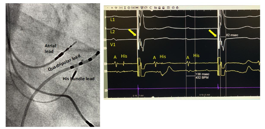
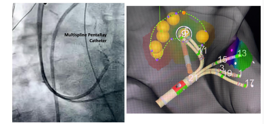
Table 2. Effect of pacing voltage on QRS parameters in patients with and without conduction system disease
| Baseline | Patients with Normal QRS Complex (n=80) | Patients with Left Bundle Branch Block (n=38) | Patients with Complete Heart Block (n=28) | |
|---|---|---|---|---|
| Ventricular activation time (msec) | 96.4 | 90.5 | 186 | __ |
| Lead 1 QRS voltage (millivolts) | 0.6 | 0.9 | 0.6 | __ |
| QRS Axis (degrees) | 30 | 34 | 6.5 ± 36 | __ |
| R wave transition (precordial V-lead) | 3.8 | 3.1 | 4.1 | __ |
In 70 of the 1299 patients, a His bundle electrogram could not be recorded. These included patients with atrioventricular conduction block (23 patients) with left bundle branch block (8 patients) and intraventricular conduction delay (13 patients). In 8 patients in whom only a right subclavian access was possible, we were unable to locate the His bundle electrogram.
In 18 patients very high pacing thresholds precluded lead placement at the site of His bundle electrogram. In these patients, pace mapping was done to locate a site with a normal QRS axis and voltage and decrease in ventricular activation time. In 21 of 70 patients there was no decrease in ventricular activation time.
Discussion
In the practice of cardiac pacing, the elephant in the room has always been the increased long-term cardiovascular morbidity and mortality associated with ventricular pacing. Notwithstanding a call made over 50 years ago, there has been no serious attempt to replace ventricular pacing with His bundle pacing. From the beginning, the focus in cardiac pacing has been on pacing the ventricles. Despite their near simultaneous beginnings over 50-years ago, cardiac pacing has remained siloed from advancements in cardiac electrophysiology where His bundle recordings are routinely obtained. It is therefore not surprising that with the renewed focus on conduction system pacing, the recording methods in use in electrophysiology laboratory have not been adapted to simplify His bundle pacing.
Our real-world cohort of 1299 consecutive patients show that, rather than having niche applicability, His bundle pacing has the makings of a front-line therapy as it addresses a wide range of pacing indications. In sinus bradycardia, where the patients with normal QRS complex are exposed to increased risk of cardiac morbidity and mortality associated with ventricular pacing, whether or not pacing avoidance algorithms are used, His bundle pacing, by maintaining the paced QRS axis, voltage and duration in normal range, allowed for programming physiologic A-V delays and normal heart rate response to exercise.
In patients with left bundle branch block, bi-fascicular block and complete heart block, His bundle pacing corrected the abnormal ventricular activation patterns with a normal QRS axis, a normal lead-1 voltage and a significant decrease in prolonged ventricular activation time. In patients with complete heart block His bundle pacing restored conduction, and the paced complex was no different than that seen in left bundle branch block, suggesting that a single mechanism, results in correction of abnormal conduction patterns.
The perception that His bundle pacing has unresolvable challenges probably relates to the difficulty in recording the His bundle electrogram. With known variability of the His bundle relative to the right atrial wall or the aortic root, we found fluoroscopy to be of little help in locating the His bundle. We found locating the His bundle recording a major challenge which contributed to long procedure times. Point-by-point mapping using the pacing lead was cumbersome and time consuming. There were no recording catheters specifically designed for mapping the His region when approached from the subclavian vein. In a small group of patients, electroanatomic mapping using the multi-spline steerable PentaRay catheter, markedly shortened the time to His bundle recording to less than 2 minutes. Displaying the His bundle electrograms on a 3-dimensional map made lead placement relatively easy. In our study, it typically required pacing at more than one His bundle electrogram site and success was achieved in 94% of patients. Our study demonstrates that technologies exist which can simplify His bundle mapping, and one can expect that simpler versions can be developed over time to increase adoption of His bundle pacing.
The term conduction system pacing is confusing as clinical data is limited to pacing the conducting bundles or the myocardium. To our knowledge pacing the distal conduction system is yet to be demonstrated in humans. Pacing the conducting bundle result in predictable morphology of the paced QRS complex. In His bundle pacing, the pacing voltage, which is much higher than voltage generated in sinus rhythm, commonly activates the superior septal connections which connect the branching His bundle directly to the ventricular septal crest. Their activation serves to provide an alternate conduction pathway. The paced complex is no different than that observed in rare individuals with fasciculoventricular preexcitation, where these pathways are able to conduct in sinus rhythm. Fasciculoventricular preexcitation is a benign condition with excellent prognosis, where other than a delta wave the QRS complex has normal axis, duration and voltage.
A low safety factor of conduction explains their dormant state, as conduction is rarely possible in sinus rhythm and explains their activation by the higher voltage of His bundle pacing. This voltage dependent conduction is typically manifest as a shorter ventricular activation time with higher pacing voltage. Activation of a parallel conducting pathway provides a rational explanation why left bundle branch block is corrected and how conduction is restored in complete heart block and why the two paced complexes are similar.
It follows that in left bundle branch pacing the paced complex must show the delay in right bundle branch and therefore the paced QRS would meet criteria for right bundle branch block. The QRS complex in fascicular pacing can be predicted from morphologies observed in fascicular ventricular tachycardias, and a narrow QRS with left axis deviation will be seen in case of left posterior fascicular bundle pacing. A pacing lead embedded in intra-ventricular septum will show QRS morphology no different than septal pacing and effect of activating nearby conducting bundle may not be discernible.
In this regard, left bundle branch area pacing which reportedly includes left bundle branch pacing, left fascicle pacing, and left ventricular septal pacing, the paced QRS is variable, cannot be distinguished from ventricular septal pacing and therefore may not meet criteria for pacing a conduction bundle. In left bundle branch area pacing an electrogram from a conduction bundle is not always recorded. It is described as easier to do and having stable electrical pacing parameters and was selected as the dormant emergent conduction system pacing method. We are reminded of Michael Crichton s warning that science is not about consensus; it is about evidence, and consensus, before science is sufficiently established, may serve only to avoid debate by claiming an issue is settled.
Ultimately, conduction system pacing must be judged on its long-term all-cause mortality and cardiovascular morbidity. Our one-year and three-year mortality was 7.2 % and 13.5 % respectively. We observed no significant difference in mortality in patients with congestive heart failure (mean left ventricular ejection fraction of 35 ± 15 %) and those without congestive heart failure (mean ejection fraction of 48±13 %). This unexpected observation is similar to our previous report of His bundle pacing in patients with narrow QRS complex, where 3-year all-cause mortality of around 10%, was not significantly different in patients with left ventricular dysfunction. Our mortality figures compare favorably with those reported in previous large randomized controlled trials where ventricular pacing is associated with increased heart failure hospitalizations and a 3-year mortality greater than 20%.
To our knowledge, the long-term mortality of left bundle area pacing, in a large, unselected population, has not been reported and serious complications associated with left bundle branch area pacing such as septal perforation, myocardial infarction, coronary artery fistula, septal hematoma, and permanent complete heart block were not observed in our study with His bundle pacing.
Summary
There is general agreement that compared to ventricular pacing, His bundle pacing is more physiological and has better outcomes. Our study is the largest single center study in which His bundle pacing replaced ventricular pacing in all patients requiring pacemakers with high success rates and low all-cause mortality. A lack of recording technology designed for His bundle pacing prolonged procedure time, however, a paced complex in normal range or, in case of conduction system disease, correction of abnormal QRS axis and voltage and a decrease in prolonged ventricular activation time was obtained in 94% of patients. The replacement of ventricular pacing with His bundle pacing has been impeded by use of generic technology and a failure to adapt basic concepts and methods used for His bundle recordings in electrophysiology procedures.
Paradigm shifts are commonly associated with increased training burden, need for inter-disciplinary collaboration, expense of upgrading infrastructure and reimbursement uncertainties. These factors may cause a delay in change but can hardly be considered unresolvable challenges.
Limitations:
Our success rate with His bundle pacing was no doubt related to our experience in electrophysiology procedures and access to resources of an electrophysiology laboratory, and therefore may not reflect the real-world experience. Our report is observational, and no causality is inferred between maintaining a normal or near normal QRS morphology and low all-cause mortality.
Conflict of interest:
None for all authors.
Funding:
This research received no specific grant from any funding agency in the public, commercial, or not-for-profit sectors.
References:
- Scherlag, BJ, Kosowsky, BD, Damato, AN. Technique for ventricular pacing from the His bundle of the intact heart. J Appl Physiol 22: 584, 1967
- Scherlag, BJ, Lau, SH. Helfant, R.H. Catheter technique for recording His bundle activity in man Circulation. 1969; 39:13-18
- Zottoli, SJ. The origins of the Grass foundation. Biol. Bull. 201:218-226 2001
- Gott VL. Critical role of physiologist John A. Johnson in the origins of Minnesota’s billion dollar pacemaker industry. Ann Thoracic Surg. 2007 Jan; 83(1):349-53. doi:10.1016/j.athoracsur.2006.10.070. PMID: 17184706
- Hopps JA. The development of the pacemaker. Pacing Clin Electrophysiol. 1981 Jan;4(1):106-8. doi: 10.1111/j.1540-8159.1981.tb03682.x. PMID: 6171784.
- Gott VL, Weirich WL, Paneth M, Lillehei CW. Control of complete heart block by use of an artificial pacemaker and a myocardial electrode. Circ Res 1958;6:410-5.
- Furman S, Robinson G. Stimulation of the ventricular endocardial surface in control of complete heart block. Ann Surg 1959; 150: 841 845
- Fisher JD. Transvenous cardiac pacing. Heart Rhythm, Volume 21, Issue 10, 1765 1766
- Kronborg MB, Varma N, Nielsen JC. How harmful is right ventricular pacing? The question revived by the BioPace trial. Europace. 2025 Mar 5;27(3):euaf005. doi: 10.1093/europace/euaf005. PMID: 40105784; PMCID: PMC11921416.
- Funck RC, Müller HH, Lunati M, De Roy L, Klein N, Meisel E, Milasinovic G, Carlson MD, Wittenberg M, Hindricks G, Blanc JJ. Biventricular vs. right ventricular pacing devices in patients anticipated to require frequent ventricular pacing (BioPace). Europace. 2025 Mar 5;27(3):euaf029.
- Ruschitzka F, Abraham WT, Singh JP, et al. Cardiac-resynchronization therapy in heart failure with a narrow QRS complex. N Engl J Med 2013; 369:1395-1405
- Lamas GA, Lee KL, Sweeney MO, Silverman R, Leon A, Yee R et al. Ventricular pacing or dual-chamber pacing for sinus-node dysfunction. N Engl J Med 2002;346:1854 62.
- Udo, Erik O. et al. Incidence and predictors of short- and long-term complications in pacemaker therapy: The FOLLOWPACE study. Heart Rhythm, Volume 9, Issue 5, 728 735
- Botto GL, Ricci RP, Bénézet JM, et al. PreFER MVP Investigators Managed ventricular pacing compared with conventional dual chamber pacing for elective replacement in chronically paced patients: results of the Prefer for Elective Replacement Managed Ventricular Pacing randomized study Heart Rhythm, 11 (2014), pp. 992-1000
- Stockburger M, Boveda S, Moreno J, et al. Long-term clinical effects of ventricular pacing reduction with a changeover mode to minimize ventricular pacing in a general pacemaker population Eur Heart J, 36 (2015), pp. 151-157
- Boriani G, Tukkie R, Manolis AS, et al. Atrial antitachycardia pacing and managed ventricular pacing in bradycardia patients with paroxysmal or persistent atrial tachyarrhythmias: the MINERVA randomized multicenter international trial Eur Heart J, 35 (2014), pp. 2352-2362
- Alanis J, Gonzalez H, Lopez E. Electrical activity of the bundle of His. J Physiol 142: 127, 1958
- Deshmukh P, Casavant DA, Romanyshyn M, Anderson K. Permanent, direct His-bundle pacing: a novel approach to cardiac pacing in patients with normal His-Purkinje activation. Circulation. 2000 Feb 29;101(8):869-77. doi: 10.1161/01.cir.101.8.869. PMID: 10694526.
- Abdelrahman M, Subzposh FA, Beer D, et al. Clinical outcomes of His bundle pacing compared to right ventricular pacing. J Am Coll Cardiol 2018; 71:2319 30.
- Bhatt AG, Musat DL, Milstein N, et al. The efficacy of His bundle pacing: lessons learned from implementation for the first time at an experienced electrophysiology center. JACC Clin Electrophysiol 2018;4:1397 406.
- Teng AE, Massoud L, Ajijola OA. Physiological mechanisms of QRS narrowing in bundle branch block patients undergoing permanent His bundle pacing. J Electrocardiol 2016;49:644 8.
- Verma N, Knight BP. Permanent His-Bundle pacing in a patient with advanced conduction system disease: What is the mechanism of QRS narrowing? Clin Case Rep 2018;6:1236-40.
- Upadhyay GA. Rozmino P, Tung R. Heart Rhythm O2, Volume 1, Issue 1, 68 – 75 His-bundle pacing is the best approach to physiological pacing
- Glikson M, Burri H, Abdin A, Cano O. et al European Society of Cardiology (ESC) clinical consensus statement on indications for conduction system pacing. EP Europace: 27 2025, 10.1093/europace/euaf05 21099-5129
- Mahmud R, Sternick EB, Sanchez-Quintana D, et al. Evidence for concealed fasciculoventricular connections as revealed by His bundle pacing. Europace. 2023;25:euad050.Issue 12, 2024, Pages 2563-2570, ISSN 1547-5271,
- Mahmud R, Lee J, Lee D, Mohan A, Phillips B, Hakes S, Talaei, F Sternick EB. Outcomes with physiologic His bundle pacing in patients with narrow QRS complex, Heart Rhythm, Volume 21,
- Mahmud R, Sanchez-Quintana D, Macias Y, De Almeida MC, Anderson RH, Sternick, EB. Correction of bundle branch block by so called nonselective His bundle pacing: The potential role of accessory connections in the ventricular crest. Heart Rhythm 2024. doi.org/10.1016/j.hrthm.2024.01.003
- Mahmud R, Jamal S, Brenda H, Kukla S. Effect of Non-Selective His Bundle Pacing on Delayed Myocardial Activation in Left Axis Deviation and Left Bundle Branch Bloc. The Journal of Innovations in Cardiac Rhythm Management 2021,12:5 Doi: 10.19102/icrm.2021.121001
- Mahmud R, Jamal S, Effect of His bundle pacing on right bundle branch block located distal to site of pacing. Journal of Electrocardiology. 2021;64: 58-65.
- Macías, Y. Tretter, J.T. Sánchez-Quintana, D. Cabrera, JA. Spicer, DE. de Almeida, MC. Anderson, RH. The atrioventricular conduction axis and the aortic root Inferences for transcatheter replacement of the aortic valve. Clin Anat. 2022, 35, 143 154.
- Mahaim I. Kent s fibers and the AV paraspecific conduction through the upper connection of the bundle of His-Tawara Am Heart J, 33 (1947), pp. 651-653
- Macías Y, Tretter JT, Anderson RH, et al. Miniseries 1 Part IV: how frequent are fasciculo-ventricular connections in the normal heart? Europace 2022;24:464 72
- Sternick EB, Gerken LM, Vrandecic MO, Wellens HJ. Fasciculoventricular pathways: clinical and electrophysiologic characteristics of a variant of preexcitation. J Cardiovasc Electrophysiol. 2003 Oct;14(10):1057-63. doi: 10.1046/j.1540-8167.2003.03206.x. PMID: 14521658.
- Cappato R. What is concealed in concealed accessory pathways? Eur Heart J. 1999 Dec;20(24):1766-7. doi: 10.1053/euhj.1999.1842. PMID: 10581131.
- Upadhyay GA, QRS morphologies in V1 and V6 during left bundle branch area pacing: assessing the patterns, EP Europace, Volume 25, Issue 9, September 2023, euad284,
- Briongos-Figuero S, Estévez-Paniagua Á, Sánchez-Hernández A, Muñoz-Aguilera R. Combination of current and new electrocardiographic-based criteria: a novel score for the discrimination of left bundle branch capture. Europace. 2023 Mar 30;25(3):1051-1059. doi: 10.1093/europace/euac276. PMID: 36691717; PMCID: PMC10062292.
- Crichton, M, Aliens cause Global Warming, 17 January 2003 speech at the California Institute of Technology (http://s8int.com/crichton.html)
“`