


Modified Quigley Technique for Lisfranc Injury Reduction
Description and Demonstration of a Novel Technique Utilizing a Modified Quigley Maneuver for Reduction of Lisfranc Injury and Midfoot Dislocations
Gregory Foote DPM, FACFAS1,4, Vikram Bala, DPM, FACFAS2,4, Maliha Arzumand, DPM1,3
- MercyOne Waterloo Medical Center, IA, USA
- Ankle and Foot Associates, Jacksonville, FL, USA
- Resident, Foot & Ankle Surgery, MercyOne Waterloo Medical Center, IA, USA
- Fellowship Trained Foot and Ankle Surgeon
OPEN ACCESS
PUBLISHED: 31 Decemeber 2024
CITATION: FOOTE, Gregory; BALA, Vikram; ARZUMAND, Maliha. Description and Demonstration of a Novel Technique Utilizing a Modified Quigley Maneuver for Reduction of Lisfranc Injury and Midfoot Dislocations. Medical Research Archives, Available at: <https://esmed.org/MRA/mra/article/view/6197>.
COPYRIGHT: © 2025 European Society of Medicine. This is an open-access article distributed under the terms of the Creative Commons Attribution License, which permits unrestricted use, distribution, and reproduction in any medium, provided the original author and source are credited.
ISSN 2375-1924
Abstract
The Lisfranc ligament joint complex is comprised of the base of the second metatarsal, the lateral facet of the medial cuneiform, the first and second tarsometatarsal joints, as well as the ligaments spanning the joint. Lisfranc injuries have been reported as one of the most commonly misdiagnosed foot injuries in both the emergency and outpatient settings. These injuries tend to involve the ligamentous structures but can also be associated with fracture and/or dislocation. The mechanism of injury can be both high and low energy. The lower energy mechanism of Lisfranc injury is commonly a result of the foot dynamic twisting or abduction, creating excessive stress on the ligaments and bones of the midfoot. When displaced, potential injury or impingement on neurovascular structures may warrant an immediate closed reduction, as it is important to achieve near anatomic alignment to prevent skin necrosis or neurovascular complications. This manuscript presents the novel use of a widely accepted distraction technique used for ankle fracture reduction, modified to allow the reduction of midfoot joint dislocations. Utilizing a gauze roll and an IV pole, both of which are readily available in the clinical and emergency department setting, this technique has proven successful in clinical practice without resultant complications. A gauze roll is affixed to the first and second digit to provide axial traction at the second metatarsal. The free ends are then attached to a point of stability and the foot is placed in gravity-assisted traction. A reduction of the Lisfranc dislocation is then obtained by traction of the second metatarsal via an indirect focus at the central point of instability within the midfoot. This injury can then be immediately splinted in this reduced position to provide stability until operative fixation can be achieved. The use of the modified Quigley maneuver is highly efficient in reducing displaced Lisfranc injuries and midfoot dislocations. With simple modifications of the previously described technique, near-anatomic reduction of midfoot dislocations can be achieved without the need for more invasive techniques currently described in literature. This technique provides the practitioner with an additional tool to safely stabilize this inherently unstable injury until more appropriate intervention can be performed.
Keywords
Lisfranc injury, midfoot dislocation, Quigley maneuver, closed reduction, foot surgery
Introduction:
Lisfranc injuries are defined as bony or ligamentous trauma causing displacement of one or more metatarsals at the tarsus complex. The term was first coined after Jaque Lisfranc de Saint-Martin, a French military surgeon and gynecologist, described the injury pattern and amputation through the midfoot. The disruption of the Lisfranc ligament is caused when excessive kinetic energy is applied, directly or indirectly, to the midfoot and is often associated with traumatic mechanisms of injury. Pain, progressive deformity, and loss of function are common sequela in the event of delayed or missed diagnosis and appropriate treatment. Studies have reported that 20% of Lisfranc injuries are missed on initial examination, particularly due to the complex anatomy and reliance on radiographs alone for subtle injuries. Non-operative treatment may be employed for less severe disruptions with < 2 mm of displacement. Surgical management is indicated in the setting of displaced fractures or those involving dislocation. Lisfranc injuries specifically involve a disruption of the interosseous ligament between the medial cuneiform and second metatarsal base, the strongest ligamentous connection in the midfoot. The dorsal midfoot ligaments tend to be the weakest in comparison to the plantar and interosseous ligaments. Due to these inherent characteristics, injuries to this area typically result in a dorsal dislocation of the forefoot upon the midfoot and may present with fractures of the osseous structures comprising the Lisfranc joint. The dorsal neurovascular bundle is typically located in close proximity to the Lisfranc joint. Acute Lisfranc fracture/dislocations should therefore be promptly reduced and stabilized to avoid neurovascular compromise to the foot. Due to the innate instability of Lisfranc injury, most require operative management; however, soft tissue compromise and severe edema may delay surgical intervention. Potential tendon interposition and bone fragmentation may impede anatomic reduction of Lisfranc injuries, which serves as an indication for open reduction with either internal fixation or arthrodesis. Current techniques described for closed reduction of midfoot and metatarsal fracture patterns include manual traction, finger traps, or a more invasive method of percutaneous wire fixation with axial traction. The key to the reduction is the second metatarsal, which should be realigned to allow the adjacent metatarsals and cuneiforms to reduce. T. B. Quigley originally described a technique in 1959 for difficult ankle fracture reduction, which was modified in 2015 for use with rolled gauze. In describing this method, the authors advocated for its use in the absence of fracture to the first or second metatarsals or cuneiforms. To date, there have been no reports utilizing the Quigley reduction maneuver for midfoot or distal foot fractures. There is a paucity of literature describing closed reduction techniques for acute traumatic midfoot dislocations and metatarsal fractures. We propose a novel technique that modifies the Quigley technique for use in Lisfranc injuries and metatarsal fracture/dislocation by principles similar to those of finger trap reduction. Specifically, we aim to establish an alternative technique for reduction of Lisfranc injuries with equipment and materials readily available in community emergency departments and urgent care settings.
Methods:
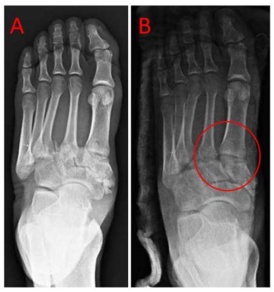
Patients presenting with midfoot dislocations in the acute setting were included in this study. Initial examination was performed on the affected foot to evaluate for any soft tissue compromise or presence of underlying peripheral vascular disease (PVD). The use of this technique is contraindicated in the setting of any digital neurovascular compromise or PVD. No numerical data was collected in this study; instead, the success of the reduction technique was assessed qualitatively through radiographic evaluation of post-reduction imaging. As evidenced by post-reduction films, the anatomic alignment of midfoot joints served as the primary measure of success using the modified Quigley technique.
Technique:
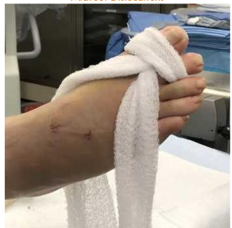
The patient is prepared for reduction with conscious sedation or anesthetizing the injured area with the use of a hematoma block. Once adequate anesthesia is confirmed, the foot is prepared for reduction. A gauze roll is unraveled and grasped at the midline. This is then brought from dorsal to plantar between the first and second digit and looped dorsally around the first two toes.
The free ends are then pulled taut and attached to an IV pole, or point of stability, at the end of the hospital bed or operating table to suspend the leg. The foot is suspended for 2-3 minutes followed by manual reduction maneuvers that can be assisted with direct fluoroscopy.
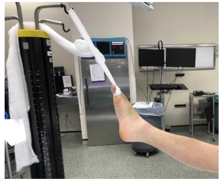
Based on the theory of ligamentotaxis, for traction to be effective, it must be balanced by counter traction provided by the ligaments and soft tissue surrounding the bone. A plantar force may be applied to the forefoot at this time to exaggerate the subluxation, recreating the mechanism of injury. In injuries that exhibit a more difficult reduction, a counterforce can be provided with weights placed around the ankle. This can assist in a supplemental downward force that can complement gravity in the linear expansion of the midfoot.

The reduced foot may then be immediately splinted in this reduced position while remaining in traction. Given the inherent instability, this injury will typically require prompt stabilization and surgical fixation for improved prognosis and anatomic reduction. In the operating room setting, percutaneous K-wire fixation may be utilized to assist in the maintenance of reduction until definitive fixation can be performed.

Discussion:
There are various methods for definitive fixation of Lisfranc injuries. However, experts agree that anatomical reduction is essential in ensuring optimal long-term outcomes. Reduction may be achieved acutely in clinical practice or community emergency department settings utilizing this modified Quigley technique. Alternatively, this technique may also be beneficial pre-operatively or intra-operatively prior to definitive fixation.
The second metatarsal is considered the “keystone” of the entire midfoot complex because of its anatomic position securely nested between the medial and lateral cuneiforms. A transverse ligamentous connection exists between the bases of the lateral four metatarsals; however, no transverse ligament exists between the first and second metatarsal bases. The joint capsule and dorsal ligaments are solely responsible for support on the dorsal surface of the Lisfranc joint. The “keystone” nesting of the second metatarsal into the cuneiform forms the crucial relationship that supports the entire tarsometatarsal articulation. However, this unique anatomy provides a “weak link” that is prone to injury when stressed. Consequently, our closed reduction approach emphasizes targeting of the second metatarsal as the focal point for achieving an effective reduction.

Utilization of this method has resulted in acceptable reduction of Lisfranc joint fracture/dislocations performed in the acute setting. Traction applied at the second digit allows for axial traction along the second metatarsal due to the stout ligamentous attachments at the metatarsophalangeal joint level. This, in turn, provides distraction of the dislocation at the tarsometatarsal level and allows re-nesting of the “keystone” of the midfoot by relocating the second metatarsal base between the medial and lateral cuneiforms. This allows for expedited stabilization of the Lisfranc or midtarsal joints while minimizing complications and soft tissue compromise. This reduction technique may be applied to many specialties to improve the quality of acute midfoot and metatarsal fracture reduction in daily practice including those in trauma, emergency medicine, orthopedics, and podiatry.
Soft tissue relaxation to restore overall foot length aids in the reduction of these difficult dislocations to the midfoot, based on the principles of ligamentotaxis. Finger traps are commonly utilized in the reduction of forearm and hand fractures for this reason. These same principles may be applied to the fracture/dislocations of the midfoot. Gravity provides the counter-traction to disimpact and aids in the realignment of the dislocation by ligamentotaxis. In 2016, Southerland et. al. described a closed reduction technique for Lisfranc injuries with the use of 7/64 or 3/32 Steinmann pins and gauze roll. This axial traction method focused on using the longitudinal distraction of the second metatarsal to restore the “keystone” of the Lisfranc joint complex. However, this technique requires use of the operating room. In contrast, utilization of the modified Quigley maneuver offers a significant advantage as it can be performed anywhere, including the emergency room or clinical settings, where these injuries are typically encountered. The use of readily available and widely accessible materials promotes the use of the modified Quigley reduction technique as a practical alternative for managing Lisfranc injuries outside of the operating room.
After immediate reduction and stabilization, nearly all Lisfranc injuries require operative treatment for definitive fixation. This, in turn, limits our long-term follow-up as to the quality of the initial reduction. However, we were able to devise that an initial reduction to near anatomic alignment showed no incidence of soft tissue or neurovascular compromise. In regard to forefoot fracture without midfoot involvement, axial traction applied to the distal aspect of the foot may allow for the reduction of shortened and displaced metatarsal fractures with this same method.
Conclusion:
This technique utilizes equipment available at all healthcare facilities, where reduction instrumentation or multiple physicians may not be readily available. This technique can also be performed with local anesthesia only. Furthermore, this can be used in anatomic reduction of these dislocations in the operative setting by use of a sterile gauze roll in preparation for fixation. Our method is inexpensive, repeatable, and effective for reducing an unstable injury to preclude soft tissue complications. This is not a proven, statistically supported procedure. Hence, further research is required to validate applicability, effectiveness, and safety in large case series before a routine recommendation is provided for the procedure.
Conflicts of Interest Statement:
The authors have no conflicts of interest to declare.
References
- Sain A, Prendergast E, Wattage K, Elkilany A, Metry A. Lisfranc Injury: Recent Trends in Management. Cureus. 2023;15(8):e43182. Published 2023 Aug 9. doi:10.7759/cureus.43182
- Kim DY, Kim JK, Kim MW, Lee KB. Irreducible Lisfranc injury by tibialis anterior tendon entrapment: A case report. Medicine (Baltimore). 2021;100(11):e24822. doi:10.1097/MD.0000000000024822
- Ponkilainen VT, Partio N, Salonen EE, et al. Outcomes after nonoperatively treated non-displaced Lisfranc injury: a retrospective case series of 55 patients. Arch Orthop Trauma Surg. 2021;141(8):1311-1317. doi:10.1007/s00402-020-03599-w
- Rosenbaum A, DiPreta J, Uhl R. Clinical Management of Lisfranc Joint Injuries. Lower Extremity Review. Web. April 2012.
- Desmond EA., Chou LB. Current Concepts Review: Lisfranc Injuries. Foot Ankle Int 2006. 27(8):653–660.
- Abdelgaid, S. M. Closed Reduction & Percutaneous Fixation of Lisfranc Joints Injuries: Possibility, Technique & Results. Clinical Research on Foot & Ankle, 01(02). 2013.
- Richter M, Wippermann B, Krettek C. Fractures and Fracture Dislocations of the Midfoot: Occurrence, Causes, and Long-Term Results. Foot Ankle Int. 2001; 22(5):392-398
- Southerland CC Jr, Smith CE, Merrill TJ. Reduction of Lisfranc Dislocations Using Second Ray Axial Traction. The Podiatry Institute. Chapter 29; 147-151.
- Alton TB, Harnden E, Hagen J, Firoozabadi R. Single Provider Reduction and Splinting of Displaced Ankle Fractures: A Modification of Quigley’s Classic Technique. J Orthop Trauma. 29:4; (2015).
- Quigley TB. A Simple Aid to the Reduction of Abduction-External Rotation Fractures of the Ankle. Am J Surg. 1959;97:488–493.
- Boparai R, Boparai RS, Kapila R, Pandher DS. Role of Ligamentotaxis in Management of Comminuted intra/juxta articular fractures. Indian J Orthop 2006;40:185-7.
- Charnley J. The Closed Treatment of Common Fractures. London: Cambridge University Press; 1950.
- Hardcastle PH, Reschauer R, Kutscha-Lissberg E, Schoffmann W. Injuries to the Tarsometatarsal Joint: Incidence, Classification and Treatment. J Bone Joint Surg. 1982;64(3):349-356
- Myerson MS, Fisher RT, Burgess AR, Kenzora JE. Fracture dislocations of the tarsometatarsal joints: End results correlated with pathology and treatment. Foot Ankle 1986;6(5):225-242.
- Arntz CT, Hansen ST Jr. Dislocations and fracture dislocations of the tarsometatarsal joints. Orthop Clin North Am 1987;18(1):105-114.
- Kuo RS, Tejwani NC, DiGiovanni CW, Holt SK, Benirschke SK, Hansen ST Jr, Sangeorzan BJ. Outcome after open reduction and internal fixation of Lisfranc joint injuries. J Bone Joint Surg Am 2000;82(11):1609-1618.
- Arntz CT, Veith RG, Hansen ST Jr. Fractures and fracture-dislocations of the tarsometatarsal joint. J Bone Joint Surg Am 1988;70(2):173-181.
- Ebraheim NA, Yang H, Lu J, Biyani A. Computer evaluation of second tarsometatarsal joint dislocation. Foot Ankle Int 1996;17(11):685-689.
- Mantas JP, Burks RT. Lisfranc injuries in the athlete. Clin Sports Med. 1994;13:719-30.
- Heckman JD. Fractures and dislocations of the foot. In: Rockwood CA, Green DP, Bucholz RD, eds. Rockwood and Green’s Fractures in adults. Vol 2. 3d ed. Philadelphia: Lippincott, 1991:2140–51.
- Wiley JJ. The mechanism of tarsometatarsal joint injuries. J Bone Joint Surg [Br]. 1971;53:474-82.
- Englanoff G, Anglin D, Hutson HR. Lisfranc fracture-dislocation: a frequently missed diagnosis in the emergency department. Ann Emerg Med. 1995;26:229-33.
- Burroughs KE, Reimer CD, Fields KB. Lisfranc injury of the foot: a commonly missed diagnosis. Am Fam Physician. 1998;58(1):118-124.
- Kammela M, Biswas D, Karuppaiah K. Gaining control over complex distal radius and ulna fractures: Tips and tricks. The Annals of The Royal College of Surgeons of England. 2023;106(3):288-290. doi:10.1308/rcsann.2023.0069
- Akhtar K, Akhtar D, Simmons, J. A readily available alternative to Chinese finger traps for fracture reduction. The Annals of The Royal College of Surgeons of England, 2013:95(2), 159–159. doi:10.1308/rcsann.2013.95.2.159