


Mydriasis in Diabetic Eye Screening: New Camera Insights
Review of the evidence for dilating eye drops in a Diabetic Eye Screening Programme following the introduction of new camera technologies.
Arya Ghatge, Peter H Scanlon1,2,3
- Gloucestershire Retinal Research Group, Department of Ophthalmology, Gloucestershire Hospitals NHS Foundation Trust, Cheltenham, UK
- Nuffield Department of Clinical Neuroscience, University of Oxford
- University of Gloucestershire
AG Orcid: 0009-0001-6323-8409. PS Orcid: 0000-0001-8513-710X
OPEN ACCESS
PUBLISHED: 31 January 2026
CITATION: Ghatge, A., and Scanlon., PH., 2026. Review of mydriasis in diabetic eye screening with new camera technology. Medical Research Archives, [online] 14(1).
COPYRIGHT: © 2025 European Society of Medicine. This is an open-access article distributed under the terms of the Creative Commons Attribution License, which permits unrestricted use, distribution, and reproduction in any medium, provided the original author and source are credited.
DOI: https://doi.org/10.18103/mra.v14i1.7165
ISSN 2375-1924
ABSTRACT
Purpose: To review the literature to determine if new technologies have reduced the need for dilation in a Diabetic Eye Screening Programme.
Methods: Two approaches were used:
- A review of the literature relating to screening for diabetic retinopathy that has been on-going by the second author (PHS) since March 2000. The results are held in Endnote which was subsequently searched using keywords.
- Our local library conducted a search using a combination of keywords and subject headings were used to formulate search strategies on Medline (Ovid), Embase (Ovid), CINAHL (EBSCO), and the Cochrane Library.
Results: A total of 133 articles were identified, separated into 117 original articles and 16 review articles. After discarding those that did not appear relevant, 76 original articles and 15 reviews remained. These were analysed to determine whether they helped to answer the main question, and 49 references have been quoted. The sensitivities and specificities of mydriatic digital photography for detection of referable diabetic retinopathy have been consistently high since the early studies with sensitivities of >80% and specificities of > 85% against recognised reference standards in the majority of studies. The specificity achieved is often above 90% if ungradable images are not counted as test positive. Non-mydriatic digital photography performs well in those in whom gradable images can be obtained. In Scotland, which has a long history of performing staged mydriasis, they currently dilate 30% of their screened population. This has risen slowly from 24% in 2002 because of increasing ageing of the population which is one of the risk factors for poor quality images. Recent studies using confocal technology have demonstrated that the percentage of ungradable images without eye drops can be reduced to under 5.1% and after staged mydriasis to under 3.4%. This will depend on the age group and the percentage of untreated cataract in the population.
Conclusions: The results of this review are encouraging that, with the development of less expensive confocal cameras, this technology is likely to be used routinely in diabetic eye screening programmes around the world using staged mydriasis.
Keywords
- diabetic retinopathy
- mydriasis
- screening
- digital photography
- confocal technology
Introduction
Screening for Diabetic Retinopathy has been taking place in the UK since the early studies lead by Taylor in the 1990’s. The initial studies used either Polaroid film or 35 mm film. In the late 1990’s screening commenced with the use of digital cameras. At that time screening was also being conducted in Iceland, Scandinavia, Sweden, and in the USA with 35 mm film and also with these early digital cameras.
Whether or not eye drops should be used on all people attending a screening programme or just those who have poor quality images (staged mydriasis) has been debated since the early programmes commenced. When National Screening Programmes were introduced in the UK with Scotland commencing in 2002, they chose a staged mydriatic approach and only one 45-degree field. In 2023, the English NHS Diabetic Eye Screening Programme commenced with a mydriatic approach for all people with diabetes and 2 x 45-degree fields. Wales and Northern Ireland also introduced programmes in 2023. Wales used the same approach as England, and Northern Ireland used routine mydriasis over the age of 55yrs with 2 x 45-degree fields.
It is important to acknowledge that a screening test does not replace the eye examination but is supposed to be an inexpensive test that, if performed well, will pick up a high percentage of those with the condition, and will only refer a small percentage as false positive.
The Scottish Screening Programme has used a staged mydriatic approach since 2002 with one 45-degree field and the English NHS DESP has used routine mydriasis and two 45-degree fields since 2003. Modern cameras have been developed over the last few years, using newer technologies, that can potentially capture a wider field of view and may require much less mydriasis even with a staged mydriatic approach. This narrative review of the literature is designed to assess the impact of the new technologies on these questions as to whether drops are still necessary and what area of retina is required to be photographed.
Methods
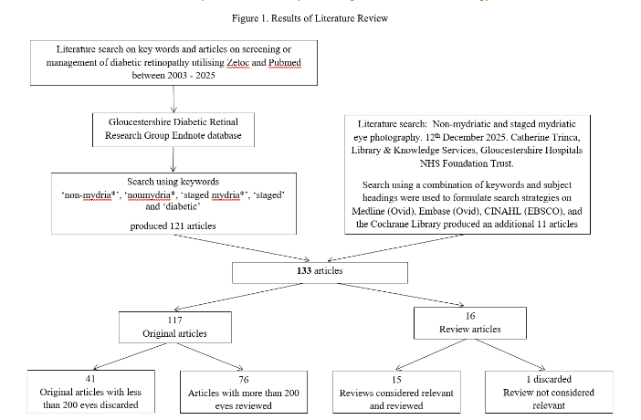
The review of the literature relating to screening for diabetic retinopathy has been on-going by the second author (PHS) since March 2000. The methodology involves a search technique for articles relating to screening or management of diabetic retinopathy utilising Zetoc, a cooperative venture between the British Library, Manchester Information and Associated Services (MIMAS) and the Joint Information Systems Committee (JISC) of the UK Higher Education Funding Council, which was available to Universities until 1st August 2022. Zetoc provided access to over 29,000 journals and more than 52 million article citations and conference papers through the British Library’s electronic table of contents. The database covered 1993 to 1st August 2022 and was updated daily.
The following subject title keywords were used: ‘retinopathy’, ‘digital’ and ‘imaging’ and ‘eye’. ‘digital’ and ‘imaging’ and ‘ophthalm’, ‘digital’ and ‘imaging’ and ‘diabet’, ‘laser’ and ‘eye’, ‘laser’ and ‘ophthalm’, ‘laser’ and ‘diabet’, ‘visual’ and ‘acuity’, ‘visual’ and ‘impairment’, ‘blindness’ and ‘diabet’, ‘diabetic’ and ‘screening’, ‘uptake’ and ‘screening’ and ‘diabet’ in title, ‘attendance’ and ‘screening’ and ‘diabet’, and/or ‘vitrectomy’ and ‘diabet’ in title.
In addition, the contents page lists of the following journals, considered to be those most likely to publish articles relevant to this topic, were reviewed each month: Acta Ophthalmologica Scandinavia, American Journal of Ophthalmology, Archives of Ophthalmology, British Journal of Ophthalmology, British Medical Journal, Clinical and Experimental Ophthalmology, Current Eye Research, Diabetes, Diabetes Care, Diabetes Metabolism Research and Reviews, Diabetes Research and Clinical Practice, Diabetes Technology and Therapeutics, Diabetic Medicine, Diabetologia, European Journal of Ophthalmology, Eye, Graefes Archive for Clinical and Experimental Ophthalmology, Investigative Ophthalmology and Visual Science, Journal of Diabetes and its complications, Journal of Medical Screening, Journal of the Eye, Lancet, Ophthalmic Surgery and Lasers, Ophthalmologica, Ophthalmology, Pediatric Diabetes, Retina, Survey of Ophthalmology.
Since Zetoc ceased on 1st August 2022, we have used PubMed as our primary search database. The database was launched in 1996 by the U.S. National Library of Medicine and now contains more than 39 million citations, primarily from MEDLINE.
In addition, our local library conducted a search using a combination of keywords and subject headings were used to formulate search strategies on Medline (Ovid), Embase (Ovid), CINAHL (EBSCO), and the Cochrane Library. Articles of interest identified with this search strategy were sourced from the local NHS Trust library or on-line from the electronic journal resource (Athens1) accessible in the Trust library.
All articles sourced were included in the extensive Endnote database that we have developed. We searched for articles on non-mydriatic and staged mydriatic photography in diabetic eye screening within this database using the search words ‘non-mydria*’, ‘nonmydria*, ‘staged mydria*’, ‘staged’ and ‘diabetic’.
Results
With the search strategy of the second author (PHS) outlined above, 121 relevant articles were identified and the search undertaken by the Gloucestershire Knowledge and Library Services identified a further 11 articles, making a total of 133 articles.
When the English Screening Programme commenced in 2003, they used the results from the Scanlon study with a sensitivity of 87.8%, specificity was 86.1% and an ungradable image rate of 3.7% with mydriatic digital photography. In the same study, the results for non-mydriatic photography were sensitivity of 86.0%, specificity was 76.7% and ungradable image rate was 19.7%. The Health Technology Board for Scotland also used data from this study to model their proposed staged mydriatic service, prior to the introduction of the Scottish Screening Programme, which they published in their proposal.
At that time, there were several studies that looked at risk factors for ungradable images in the screening environment. Age, duration of diabetes, untreated cataract, ethnicity and poor near vision were the commonly reported risk factors. Some studies reported ungradable image rates < 12% but the average age of the study population was usually under 55 years. In 2010, Dervan reported targeted mydriasis strategies using smaller pupil size, denser nuclear colour, older age, poorer best-corrected visual acuity, cortical lens opacity and posterior subcapsular lens opacity which were associated with the need for dilation (P<0.001 in all).
If the ungradable images are reported as test positive this reduces the specificity of the test. However, some studies reported the sensitivity and specificity of those with gradable images and so the specificity is not reported as reduced.
In a study comparing an Ophthalmologists examination and 2-field digital imaging to seven field stereophotography, the ophthalmologist’s examination gave a sensitivity of 87.4% (confidence interval 83.5 to 91.5), a specificity of 94.9% (91.5 to 98.3), and a kappa statistic of 0.80. Two field mydriatic digital photography gave a sensitivity of 80.2% (75.2 to 85.2), specificity of 96.2% (93.2 to 99.2), and a kappa statistic of 0.73.
In 2015, the use of a smartphone was validated in a study against standard seven-field digital fundus photography with a Carl Zeiss fundus camera. In this study the smartphone was fixed, and the patient used a standard headrest which kept the head in a fixed position. Since that time, there have been reports of validation of handheld devices that are not fixed and the patient is sitting without a headrest. We have found it easier to use devices where the patients head is in a fixed position and the camera is also, which removes the concerns of hand and head movement.
Many of the recent reports have compared grading by General Practitioners or non – ophthalmologist personnel compared to grading by Ophthalmologists with a further programme capturing images in a remote setting with grading by retinal specialists. In Southern Israel, when non-mydriatic fundus photography was compared with dilated fundus examination by an ophthalmologist, sensitivity of 99.3 %, specificity of 88.3 %, and positive predictive value of 85.3 % were found. The ungradable image rate was 14.4%.
In 2018, Piyasena et al reviewed six thousand six hundred forty-six titles and abstracts, and data were extracted from 122 potentially eligible full reports. The highest sensitivity was observed in the mydriatic greater than two field strategy (92%, 95% CI 90–94%). The highest specificity was observed in greater than two field methods (94%, 95% CI 93–96%) where mydriasis did not affect specificity. Overall, there was no difference in sensitivity between non-mydriatic and mydriatic methods (86%, 95% CI 85–87) after exclusion of ungradable images.
In 2021, Kanclerz et al concluded that regardless of the type of the device used, retinal photographs should be taken on eyes with dilated pupils, unless contraindicated, as this setting decreases the rate of ungradable images.
In 2024, Abou Taha et al conducted a narrative review of present and future screening programs for diabetic retinopathy. They concluded that many nations use 2–4 fields fundus images, proven effective with 80–98% sensitivity and 86–100% specificity compared to the traditional seven-field evaluation for DR.
In 2024, an Ophthalmic Technology Assessment Report by the American Academy of Ophthalmology entitled ‘Effectiveness of Conventional Digital Fundus Photography-based Teleretinal Screening (TS) for Diabetic Retinopathy and Diabetic Macular Oedema’ reported that thirty percent of non-mydriatic digital images and 10% of mydriatic images were deemed ungradable. For ETDRS level 35 or more, sensitivity of TS (i.e., the likelihood that those who have true threshold disease will show positive results on TS) in detecting referable DR or DME was poor to moderate (38%-71%), whereas specificity was moderate to high (70%- 96%).
In the UK, the English NHS Diabetic Eye Screening Programme has a standard that the ungradable image rate within a programme should be below 8%. This does depend on the proportion of treated cataracts in the population.
With respect to the ungradable image rates for staged mydriatic photography with conventional digital cameras, the Scottish Screening Programme has been using staged mydriasis since it commenced in 2002 and the proportion of people with diabetes requiring dilation has slowly increased with the increasing age of the population. In 2004, Murgatroyd reported an ungradable image rate without mydriasis of 26% which was brought down to 5% with mydriasis. In 2020, Styles reported that, in the Scottish Screening Programme the overall use of dilating drops was 30% but varied greatly with age group. 33% of the 65–74-year-olds required dilation and 8.9% of the 35-44 age group. Scotland has a relatively white Caucasian population and there have been reports of higher levels of dilation being required in those with more pigmented pupils e.g. people of Asian or Afro-Caribbean origin.
What is exciting is that there are signs that the new modern cameras that use scanning confocal or similar technology such as broad line fundus imaging are able to capture good quality images at much smaller pupil diameters (2.5 – 3.0 mm) and hence there is a much greater possibility of screening the majority of people with diabetes without eye drops.
In an early screening study of the Optos camera in 2010, Wilson reported that ungradable image rates for undilated wide-field scanning laser ophthalmoscopy were 10.8%, and the type of lesion identified by each imaging modality was also assessed. Single-field photography was able to identify microaneurysms in 95.9% of cases that slit lamp had done so, whereas WSLO achieved this in only 79.2% (p < 0.001), a difference that appeared to be due to the lower resolution.
However, the camera has shown improvements since that time both in resolution and in ungradable image rates. In 2014, Silva reported results from an Ocular Telehealth Programme performed on 1,633 and 2,170 consecutive patients, in which the ungradable rate per patient for DR (2.9 vs. 9.9%, P < 0.0001) and DME (3.8 vs. 8.8%, P < 0.0001) was lower with ultra-wide field (UWF) imaging than with non-mydriatic digital fundus photography (NMFP). In 2016, he reported on thirty-five thousand fifty-two eyes (17 526 patients) imaged using NMFP and 16 218 eyes (8109 patients) imaged using UWF imaging. The ungradable rate per patient for DR and DME was significantly lower with UWF imaging compared with NMFP (DR, 2.8% vs. 26.9% [P < 0.0001]; DME, 3.8% vs. 26.2% [P < 0.0001]). The only weakness of these studies was that the photography was consecutive and so the people with diabetes had not been photographed by the same camera on the same day.
Scanlon reported the ungradable image rates on two groups of patients using staged mydriatic photography. In the CONCORDIA study paper 1, both the Zeiss Clarus and the Optos using staged mydriasis on 1497 people with diabetes detected high levels of any and referable diabetic retinopathy when compared to the standard two-field mydriatic digital imaging. The ungradable image rates without eye drops were 3.3% for the Clarus and 5.1% with the Optos and this improved to 1.7% and 3.4% respectively with staged mydriasis. In the CONCORDIA study paper 2, the Eidon camera using staged mydriasis on 1050 people with diabetes also detected high levels of any and referable diabetic retinopathy when compared to the standard two-field mydriatic digital imaging. The ungradable image rate for the Eidon without eye drops was 4.2% and 1.7% with staged mydriasis.
A smaller and less expensive confocal camera, which uses similar technology to the Eidon camera and has a 45 degree field similar to digital cameras in the English Screening Programme, has recently become available (DRS Plus from Centervue) and the only report of its use in diabetic retinopathy screening suggested that the ungradable image rate according to an artificial intelligence system was 2.58%.
Discussion
In England, the current recommended screening process uses mydriatic drops to dilate the pupils of people with diabetes. The drops have been identified as a barrier to attendance in several studies. Individuals complain that the drops are painful and the blurring that results means that they are unable to drive after the appointment and it is often 6-8 hours before they can resume their normal work. The eye drops also take at least 15 minutes to take full effect, which means longer appointment and waiting times.
Untreated cataract is the principal cause of poor-quality images but, age, duration of diabetes, and ethnicity have been commonly reported risk factors. With respect to ethnicity, it is felt to be the iris pigmentation and possibly retinal pigmentation causing the problem as a higher flash intensity may be required with conventional digital cameras to obtain an adequate photograph, with longer pupil recovery times for the second eye.
Cameras using the latest technology mostly use confocal light. The Optos camera uses red, green and blue laser light. The Eidon and the DRS plus camera both use white light from an LED source. The Zeiss Clarus, which uses a similar technique described as broad line fundus imaging, uses red, green and blue LED light. All of these cameras have been shown to be capable of a significant reduction in poor quality image rate using staged mydriasis with wider fields than a single 45-degree field.
For routine diabetic retinopathy screening, we need inexpensive cameras that can detect over 85% of the retinopathy and will only refer in a small number of false positives (e.g. 1 in 10). With respect to the field of view required in England, the English NHS Diabetic Eye Screening Programme has demonstrated success in reducing the incidence of new blindness in the working age group and this has continued to drop. Hence, the English Programme would be looking to cover at least the area of the two 45-degree fields that it currently captures. The English NHS Diabetic Eye Screening Programme currently dilates 2.6 million people per year and, if this could be reduced to under 132,600 people (5.1%), this would be a major advance for the programme. We are currently awaiting the results of a study (CONCORDIA 2) that has been designed to determine whether these cameras work as well in people with diabetes of Asian and Afro-Caribbean background as they do in those of White Caucasian background.
The only problem with the early confocal cameras was the expense but it is becoming increasingly likely that inexpensive options will become available so that they can be used routinely for diabetic retinopathy screening.
Conclusion
The technology of cameras is changing rapidly and the possibility of being able to screen the vast majority of people with diabetes without eye drops is becoming increasingly likely over the next few years.
Conflict of interest statement
Arya Ghatge declares no conflict of interest. Peter Scanlon declares that, In the last 3 years, he has received speaker’s fees and expenses from Bayer Ltd, fees for consultancy and expenses from Boehringer Ltd and his department has received unrestricted research grants from Bayer, Zeiss, Centervue and Optos.
Funding statement
Funding: No funding was provided for writing this article.
Acknowledgements
Library Literature search: Non-mydriatic and staged mydriatic eye photography. 12th December 2025. Catherine Trinca, Library & Knowledge Services, Gloucestershire Hospitals NHS Foundation Trust.
References
- Taylor R. Practical community screening for diabetic retinopathy using the mobile retinal camera: report of a 12 centre study. British Diabetic Association Mobile Retinal Screening Group. Diabetic medicine : a journal of the British Diabetic Association. Nov 1996;13(11):946-52.
- Jacob J, Stead J, Sykes J, Taylor D, Tooke JE. A report on the use of technician ophthalmoscopy combined with the use of the Canon non-mydriatic camera in screening for diabetic retinopathy in the community. Diabetic medicine : a journal of the British Diabetic Association. May 1995;12(5):419-25.
- Harding SP, Broadbent DM, Neoh C, White MC, Vora J. Sensitivity and specificity of photography and direct ophthalmoscopy in screening for sight threatening eye disease: the Liverpool Diabetic Eye Study. BMJ. Oct 28 1995;311(7013):1131-5.
- Scanlon PH, Malhotra R, Thomas G, et al. The effectiveness of screening for diabetic retinopathy by digital imaging photography and technician ophthalmoscopy. Diabetic medicine : a journal of the British Diabetic Association. Jun 2003;20(6):467-74.
- Stefansson E. Prevention of diabetic blindness. Br J Ophthalmol. Jan 2006;90(1):2-3.
- Bek T. Diabetic retinopathy: a review of the Aarhus approach to studies on epidemiology, computerised grading, and the pathophysiology of the disease. Horm Metab Res. Apr 2005;37 Suppl 1:35-8.
- Agardh E, Agardh CD, Hansson-Lundblad C. The five-year incidence of blindness after introducing a screening programme for early detection of treatable diabetic retinopathy. Diabetic medicine : a journal of the British Diabetic Association. Jul 1993;10(6):555-9.
- Pugh JA, Jacobson JM, Van Heuven WA, et al. Screening for diabetic retinopathy. The wide-angle retinal camera. Diabetes care. Jun 1993;16(6):889-95.
- Lin DY, Blumenkranz MS, Brothers RJ, Grosvenor DM. The sensitivity and specificity of single-field nonmydriatic monochromatic digital fundus photography with remote image interpretation for diabetic retinopathy screening: a comparison with ophthalmoscopy and standardized mydriatic color photography. Am J Ophthalmol. Aug 2002;134(2):204-13.
- Herbert HM, Jordan K, Flanagan DW. Is screening with digital imaging using one retinal view adequate? Eye. May 2003;17(4):497-500.
- Facey K, Cummins E, Macpherson K, Morris A, Reay L, Slattery J. Organisation of Services for Diabetic Retinopathy Screening. 2002:1-224.
- NSC. A National Screening Programme for Sight-Threatening Diabetic Retinopathy. www.nscretinopathy.org.uk.
- Chew EY. Screening options for diabetic retinopathy. Curr Opin Ophthalmol. Dec 2006;17(6):519-22.
- Higgs ER, Harney BA, Kelleher A, Reckless JP. Detection of diabetic retinopathy in the community using a non-mydriatic camera. Diabetic medicine : a journal of the British Diabetic Association. Jul 1991;8(6):551-5.
- Scanlon PH, Foy C, Malhotra R, Aldington SJ. The influence of age, duration of diabetes, cataract, and pupil size on image quality in digital photographic retinal screening. Diabetes care. Oct 2005;28(10):2448-53. doi:10.2337/diacare.28.10.2448
- Klein R, Klein BE, Neider MW, Hubbard LD, Meuer SM, Brothers RJ. Diabetic retinopathy as detected using ophthalmoscopy, a nonmydriatic camera and a standard fundus camera. Ophthalmology. Apr 1985;92(4):485-91.
- Murgatroyd H, Cox A, Ellingford A, Ellis JD, Macewen CJ, Leese GP. Can we predict which patients are at risk of having an ungradeable digital image for screening for diabetic retinopathy? Eye. Mar 2008;22(3):344-8.
- Bursell SE, Cavallerano JD, Cavallerano AA, et al. Stereo nonmydriatic digital-video color retinal imaging compared with Early Treatment Diabetic Retinopathy Study seven standard field 35-mm stereo color photos for determining level of diabetic retinopathy. Ophthalmology. Mar 2001;108(3):572-85.
- Massin P, Erginay A, Ben Mehidi A, et al. Evaluation of a new non-mydriatic digital camera for detection of diabetic retinopathy. Diabetic medicine : a journal of the British Diabetic Association. Aug 2003;20(8):635-41.
- Scanlon PH, Malhotra R, Greenwood RH, et al. Comparison of two reference standards in validating two field mydriatic digital photography as a method of screening for diabetic retinopathy. Br J Ophthalmol. Oct 2003;87(10):1258-63.
- Rajalakshmi R, Arulmalar S, Usha M, et al. Validation of Smartphone Based Retinal Photography for Diabetic Retinopathy Screening. vol 10. 2015:e0138285.
- Salongcay RP, Aquino LAC, Salva CMG, et al. Comparison of Handheld Retinal Imaging with ETDRS 7-Standard Field Photography for Diabetic Retinopathy and Diabetic Macular Edema. Ophthalmology Retina. Jul 2022;6(7):548-556. doi:10.1016/j.oret.2022.03.002
- Jacoba CMP, Salongcay RP, Aquino LAC, et al. Comparisons of handheld retinal imaging devices with ultrawide field images for determining diabetic retinopathy severity. Acta Ophthalmol. Feb 27 2023;doi:10.1111/aos.15651
- Piyasena M, Yip JLY, MacLeod D, Kim M, Gudlavalleti VSM. Diagnostic test accuracy of diabetic retinopathy screening by physician graders using a hand-held non-mydriatic retinal camera at a tertiary level medical clinic. BMC ophthalmology. Apr 8 2019;19(1):89. doi:10.1186/s12886-019-1092-3
- Gajiwala UR, Pachchigar S, Patel D, et al. Non-mydriatic fundus photography as an alternative to indirect ophthalmoscopy for screening of diabetic retinopathy in community settings: a comparative pilot study in rural and tribal India. BMJ Open. Apr 8 2022;12(4):e058485. doi:10.1136/bmjopen-2021-058485
- Sengupta S, Sindal MD, Besirli CG, et al. Screening for vision-threatening diabetic retinopathy in South India: comparing portable non-mydriatic and standard fundus cameras and clinical exam. Clinical Study. Eye. 09/15/online 2017;32:375. doi:10.1038/eye.2017.199
- Schwartz S, Harasawa M, Baldivieso V, Sabel AL, Mandava N, Quiroz-Mercado H. Nonmydriatic fundus camera for diabetic retinopathy screening in a safety net hospital: effectiveness, prevalence, and risk factors. European journal of ophthalmology. Mar-Apr 2015;25(2):145-52. doi:10.5301/ejo.5000515
- Mizrachi Y, Knyazer B, Guigui S, et al. Evaluation of diabetic retinopathy screening using a non-mydriatic retinal digital camera in primary care settings in south Israel. saved in research folder International Ophthalmology. 2014;34:831-837. doi:10.1007/s10792-013-9887-3
- Piyasena M, Murthy GVS, Yip JLY, et al. Systematic review and meta-analysis of diagnostic accuracy of detection of any level of diabetic retinopathy using digital retinal imaging. Systematic reviews. Nov 7 2018;7(1):182. doi:10.1186/s13643-018-0846-y
- Kanclerz P, Tuuminen R, Khoramnia R. Imaging Modalities Employed in Diabetic Retinopathy Screening: A Review and Meta-Analysis. Diagnostics (Basel, Switzerland). Sep 29 2021;11(10) doi:10.3390/diagnostics11101802
- Abou Taha A, Dinesen S, Vergmann AS, Grauslund J. Present and future screening programs for diabetic retinopathy: a narrative review. International journal of retina and vitreous. Feb 3 2024;10(1):14. doi:10.1186/s40942-024-00534-8
- Weng CY, Maguire MG, Flaxel CJ, et al. Effectiveness of Conventional Digital Fundus Photography-Based Teleretinal Screening for Diabetic Retinopathy and Diabetic Macular Edema: A Report by the American Academy of Ophthalmology. Ophthalmology. 2024;doi:10.1016/j.ophtha.2024.02.017
- NHSE. NHS Diabetic eye screening pathway standards from 1st October 2024. Accessed 15/12/2025, https://www.gov.uk/government/publications/diabetic-eye-screening-programme-standards/nhs-diabetic-eye-screening-pathway-standards-from-1st-october-2024-public-facing-guidance-information
- Murgatroyd H, Ellingford A, Cox A, et al. Effect of mydriasis and different field strategies on digital image screening of diabetic eye disease. Br J Ophthalmol. Jul 2004;88(7):920-4.
- Styles C, Lee N, Black M, Ah-See K. Use of Dilating Drops in the Scottish Diabetic Retinopathy Screening Programme. presented at: European Association for the Study of Diabetic Eye Complications (EASDec); 2020;
- Gupta V, Bansal R, Gupta A, Bhansali A. Sensitivity and specificity of nonmydriatic digital imaging in screening diabetic retinopathy in Indian eyes. Indian J Ophthalmol. Aug 2014;62(8):851-6. doi:10.4103/0301-4738.141039
- Gajwani P, Zhao D, Guallar E, et al. Ungradable non mydriatic fundus photography in community eye screening. presented at: ARVO; 2019; Vancouver. Accessed 30/12/23. https://iovs.arvojournals.org/article.aspx?articleid=2746784
- Wilson PJ, Ellis JD, MacEwen CJ, Ellingford A, Talbot J, Leese GP. Screening for diabetic retinopathy: a comparative trial of photography and scanning laser ophthalmoscopy. Ophthalmologica Journal international d’ophtalmologie International journal of ophthalmology Zeitschrift fur Augenheilkunde. 2010;224(4):251-7.
- Silva PS, Cavallerano JD, Sun JK, Noble J, Aiello LM, Aiello LP. Nonmydriatic ultrawide field retinal imaging compared with dilated standard 7-field 35-mm photography and retinal specialist examination for evaluation of diabetic retinopathy. American Journal of Ophthalmology. 2012;154(3):549-559.e2.
- Soliman AZ, Silva PS, Aiello LP, Sun JK. Ultra-wide field retinal imaging in detection, classification, and management of diabetic retinopathy. Seminars in Ophthalmology. 2012;27(5-6):226-232.
- Aiello LP, Odia I, Glassman AR, et al. Comparison of Early Treatment Diabetic Retinopathy Study Standard 7-Field Imaging With Ultrawide-Field Imaging for Determining Severity of Diabetic Retinopathy. JAMA ophthalmology. Jan 1 2019;137(1):65-73. doi:10.1001/jamaophthalmol.2018.4982
- Silva PS, Horton MB, Clary D, et al. Identification of Diabetic Retinopathy and Ungradable Image Rate with Ultrawide Field Imaging in a National Teleophthalmology Program. Ophthalmology. Jun 2016;123(6):1360-7. doi:10.1016/j.ophtha.2016.01.043
- Fonda SJ, Bursell SE, Lewis DG, Clary D, Shahon D, Silva PS. Prevalence of Diabetic Eye Diseases in American Indians and Alaska Natives (AI/AN) as Identified by the Indian Health Service’s National Teleophthalmology Program Using Ultrawide Field Imaging (UWFI). Ophthalmic epidemiology. Dec 2022;29(6):672-680. doi:10.1080/09286586.2021.1996611
- Scanlon PH, Gruszka-Goh M, Javed U, et al. The Scanning CONfoCal Ophthalmoscopy foR DIAbetic eye screening (CONCORDIA) study paper 1. Eye (London, England). Dec 2024;38(18):3539-3546. doi:10.1038/s41433-024-03360-2
- Scanlon PH, Gruszka-Goh M, Javed U, et al. The scanning CONfoCal Ophthalmoscopy foR DIAbetic eye screening (CONCORDIA) study paper 2. Eye (London, England). Oct 11 2024; doi:10.1038/s41433-024-03361-1
- Piatti A, Rui C, Gazzina S, et al. Diabetic retinopathy screening with confocal fundus camera and artificial intelligence – assisted grading. European journal of ophthalmology. 03// 2025;35(2):679-688. doi:10.1177/11206721241272229
- Graham-Rowe E, Lorencatto F, Lawrenson JG, et al. Barriers to and enablers of diabetic retinopathy screening attendance: a systematic review of published and grey literature. Diabetic medicine : a journal of the British Diabetic Association. Oct 2018;35(10):1308-1319. doi:10.1111/dme.13686
- Hipwell AE, Sturt J, Lindenmeyer A, et al. Attitudes, access and anguish: a qualitative interview study of staff and patients’ experiences of diabetic retinopathy screening. BMJ Open. 2014;4(12):e005498. doi:10.1136/bmjopen-2014-005498
- Liew G, Michaelides M, Bunce C. A comparison of the causes of blindness certifications in England and Wales in working age adults (16-64 years), 1999-2000 with 2009-2010. BMJ Open. Feb 12 2014;4(2):e004015. doi:10.1136/bmjopen-2013-004015
- Scanlon PH. The contribution of the English NHS Diabetic Eye Screening Programme to reductions in diabetes-related blindness, comparisons within Europe, and future challenges. Acta diabetologica. Apr 2021;58(4):521-530. doi:10.1007/s00592-021-01687-w
- ISRCTN. Evaluating a new camera for diabetic eye disease screening in Afro-Caribbean and Asian populations without pupil dilation. Accessed 16/12/2025, https://www.isrctn.com/ISRCTN18020816