


Session: Advancing Knowledge in Diabetes
Cardiovascular disease causes 3.9 million deaths in Europe each year. Risk factors such as obesity and diabetes have increased in recent years, resulting in the need for new approaches to reducing the burden of cardiovascular disease. ESMED Congress 2021 aims to advance cardiovascular knowledge in three key areas: Prevention strategies, Early detection, and Treatment.
Chaired by Valery Shestopalov, University of Miami
Featured Presentations:

Lisandra de Castro Brás, East Carolina University, Brody School of Medicine
Rationale: Previously, a peptide mimetic of a collagen-derived matricryptin – p1159 – was shown to reduce left ventricular (LV) dilation and fibrosis after 7 days delivery in a mouse model of myocardial infarction (MI). This suggested p1159 long-term treatment post-MI could have beneficial effects and reduce or prevent adverse LV remodeling.
Objective: Test the potential of p1159 to reduce adverse cardiac remodeling in a chronic MI model and elucidate p1159’s mode-of-action.
Methods and Results: Using a permanent occlusion MI rodent model, an osmotic mini-pump was inserted s.c. 3h post-MI to continuously deliver p1159 or vehicle solution. Serial echocardiography was performed weekly and animals were followed up to 28 days. To assess peptide effects on scar vascularization, a cohort of mice was injected with Griffonia simplicifolia isolectin-B4 Dylight594 2h prior to euthanasia. To investigate p1159’s mode-of-action, LV fibroblasts from naïve animals were isolated and treated with increasing doses of p1159. We found that p1159 increases cardiac fibroblast migration by activating RhoA pathways via the membrane receptor integrin alpha 4. Fibroblast migration is an essential step in cardiac healing. In vivo, p1159 significantly improved cardiac function post-MI (2-fold increase in EF compared to controls) by inducing the formation of a compliant and organized infarct scar, which promoted LV contractility and preserved the structural integrity of the heart. Specifically, infarcted scars from p1159-treated animals displayed collagen fibers aligned parallel to the epicardium, to resist circumferential stretching, with reduced levels of cross-linking, and improved tissue perfusion.
Conclusion: Our data indicate p1159 treatment reduced adverse LV remodeling post-MI in a rodent model by modulating the deposition, arrangement, and perfusion of the fibrotic scar.
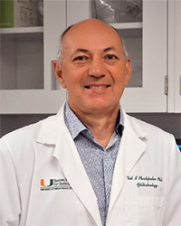
Role of inflammasome in pathophysiology of the retina in elevated intraocular pressure
Valery Shestopalov, University of Miami
Systemic, intracranial and intraocular hypertension is ammajor systemic risk factor for stroke, neurological and cardiological diseases. Spiking pressure elevation is the most dangerous as it is increasingly difficult to detect and treat. This study is focused on the role of inflammasome pathway in facilitating retinal ganglion cell (RGC) dysfunction and death in response to ocular hypertension (OHT)-induced stress and glaucoma.
Glaucoma was induced by intracameral injection of Ad5-MYOC vector that produced a long-lasting IOP elevation. Inflammasome activation was detected by IL-1β production, changes in Casp1, ASC, NLRP1 and GsdmD gene expression and protein accumulation using RT-PCR, Western blot, and immunohistochemistry. Wild type and knockout mouse strains deficient for Casp1 and NLRP1 proteins were used to determine the role of key inflammasome components in ocular hypertension glaucoma. IL-1β cytokine production was assessed with sandwich ELISA; gene expression changes in the retina, by RT-PCR. Changes in RGC functionality was assessed by PERG recordings; RGC loss was assessed at 6 wks post-OHT induction by direct counts in retinal flat mounts.
Activation of the inflammasome became evident in the inner retinal layer and co-localized with RPBMS+ RGCs in C57Bl6 (WT) mice at 2-4 wks after glaucoma induction. This correlated in time with RGC dysfunction, where PERG amplitude was reduced 55.8% in WT eyes with chronic OHT glaucoma. RGC density analysis in these animals showed a loss of 32.3±9.2 % in the central and 28.4±9.2 % in peripheral retina at 6 wks post OHT-induction. Mice deficient for key inflammasome complex proteins NLRP1, Casp1 and GsdmD, had changes in PERG amplitude and RGC not significantly different from zero: a 7.4% reduction in NLRP1KO and 2.35 % inrease in Casp1KO eyes. Mice deficient for Casp1 and Nlrp1 genes had no statistically significant changes in RGC density: a 7.3 ±3.0 % and 0.3 ±2.6 % decline in the central retina, 0.0 ±5.6% and 0.0 ±11.2% in the peripheral retina, respectively. Immunohistochemistry data in retinal cross-sections showed absence of Casp1 and dramatic reduction in and GsdmD immunolabeling labeling in RGCs of glaucomatous retinas from the eyes of knockout strains.
Our results show the key role of inflammasome and RGC pathology in the retina exposed to IOP elevation. Our results show that RGC dysfunction and neurotoxicity triggered by the inflammasome depends on its endpoint product, gasderminD. Drugs inhibitors of inflammasome and gasderminD could protect vision in ocular hypertension glaucoma and, potentially brain neurons during intracranial pressure spikes.

Hermann Körperich, Heart and Diabetes Center NRW, Univ. Hospital of the Ruhr-University Bochum, Bad Oeynhausen, Germany

Autophagy in atherosclerosis: a potential drug target for plaque stabilization
Wim Martinet, University of Antwerp
According to the European Society of Cardiology and Eurostat, (cardio)vascular disease remains the leading cause of mortality and a major cause of morbidity in our western society. Even though recent statistics indicate that mortality due to vascular disease is now decreasing in nearly all European countries, there are still more than 6 million new cases in the EU every year. The majority of (cardio)vascular diseases relates to the development of atherosclerotic plaques in medium/large-sized arteries, such as coronary arteries. High cholesterol, smoking, diabetes mellitus and obesity are major risk factors of vascular disease. While high cholesterol is relatively well managed by preventive treatment with statins and other hypolipidemic drugs, the impact of other factors such as the prevalence of overweight/obesity and diabetes have increased considerably in recent decades. Besides being a risk factor for atherosclerosis, diabetes causes severe microvascular complications. Therefore, new strategies to treat both macro- and microvascular pathologies are desperately needed.Growing in vitro and preclinical evidence indicates that (basal) autophagy in vascular smooth muscle cells (VSMCs) and endothelial cells (ECs) plays an essential role in the maintenance of (cardiovascular) health. Autophagy is an evolutionary conserved physiological process that maintains intracellular homeostasis by sequestrating unnecessary or dysfunctional cellular components in double membrane structures, called autophagosomes. The latter fuse with lysosomes, where the contents are degraded by acid hydrolases. Since the autophagic process allows recycling of macromolecules and energy, basal autophagy represents a reparative and life-sustaining process that is strictly regulated by a number of highly conserved AuTophaGy-related (ATG) genes. It should be noted that autophagy is stimulated by many stress-related stimuli in the arterial wall to protect VSMCs and ECs against cell death and the initiation of vascular disease, in particular atherosclerosis. However, autophagic activity declines with age, thereby contributing to the accumulation of damaged macromolecules and organelles during aging and worsening of aging-associated diseases including atherosclerosis. Indeed, autophagy defects in VSMCs or myeloid cells results in accelerated neointima formation and atherosclerosis. Most interestingly, arterial stiffness and EC dysfunction related to arterial aging are normalized by enhancing autophagy with specific drugs. In addition, pharmacological activation of autophagy reduces the progression and destabilization of atherosclerotic plaques. Collectively, these data support the concept that basal autophagy in vascular cells is atheroprotective and that induction of (basal) autophagy could be a game-changer in the treatment of unstable atherosclerotic lesions.

What does a prospective twin study tell us about DNA hydroxymethylation and cardiovascular death?
Jun Dai, Des Moines University College of Health Sciences