


Curcumin vs Nano Curcumin: Antioxidant Effects in SE
Comparative protective evaluation of curcumin and nano curcumin on the oxidative stress and other indices in the blood of experimental status epilepticus animal model
Mohammad Ahmad¹, Homood Alharbi¹, Ahmad Alsaaloun¹, Abdulrahman Alshehry¹
- Department of Medical-Surgical Nursing, College of Nursing, King Saud University, Riyadh – 11421, Saudi Arabia.
OPEN ACCESS
PUBLISHED: 31 January 2025
CITATION: Ahmad, M., Alharbi, H., et al., 2025. Comparative protective evaluation of curcumin and nano curcumin on the oxidative stress and other indices in the blood of experimental status epilepticus animal model. Medical Research Archives, [online] 13(1).
https://doi.org/10.18103/mra.v13i1.6240
COPYRIGHT: © 2025 European Society of Medicine. This is an open-access article distributed under the terms of the Creative Commons Attribution License, which permits unrestricted use, distribution, and reproduction in any medium, provided the original author and source are credited.
DOI https://doi.org/10.18103/mra.v13i1.6240
ISSN 2375-1924
ABSTRACT
Background: Curcumin possesses antioxidant properties against oxidative damage, exerting powerful oxygen-free radical scavenging effects. The present study compares the protective effects of pre-treatment with curcumin and nanoparticles of curcumin on lithium pilocarpine-induced status epilepticus (SE) for oxidative damages and other blood indices in adult male rats.
Methods: The pretreated rats with curcumin and nano curcumin were induced with lithium pilocarpine-induced SE, and the non-enzymatic oxidative stress indices like lipid peroxides (TBARS), reduced total glutathione (GSH), as well as the enzymatic oxidative stress indices like catalase (CAT), glutathione S-transferase (GST), and superoxide dismutase (SOD), were estimated together in the blood cells with some blood indices in the serum.
Results: SE was able to induce a significant increase in TBARS and CAT and a substantial decrease in GSH, GST, and SOD levels in the blood cell tissues. The curcumin and nano curcumin pretreated groups significantly attenuated these oxidative stress indices. Furthermore, the blood indices like counts of red and white blood cells, platelets, hemoglobin, and packed cell volume were also depleted due to SE. Still, they were unaffected in the curcumin and nano curcumin pretreated groups. The serum enzyme levels of alkaline phosphatase, creatinine kinase, and other serum indices like high-density lipoprotein, urea, and glucose were decreased in the SE group. However, pretreatment with curcumin and nano curcumin ameliorated these serum indices.
Conclusion: The results indicate that oxidative stress indices in blood cells and other serum indices can be potential biomarkers for SE. Curcumin and nano curcumin may act as natural antioxidants for SE in the effective order of nano curcumin > curcumin. Further studies are required to ascertain the possibility of nano curcumin as an antiepileptic natural product.
Introduction
Recent studies have reported the protective effects of curcumin (Cur) against oxidative damage with antioxidant and anticonvulsant properties.¹–³ Status epilepticus (SE) is a neurodegenerative condition causing neuronal injuries in the brain⁴ due to neurochemical imbalances in affected brain regions. Such chemical imbalances have been evidenced by the excessive generation of free radicals, suggesting oxidative stress.⁵ Lithium (Li)/pilocarpine (Pc)-induced SE in rodent models has provided information regarding oxidative stress-related epileptic activity.³,⁶,⁷ The Li–Pc model of SE reproduces most clinical, temporal, and neuropathological features of SE.⁸
Blood-brain barrier (BBB) leakage is one of the earliest characteristic pathophysiological disturbances during SE and might, therefore, play an essential role in the development of SE.⁹ Initial studies in various SE animal models indicated that BBB leakage can be easily detected during the first 10 days after pharmacologically induced SE.¹⁰,¹¹ There is ample evidence that SE-induced BBB disruption and inflammation play an important role in the development of epilepsy and the progression of seizure activity. BBB-SE-associated oxidative stress is accelerated by different pathological processes that include inflammation and angiogenesis.⁹
Although the anticonvulsant effect of several compounds having antioxidant properties has been demonstrated in various studies,⁶,¹² the unsatisfactory pharmacotherapy of SE necessitates the search for alternative natural resources that can target the different underlying mechanisms of SE pathology and reduce disease occurrence and/or progression. Among the natural resources, many studies on Cur effects on SE have been undertaken.⁷,¹³,¹⁴ Still, no study has been conducted on the comparative protective effect of Cur and its nanoparticle form as nano curcumin (NCur) pretreatment on the oxidative stress of the blood cells and other serum indices in experimentally induced SE.
In light of the above, this study explores the comparative neuroprotective effects of Cur and NCur on Li–Pc-induced SE in rats to understand hematological indices and oxidative stress and other blood and serum indices that may have a correlative perspective with SE. Furthermore, the present research work can indirectly suggest the importance of screening natural food products that are beneficial as neurotherapeutic agents in ameliorating SE using oxidative stress in the blood cells and other indices in blood and serum as biomarkers for SE.
Methodology
Experimental Animals
Male Sprague Dawley rats (weighing 200–250 g, 2 months old) were used in this study. The animals were maintained under controlled conditions at 22 ± 1°C, humidity at 50–60% with 12 hours of light-dark diurnal cycle with free access to food and water except during experimental handling. All experimental procedures were conducted according to the institutional guidelines for the care and use of laboratory animals by the local animal care and ethics committee institutions.
Induction of Status Epilepticus and Treatment Groups
Animals were randomly assigned into seven groups. Groups 1, 2, 3, and 4 served as controls receiving saline, Li (3 mEq/ml/kg, i.p.), 50% dimethyl sulfoxide (DMSO), and Pc (20 mg/ml/kg, s.c.), respectively. SE was induced in groups 5, 6, and 7 by administering an aqueous (saline) solution of Li (BDH Laboratory Supplies, Poole, England, in a dose as control), followed by (20 h later) Pc (Sigma Chemical Co., St. Louis, MO, USA, in the dose as used for control). Group 5 served as the experimental control of the SE group, and groups 6 and 7 served as Cur (group 6) and NCur (group 7) test groups. Cur (Sigma, USA), was dissolved in 50% DMSO and was given to group 6 at a dose of 50 mg/kg body weight/ml orally. The prepared NCur was given to group 7 orally at a dose of 5 mg/kg body weight for seven days before administering Li and Pc for the induction of SE. Our pilot studies used a range of doses (low, medium, and high) to determine the effects of Cur and NCur. However, Cur (50 mg/kg) and NCur (5 mg/kg) are the best-observed doses used in this study. After the Pc injections, the animals (n = 20 in per group) were observed for convulsive behavioral alterations, which developed progressively into SE within 1–2 h. Data from behavioral observations are not included in this study. Animals not reaching the SE level were discarded.
Preparation of Curcumin Nanoparticles of Curcumin
Curcumin nanoparticles were prepared using dichloromethane under the ultrasonication method as described elsewhere.¹⁶ In brief, curcumin (0.30 mmol) was taken in dichloromethane (50 mL), and 1 mL of the prepared solution was added dropwise to boiling water (50 mL) at a flow rate of 0.1 mL/min within 3 minutes at an ultrasonic power of 100 W with a frequency of 30 kHz. After sonication for 15 mins, the contents were centrifuged at 5000 rpm for 20 mins at room temperature, and the solution was concentrated under reduced pressure at 50°C to obtain a powder. Furthermore, ¹H NMR and the lyophilized powder’s ultraviolet (UV) spectra confirmed it to be NCur.¹⁶
Collection of Blood Samples for Assays
After inducing SE, the animal’s blood samples were collected under light anesthesia from the retro-orbital sinus plexuses of their eyes with the help of capillary tubes.¹⁷ Some assays were done in the whole blood, and for assays in the serum, the collected blood was allowed to clot and centrifuged at 2500 rpm in a refrigerator centrifuge at 10°C for 15 min. The clear serum was isolated and stored at 0°C until required for serum assays. The pellets of blood cells were used to prepare the erythrocyte lysates for measuring the oxidative stress indices in the erythrocyte membrane tissue.
Analysis of Whole Blood
The automated parameter hematology analyzer (T 450, USA) measured the red blood cell count, hemoglobin content, packed cell volume, total white blood cell count, and blood platelets.
Preparation of Erythrocyte Lysates
Erythrocyte lysates were prepared using this method.¹⁸ Erythrocyte pellets were obtained from the samples by centrifugation at 2500 rpm for 15 min at room temperature. The plasma and buffy coats were then removed, and the erythrocytes were washed twice in saline and stored at 20°C for 15 min. Lysed erythrocytes were prepared by thawing frozen samples and adding 3 volumes of ice-cold distilled water. Cell membranes were removed by centrifugation at 1000 g for 20 min.
Determination of Nonenzymatic Oxidative Stress Indices in Blood Cells
Thiobarbituric acid-reactive substances. Lipid peroxides were determined spectrophotometrically as thiobarbituric acid-reactive substances (TBARS) according to the method of Ohkawa et al.¹⁹ Tissue lipid peroxide levels were quantified using an extinction coefficient of 1.56 × 10⁵ M⁻¹ cm⁻¹ and expressed as nanomoles of TBARS formed per g tissue weight. The results are expressed as nmol/g wet weight.
Glutathione. Reduced glutathione (GSH) level was measured enzymatically by a slightly modified method of Mangino et al.²⁰ The slope of the change in absorbance was used to quantify total GSH by comparing the slope of the samples with a standard curve prepared with pure glutathione (Sigma). The specific activity is expressed into μmol/g tissue weight.
Determination of Enzymatic Oxidative Stress Indices in Blood Cells
Glutathione-S-transferase. Glutathione S-transferase (GST) was estimated by Habig et al.²¹ using 1-chloro-2,4-dinitrochlorobenzene (CDNB) as substrate at 340 nm. The GST activity is expressed as U/g tissue weight.
Catalase. Catalase (CAT) activity was measured using the Aebi method²², which tracks the decomposition of hydrogen peroxide by measuring the decrease in extinction of H₂O₂ at 240 nm. The activity of CAT is expressed as the rate constant of first-order reaction K per gram tissue weight.
Superoxide Dismutase. Superoxide dismutase (SOD) activity was estimated by the method of Misra and Fridovich.²³ Activity is expressed as the amount of enzyme that inhibits the oxidation of epinephrine by 50%, equal to U per gram tissue weight.
Serum Analysis
The clear serum was used for the estimation of various blood chemical parameters, including some serum enzymes like alkaline phosphatase (ALP), alanine transaminase (ALT), aspartate transaminase (AST), creatine kinase (CK), and some other serum entities like high-density lipoprotein (HDLP), urea, glucose, and potassium ion (K), by the auto analyzer apparatus (Reflotron Plus, Roche, Germany) using commercially available Reflotron kits (Roche Diagnostics, Germany) for all serum parameters. The principles of the tests of each analyte were based on the methods as follows: ALP,²⁴ ALT,²⁵ AST,²⁶ CK,²⁷ HDLP,²⁸ urea and glucose,²⁹ and K.³⁰
Statistical Analysis
The data were analyzed using Bartlett’s test for equal variance and Gaussian-shaped distribution for normality using the Kolmogorov-Smirnov goodness-of-fit test. As the data passed the normality test (P > 0.10), group means were compared with one-way ANOVA with post hoc testing using the Tukey-Kramer Multiple Comparisons Test or Student-Newman-Keuls Multiple Comparisons Tests. All results were expressed as means ± SEM, and the significance was defined as P < 0.05 for all analyses.
Results
Characterization of Curcumin Nanoparticles
A co-TLC of NCur with Cur was performed to rule out any chemical modification or degradation. In our earlier study,¹⁶ ¹H NMR spectrum was recorded; however, for easy reference, the results from our earlier study¹⁶ are given in Figures 1a and 1b. The TLC profile and NMR data suggested that both had an identical chemical structure. The particle size analysis and distribution of the NCur were performed using the TEM and SEM analysis, respectively, and the results agree with those of our earlier study.¹⁶ The results are shown in Figures 2a and 2b, respectively. As reported earlier, the dry powder form of NCur was found to have good physical and chemical stability and was readily dispersible in water.¹⁶
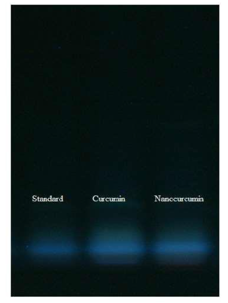
Figure 1a. Thin layer chromatography (TLC) data of Curcumin (Cur) and Nano curcumin (NCur).
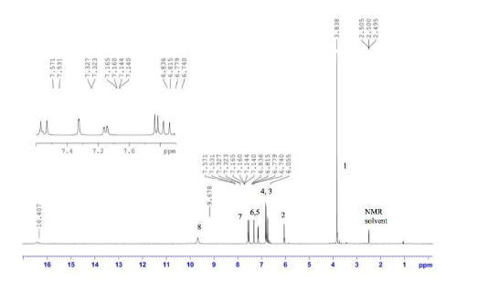
Figure 1b. Nuclear Magnetic Resonance (NMR) data from Cur and NCur.
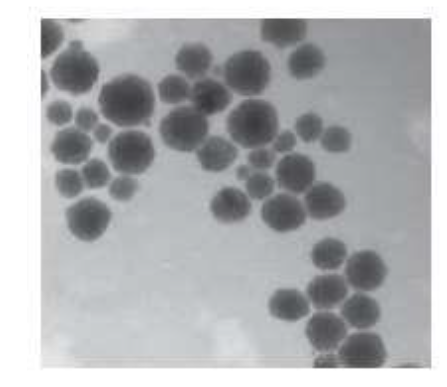
Figure 2a. Transmission Electron Microscopy (TEM) data of NCur.
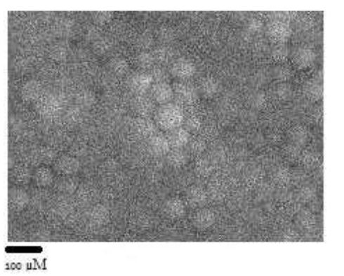
Figure 2b. Scanning Electron Microscopy (SEM) data of NCur.
Note: Figures 1a, 1b, 2a, and 2b are taken from Ahmad et al.¹⁶ to suggest that our present findings are the same as those reported earlier.
Analysis of Blood Parameters
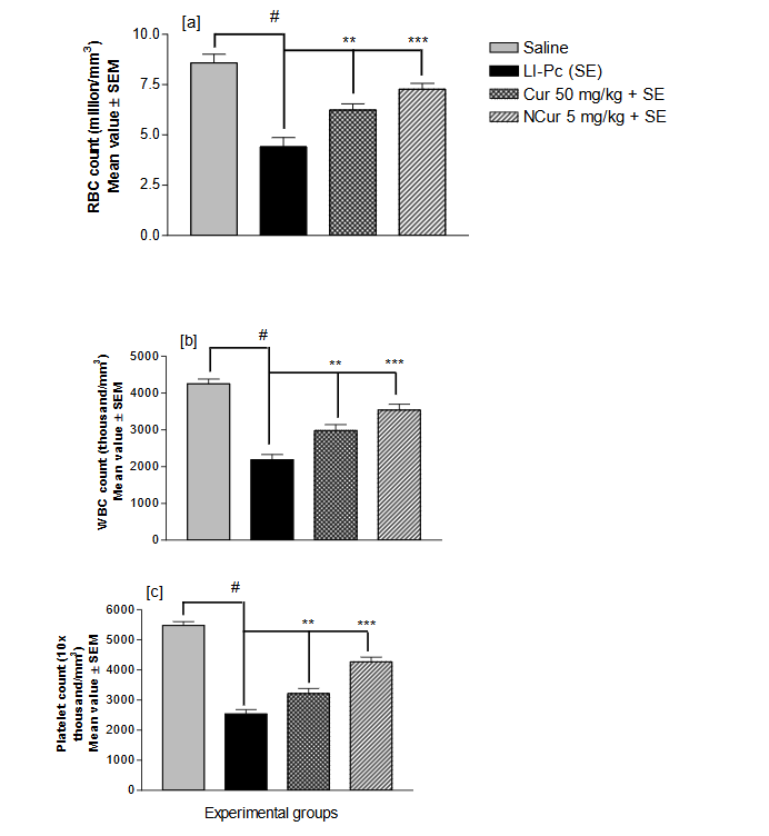
Induction of SE significantly reduced blood parameters like red blood cell count, white blood cell count, platelet count (Figure 3 a, b, and c, respectively), hemoglobin content, and packed cell volume (Figure 4 a and b, respectively) compared to the control group. Pretreatment with Cur and NCur ameliorated all the observed blood parameters in the order NCur > Cur (Figures 3 and 4).
The Li, DMSO, and PC control results are not included in these figures, as they were not different from the saline control.
Oxidative Stress Indices in Blood Cells
Nonenzymatic Oxidative Stress Indices
Thiobarbituric acid-reactive substances. The lipid peroxidation level (TBARS) in the erythrocyte membranes was markedly (P < 0.001) increased after Li-PC (SE) treatment as compared to the control group (Figure 5a). Pretreatment with Cur and NCur significantly (P < 0.001) attenuated Li-PC induced increase in TBARS in the order NCur > Cur (Figure 5a) as compared to the Li-PC (SE) group.
Glutathione. A highly significant (P < 0.001) depletion of glutathione (GSH) was observed in the erythrocyte membrane tissue of the Li-PC (SE) group (Figure 5b). Pretreatment with Cur and NCur significantly attenuated this depletion of GSH in the order NCur > Cur (Figure 5b) as compared to the Li-PC (SE) group.
Figure 3
Caption:
Figure 3. The protective effect of pre-treatment with curcumin (Cur) and nano curcumin (NCur) on the counts of (a) Red Blood Cells (RBC), (b) White Blood Cells (WBC), and (c) Platelets levels in the blood of the status epilepticus (SE)-induced rats. The two antioxidants were effective in the order NCur > Cur.
SE-induced group differed significantly (p < 0.001) from the Saline control group.
NCur*** and Cur** significantly differed from the SE-induced group at p < 0.001 and p < 0.01, respectively, by Newman-Keuls student’s test after one-way ANOVA.
Figure 4
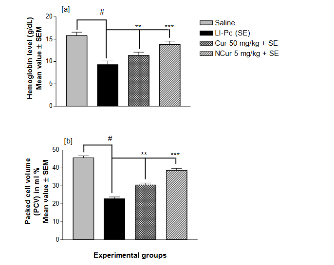
Figure 4. The protective effect of pre-treatment with curcumin (Cur) and nano curcumin (NCur) on (a) hemoglobin (Hb), and (b) packed cell volume (PCV) in the blood of the status epilepticus (SE)-induced rats. The two antioxidants were effective in the order NCur > Cur.
Statistical significance is the same as in Figure 3.
Figure 5
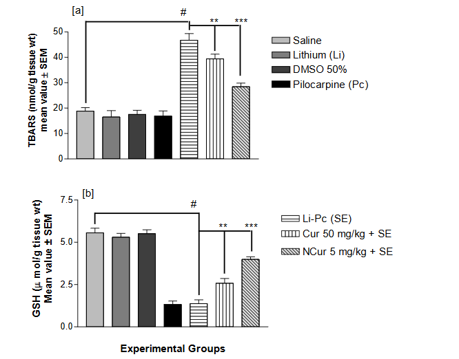
Figure 5. The protective effect of pre-treatment with Cur and NCur on the non-enzymatic oxidative stress indices (a) TBARS (lipid peroxidation), and (b) GSH (glutathione) content in the blood cells of the SE-induced rats. The two antioxidants were effective in the order NCur > Cur.
Statistical significance is the same as in Figure 3.
ENZYMATIC OXIDATIVE STRESS INDICES
Glutathione-S-Transferase.
A highly significant (P < 0.001) depletion of glutathione-S-transferase (GST) was observed in the Li-Pc (SE) group (Figure 6a). Pretreatment with Cur and NCur significantly attenuated this depletion of GST in the order NCur > Cur (Figure 6a) as compared to the Li-Pc (SE) group.
Catalase.
The catalase (CAT) level was markedly (P < 0.001) increased after SE induction as compared to the control group (Figure 6b). Pretreatment with Cur and NCur significantly (P < 0.001) attenuated Li-Pc-induced increase in CAT in the order NCur > Cur (Figure 6b) as compared to the Li-Pc (SE) group.
Superoxide Dismutase.
The superoxide dismutase (SOD) level was significantly (P < 0.001) decreased in the SE-induced group as compared to the control group (Figure 6c). Pretreatment with Cur and NCur significantly (P < 0.001) attenuated Li-Pc-induced decrease in SOD in the order NCur > Cur (Figure 6c) as compared to the Li-Pc (SE) group.
SERUM ANALYSIS
Among the blood serum enzyme parameters (Figures 7 and 8), SE induction inhibited the level of enzymes ALP (Figure 7a), ALT (Figure 7b), and AST (Figure 7c), HDLP, Urea, and Glucose (Figure 8a), and CK (Figure 8b) significantly as compared to the control. Pretreatment with Cur and NCur significantly attenuated the observed serum parameters in NCur > Cur. However, the level of K remained unaffected (Figure 8c).
The Li, DMSO, and Pc control results are not included in these figures, as they were not significantly different from the saline control.
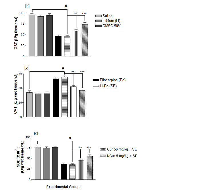
Figure 6. The protective effect of pre-treatment with Cur and NCur on the enzymatic oxidative stress indices (a) GST (glutathione-S-transferase), (b) CAT (catalase), and (c) SOD (superoxide dismutase) content in the blood cells of the SE-induced rats. The two antioxidants were effective in the order NCur > Cur.
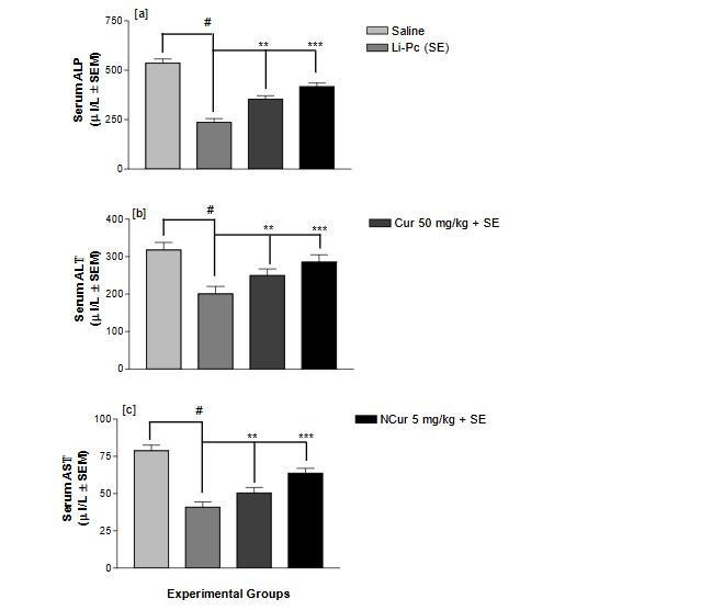
Figure 7. The protective effect of pre-treatment with Cur and NCur on serum enzymes (a) ALP (alkaline phosphatase), (b) ALT (alanine transaminase), and (c) AST (aspartate transaminase) in the serum of the status epilepticus (SE)-induced rats. The two antioxidants were effective in the order NCur > Cur.
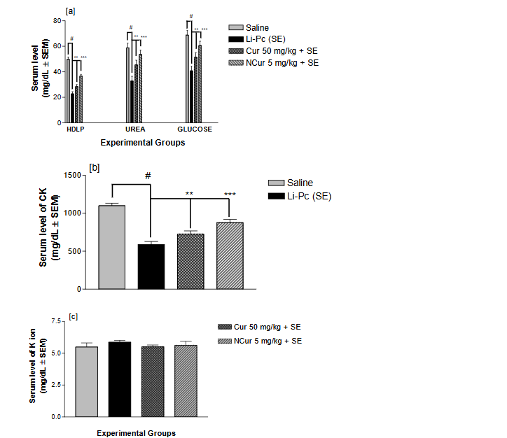
Figure 8. The protective effect of pre-treatment with Cur and NCur on some serum entities like (a) HDLP (high-density lipoproteins), urea, and glucose, (b) CK (creatinine kinase), and (c) K⁺ (potassium ion) in the serum of the status epilepticus (SE)-induced rats. The two antioxidants were effective in the order NCur > Cur. However, K⁺ remained unaffected by the two antioxidants (Figure 8c).
Discussion
It is evident that in the recent past, researchers have been globally involved in evaluating and screening natural products for their antioxidant properties. In this study, we assessed the effect of Cur and NCur on various blood parameters, including oxidative stress indices in the SE-induced model. From the results, it was inferred that the natural product Cur and its nanoparticles, NCur could be a good antioxidant in the order NCur > Cur in attenuating the harmful effects of oxidative stress and other blood-related parameters caused by SE in rats.
One of the earliest characteristic pathophysiological disturbances during SE has been related to blood-brain barrier (BBB) leakage, which might play an essential role in the development of epileptic seizures. It was presumed that such BBB could have some indication of SE-induced disruptions in the blood of the rat model. Thus, we studied the oxidative stress indices and other blood-related parameters in the blood of the SE-induced rats with an insight to compare the protective effect of Cur and NCur on all observed parameters in the blood.
The biochemical results of our study further indicate a significant disruption in the oxidative indices (enzymatic and non-enzymatic) levels in the blood cells of the rats treated with Li-Pc, suggesting a preliminary and considerable level of oxidative stress. Antioxidants could arrest the seizure genesis caused by excitotoxic agents.³¹ Potent antioxidant activities have been reported for Cur.³,⁶,³²–³⁴ Although Cur has been reported to have neuroprotective role in SE-induced models, to the author’s best knowledge, no precedent has been reported for Cur and NCur in a comparative evaluation. In the SE groups
of this study, a significant disruption in levels of oxidative stress indices in the blood cells was observed. Compared to the respective SE group, pretreatment with Cur and NCur significantly attenuated Li-Pc induced oxidative stress-related indices (enzymatic and non-enzymatic) in the blood cells.
This study was also designed to assess the antioxidative defense system in the RBCs of SE-induced rats and the ameliorating effect of Cur and NCur on such oxidative stress. Reduced glutathione (GSH) is central to cellular antioxidant defenses and an essential cofactor for antioxidant enzymes.³⁵ Under oxidative stress, glutathione is consumed by the glutathione-related enzymes to detoxify peroxides produced because of increased TBARS³⁶,³⁷, and elevation in TBARS is a consequence of depleted GSH stores, which are otherwise capable of moderating the amount of TBARS. Similar disturbances in the antioxidant barrier (involving glutathione peroxidase and superoxide dismutase) have been shown in the rat serum and tissues where depletion of the other accompanied the increment of one enzyme.³⁸
Phosphatases, including ALP, are critical enzymes in biological processes responsible for detoxification, metabolism, and biosynthesis of energetic macromolecules for different essential functions. Any interference in these enzymes leads to biochemical impairment, tissue lesions, and cellular function lesions.³⁹ It was suggested that interference in the activities of ALP enzymes in serum might be due to altered plasma membrane permeability or cellular necrosis, thus showing the stress condition of the treated animals. Blood serum enzymes like ALP, AST, and ALT can be used as biomarker enzymes for detecting SE.
Besides the soluble enzymes of blood serum like AST, ALT, and ALP, the other observed blood chemistry indices may also be suitable bioindicators for detecting SE due to Li-Pc. As blood is the overall reflector of body health, the altered glucose, urea, and HDLP levels in treated rats suggest some damage due to SE.
It should be noted that most earlier studies on SE in experimental animal models have investigated for their behavioral changes, cognitive retention, and brain damage in the hippocampus and striatum areas using histopathological parameters (looking for neuronal structural changes) and/or biochemical alterations (measuring oxidative stress indices, neurotransmitters).³,⁵,⁷,⁸,¹²,¹⁵,¹⁶,³³ Using noninvasive methods for studying various biochemical indices in the blood of experimental animal models could be yet another reliable parameter for studying SE.
Conclusion:
It can be concluded at preliminary level that Cur and NCur have promising anticonvulsant and antioxidant activity against SE-induced rat blood cells in the order NCur > Cur. The oxidative stress indices in the blood cells and other indices in the blood and serum of SE-induced rats can be considered bioindicators for SE. However, further studies on these lines are needed.
Conflict of interest
The authors declare that they have no conflict of interest.
Funding statement
This research had no funding.
Acknowledgment
The authors are thankful to the Deanship of Scientific Research, College of Nursing Research Center at King Saud University for supporting this research.
References
- Farombi EO, Ekor M. Curcumin attenuates gentamicin-induced renal oxidative damage in rats. Food Chem Toxicol. 2006; 44:1443-48.
- El-Demerdash FM, Yousef MI, Radwan FM. Ameliorating effect of curcumin on sodium arsenite-induced oxidative damage and lipid peroxidation in different rat organs. Food Chem Toxicol. 2009; 47:249-54.
- Du P, Tang HY, Li X, Lin HJ, Peng WF, Ma Y, Fan W, Wang X. Anticonvulsive and antioxidant effects of curcumin on pilocarpine-induced seizures in rats. Chin Med J. 2012;125:1975-9.
- Chen S, Fujita S, Koshikawa N, Kobayashi M. Pilocarpine-induced status epilepticus causes acute interneuron loss and hyper-excitatory propagation in rat insular cortex. Neuroscience. 2010;166: 341-53.
- Freitas RM. Investigation of oxidative stress involvement in hippocampus in epilepsy model induced by pilocarpine. Neurosci Lett. 2009; 462: 225-29.
- Aboul Ezz HS, Khadrawy YA, Noor NA. The neuroprotective effect of curcumin and Nigella sativa oil against oxidative stress in the pilocarpine model of epilepsy: a comparison with valproate. Neurochem Res. 2011; 36: 2195-204.
- Ahmad M. Protective effects of curcumin against lithium–pilocarpine induced status epilepticus, cognitive dysfunction and oxidative stress in young rats. Saudi J Biol Sc. 2013; 20:155-62.
- Tariq M, Ahmad M, Moutaery KA, Deeb SA. Pentoxifylline ameliorates lithium–pilocarpine induced status epilepticus in young rats. Epilepsy Behav. 2008; 12: 354-65.
- Gorter JA, van Vliet EA, Aronica E. Status epilepticus, blood-brain barrier disruption, inflammation, and epileptogenesis. Epilepsy Behav. 2015; 49: 13-16.
- Pont F, Collet A, Lallement G. Early and transient increase of rat hippocampal blood-brain barrier permeability to amino acids during kainic acid-induced seizures. Neurosci Lett. 1995; 184:52-54.
- Roch C, Leroy C, Nehlig A. Magnetic resonance imaging in the study of the lithium–pilocarpine model of temporal lobe epilepsy in adult rats. Epilepsia. 2002; 43:325-35.
- Freitas RL, Santos IM, De Souza GF, Tome Ada R, Saldanha GB, Freitas RM. Oxidative stress in rat hippocampus caused by pilocarpine-induced seizures is reversed by buspirone. Brain Res Bull. 2010; 81:505-09.
- Choudhary KM, Mishra A, Poroikov VV, Goel RK. Ameliorative effect of Curcumin on seizure severity, depression like behavior, learning and memory deficit in post-pentylenetetrazole-kindled mice. Eur J Pharmacol. 2013; 15: 33-40.
- Adami R, Bottai D. Curcumin and neurological diseases. Nutr Neurosci. 2020; 22: 1-21.
- Persinger MA, Bureau YRJ, Kostakos M, Peredery O, Falter H. Behaviors of rats with insidious multifocal brain damage induced by seizures following single peripheral injections of lithium and pilocarpine. Physiol Behav. 1993; 53: 849-56.
- Ahmad M, Abu-Taweel GM, Hidayathulla S. Nano-composites chitosan-curcumin synergistically inhibits the oxidative stress induced by toxic metal cadmium. Int J Biol Macro. 2017; 108: 1-28.
- Sorg DA, Buckner B. A simple method of obtaining venous blood from small laboratory animals. Proc Sec Exp Med. 1964;115: 1131-32.
- Ceballos-Picot I, Triever JV, Nicole A, Silnet PM, Thevenin M. Age-correlated modifications of copper-zinc superoxide dismutase and glutathione-related enzyme activities in human erythrocytes. Clin Chem. 1992; 38: 66-70.
- Ohkawa H, Ohishi N, Yagi K. Assay for lipid peroxides in animal tissues by thiobarbituric acid reaction. Anal Biochem. 1979; 95: 351-58.
- Mangino MJ, Murphy MK, Glabar GG. Protective effects of glycine during hypothermic renal ischemic reperfusion injury. Am J Physiol. 1991; 261: F841-48.
- Habig WH, Pabst MJ, Jakoby WB. Glutathione S-transferases. The first enzymatic step in mercapturic acid formation. J Biol Chem.1974; 249: 7130-39.
- Aebi H. Catalase, in Methods of Enzymatic Analysis, H. U. Bergmeyer, Ed., 1972; vol. 2, Academic Press, New York, NY, USA.
- Misra HP, Fridovich I. The role of superoxide anion in the autoxidation of epinephrine and a simple assay for superoxide dismutase, J Biol Chem. 1972; 247: 3170-5.
- Schumann G, Dominick HC, Hellmann D, Klauke R, MöckeschM, Stekel H, von Schenck H, Kraft M, Nagel R, Hänseler E. Alkaline phosphatase activity: new assay for the Reflotron system. Results of the evaluation in eight clinical laboratories. Clin Chem Lab Med. 2001; 39: 71-8.
- Deneke U, Rittersdorf W. Evaluation of the refloquant GPT (ALT) reagent carriers with reflotron. Clin Chem. 1984; 30: 1009.
- Deneke U, Rittersdorf W, Werner W. Performance data of Reflotron-GOT (AST) dry chemistry test for Reflotron. Clin Chem. 1985; 31: 921.
- Horder M, Jorgensen PJ, Hafkenscheid JC, Carstensen CA, Bachmann C, Bauer K, Neuwald C, Rosalki SB, Foo AY, Vogt W. Creatine kinase determination: a European evaluation of the creatine kinase determination in serum, plasma and whole blood with the Reflotron system. Eur J Clin Chem Clin Biochem. 1991; 29: 691-6.
- Kinlay S. Comparison of Reflotron and laboratory cholesterol measurements. Med J Aust. 1988; 149: 126-9.
- Price CP, Koller PU. A multicentre study of the new Reflotron system for the measurement of urea, glucose, triacylglycerols, cholesterol, gamma-glutamyltransferase and haemoglobin. J Clin Chem Clin Biochem. 1988; 26: 233-50.
- Paschen K, Hagemann P, Lang HR, Cortes M, Weidemann G, Tof-faletti J, Burritt M, Leinberger R, Tritschler W, Ramstetter E. Determination of potassium by dry reagent carrier technology: a multicentre evaluation. Eur J Clin Chem Clin Biochem. 1993; 31: 545-52.
- Schulz JB, Henshaw DR, Siwek D, Jenkins BG, Ferrante RJ, Cipolloni PB, Kowall NW, Rosen BR, Beal MF. Involvement of free radicals in excitotoxicity in vivo. J Neurochem. 1995; 64: 2239-47.