


Effective Virus Removal in Biologicals: Virus Filters
Virus Retentive Filters – Effective Virus Removal in the Manufacturing Process of Biologicals
Albrecht Gröner1
- PathoGuard Consult, Seeheim-Jugenheim, Germany
OPEN ACCESS
PUBLISHED:
CITATION: GRONER, Albrecht. Virus Retentive Filters – Effective Virus Removal in the Manufacturing Process of Biologicals. Medical Research Archives, [S.l.], v. 12, n. 12, dec. 2024. Available at: <https://esmed.org/MRA/mra/article/view/6084>.
COPYRIGHT: © 2025 European Society of Medicine. This is an open-access article distributed under the terms of the Creative Commons Attribution License, which permits unrestricted use, distribution, and reproduction in any medium, provided the original author and source are credited.
DOI: https://doi.org/10.18103/mra.v12i12.6084
ISSN 2375-1924
Abstract
In the manufacturing process of biologicals virus reduction steps – inactivation or removal – have to be implemented to assure a high margin of virus safety of these products. Orthogonal mechanisms of virus clearance should be integrated in the manufacturing process in order to inactivate / remove viruses/virus aggregates having been able to escape to a certain degree the reduction capacity of the previous virus clearance step. Virus retentive filters as an orthogonal virus clearance step are frequently implemented in the manufacturing process of biologicals as the virus removal capacity of virus retentive filters is based on size exclusion. Only the size of a virus impacts the removal capacity and not virus properties as enveloped/ non-enveloped or RNA / DNA viruses and their resistance to physiochemical treatment. Viruses larger than the mean pore size of a virus retentive filter are removed from the feed stream and the desired protein – if smaller than the pore size of the filter membrane – will pass the filter and can be collected in the filtrate without / with very low virus contamination. Depending on the filter pore size, virus retentive filters are grouped in large and small virus retentive filters i.e., filters removing large viruses as retroviruses and small viruses as picornaviruses and, especially, parvoviruses. Data of virus reduction factors from 89 publications, resulting in a total of close to 500 virus clearance studies for different viruses, product intermediates and large and small virus retentive filters are assessed. The virus clearance capacity of these filters can depend on the membrane layout and chemistry, the volumetric throughput of product intermediate as well as of buffer flush and transmembrane pressure including pressure/flow interruption and flow decay. These parameters, when disclosed in published data, show filter brand specific differences but, having the above-mentioned parameters for each filter optimised, effective virus removal could mostly be demonstrated in virus validation studies for each filter brand.
Keywords
- Virus Retentive Filters
- Virus Removal
- Biologicals
- Manufacturing Process
1.Introduction
Virus safety of biologicals derived from cell cultures (biotechnology products) or plasma (plasma-derived medicinal products (PDMPs)) is based on the three complementary approaches (i) selecting and testing the source and raw materials for the absence of undesirable infectious viruses, (ii) testing the product at appropriate steps of production to demonstrate the absence of contaminating infectious viruses, i.e., the starting material of these biologicals as unprocessed bulk of cell culture derived products and plasma pools for further manufacturing, and (iii) testing the capacity of the production process to inactivate and/or to remove viruses potentially present in the source and raw materials. This clearance capacity of the manufacturing process is documented in virus validation studies resulting in virus reduction factors (LRF = reduction factor in log10). Dedicated virus clearance steps, i.e., manufacturing steps incorporated in the manufacturing process predominantly for virus clearance and not for the purification and/or concentration of the drug substance, demonstrate commonly an effective virus clearance capacity in such virus validation studies for a wide range of viruses. Generally, a manufacturing process contains two dedicated virus clearance steps with different mode of actions, so-called orthogonal steps, as solvent/detergent treatment, pasteurisation (heat treatment in aqueous solution at 60°C for 10 h), dry heat treatment of lyophilised product (commonly either 100°C of 30 min or 80°C for 72 hours), low pH treatment (only for plasma-derived immunoglobulins and monoclonal antibodies), or virus filtration.
In order to achieve a finished product with a sufficiently high margin of virus safety, the virus clearance capacity of the manufacturing process should definitely exceed the potential amount of viruses in the starting material. As pointed out in different guidelines, the potential virus load in that volume of the starting material required to produce one dose of product should be removed by an excess of 6 log10, i.e., less than one virus particle is to be expected in 1 million vials thus meeting the sterility assurance level also for pathogens not replicating in a cell-free environment. If the overall virus clearance capacity of the dedicated virus clearance steps, documented in virus clearance studies with inherent limitation of the LRF due to e.g., limit of the amount of virus to be added in the virus spike preparation and / or limit of the detection of the in vitro assay will not result in a sufficiently high LRF, further manufacturing steps for the purification and concentration of the drug substance, e.g., chromatography steps, will have to be validated for virus clearance capacity to achieve a high enough overall virus reduction factor.
Virus retentive filters are one of the dedicated virus clearance steps implemented in the manufacturing process of biologicals. The virus clearance capacity is based primarily on size exclusion, i.e., viruses larger than the mean pore size of the filter are retained; the drug substance, the desired therapeutic protein, passes the filter with high yield when the size of the protein – not aggregated – is smaller than the virus to be removed. Detailed information on the use of virus retentive filtration can be found in the PDA Technical Report No. 41 (rev 2022) and, as virus retentive filtration is an established method, in the ICH guideline Q5A(R2), Annex 5: Examples of prior knowledge including in-house experience to reduce product-specific validation effort. Besides removing adventitious (exogenous) and endogenous (cell culture-derived) viruses from the drug substance during its production, virus retentive filters can also be employed to minimise the risk of virus contamination of cell cultures by viruses present in the raw material, e.g., cell culture medium and its compounds. The first commercially available virus retentive filter was produced by Asahi Kasei and launched in 1989: Planova 35N with mean pore sizes of 35 ± 2 nm, followed by Planova 15N with mean pore sizes of 15 ± 2 nm. These filters were evaluated to be implemented in the PDMP Factor IX and Factor XI with very good virus removal capacity and no detectable differences in the drug products associated with the virus filtration process. The first commercial virus filtered product, licensed in Europe, was a four-factor human prothrombin complex concentrate (PCC); the Planova 35N filter removed all large viruses studied effectively, i.e., by more than 4 log10 whereas the small picornavirus poliovirus was not significantly removed. Further virus retentive filters by Asahi Kasei as well as Millipore, PALL, and Sartorius are meanwhile on the market with different membrane composition and structures.
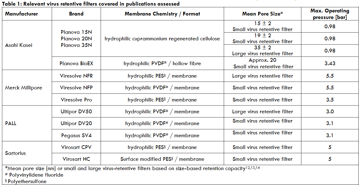
These filters are grouped in small and large virus retentive filters based on the removal capacity for the bacteriophage PP7, a ~30 nm Pseudomonas phage, (> 4 LRF) for small virus retentive filters and the bacteriophage PR 772, a ~64 to 82 nm E. coli phage, (> 6 LRF) for large virus retentive filters.
The virus removal capacity of the virus retentive filters was assessed in virus clearance studies, based on a valid downscale of the manufacturing process, by spiking the product intermediate with a defined amount of virus prior to filtration and assessing quantitatively the virus reduction capacity of the virus retentive filtration step as the difference in the spiked starting material and final sample (when a buffer flush is used according to the manufacturing process, the final sample is the pool of the filtrate plus the buffer flush). The amount of virus in the filtrate is quantified either employing an in vitro cell culture infectivity assay detecting virus replication due to infectious virus in the virus stock used for spiking or by a NAT (nucleic acid amplification test) such as PCR (polymerase chain reaction) detecting virus genome sequences in the filtrate. Polymerase chain reaction does not differentiate between infectious and non-infectious viruses. Therefore, the size of the amplicon generated by PCR should be large enough to represent infectious virus in the virus stock. Furthermore, prior to PCR assays, samples of the virus stock as well as of the filtrate to be employed in the PCR assay have to be treated by nucleases prior to capsid dissolution to remove free DNA and RNA, respectively. When performing virus clearance studies, the volume added to the product intermediate should not exceed 10% according to guidelines in order not to change the properties of the intermediate too much. Furthermore, the virus spike should be of appropriate purity and monodisperse to (i) avoid blocking of the filter by impurities from the virus spike and (ii) document a too high virus clearance capacity due to the removal of virus aggregates by the virus retentive filter. In order to remove impurities blocking the filter, either from the product intermediate itself or the virus spike, prefiltration of the feed stream is often applied; when the virus spike is of high purity, spiking the product intermediate is often performed after prefiltration.
Virus filtration can be performed in two ways, either as dead-end filtration or tangential filtration; in the early days of virus filtration implementation, tangential flow was mostly used in order to avoid blocking of the filter (using the known principle of ultrafiltration); a disadvantage of tangential filtration is that a certain amount of product intermediate is lost in the system. In the meantime, commonly dead-end filtration is applied and a buffer flush at the end of the filtration is applied to recover most of the drug substance.
2.Materials and Methods
2.1. DATA COLLECTION
Publicly available data of virus reduction factors attained by virus retentive filters were compiled by searching PubMed for the terms “virus filtration, biologicals / virus retentive filters, biologicals / nanofiltration, biologicals” and the papers were checked for results of virus reduction factors. In the majority of publications, no detailed information on the virus filtration parameters used were disclosed as volume / filter area, pressure, intermediate composition as protein concentration, pH, conductivity, flux (with potential flux decay), flow interruption etc., but it was stated that the scaled-down laboratory system represents closely the manufacturing process. Data for virus reduction factors from 89 publications, resulting in a total of close to 500 virus clearance studies for different viruses, product intermediates and virus retentive filters; 27 package inserts for plasma-derived medicinal products licensed by the FDA are not included in the assessment as commonly the filter type is not disclosed; for cell culture-derived products virus clearance factors are not required by the FDA and, thus, no virus clearance factors for this product class are disclosed in package inserts.
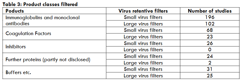
The virus clearance capacity of filtration processes were studied with the following product intermediates: Human immunoglobulins applied intravenously or subcutaneously, as well as hyperimmunoglobulin preparations and monoclonal antibodies, inhibitors include alpha-1-proteinase inhibitor, antithrombin III, and C1 esterase inhibitor, and the coagulation factor concentrates Factor VIII, IX, XI, XIII, thrombin, prothrombin complex concentrates, anti-inhibitor coagulant complex, von Willebrand factor, fibrinogen including the recombinant Factor IX and VIII, and a range of other intermediates as model proteins (e.g., human serum albumin (HSA), bovine serum albumin (BSA)), non-disclosed proteins and filters, different buffers, and some plasma- and cell culture-derived proteins.
Small and large virus retentive filters were employed in the production of the following products depending on the size of the desired protein to be filtered.
2.2. STATISTICAL ANALYSIS
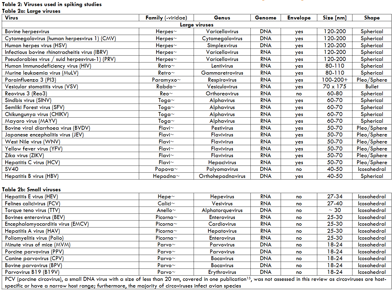
Viruses studied were grouped according to their size. Virus reduction factors (LRFs) were analysed considering small and large viruses filtered through small and large virus retentive filters as well as filters used in series (2*15N and 2*20N). LRF were differentiated between no infectious virus or PCR signal detected in the filtrate (virus titre below the Limit of Detection (LoD) of the assays) and infectious virus or PCR signal detected in the filtrate (i.e., above LoD of the assays). Furthermore, under these conditions, the capacity of a virus filtration step to remove viruses was differentiated between a so-called effective step (LRF ≥ 4) and a manufacturing step contributing to the virus safety of a defined product (LRF < 4). As all LRF below LoD can not be defined appropriately (LRF ≥ 4.3 may be e.g., 4.4 or 7.9), all data with LRF below LoD were not used in a statistical analysis but only LRF above LoD. An unpaired t-test was applied to compare equality of two means; prior to performing the t-test, the variances of both samples had to be equal / homogeneous and if the variance was not equal / homogeneous, the Welch’s t-test was performed (unpaired samples with different variances).
3. Results
3.1 SMALL VIRUS RETENTIVE FILTERS
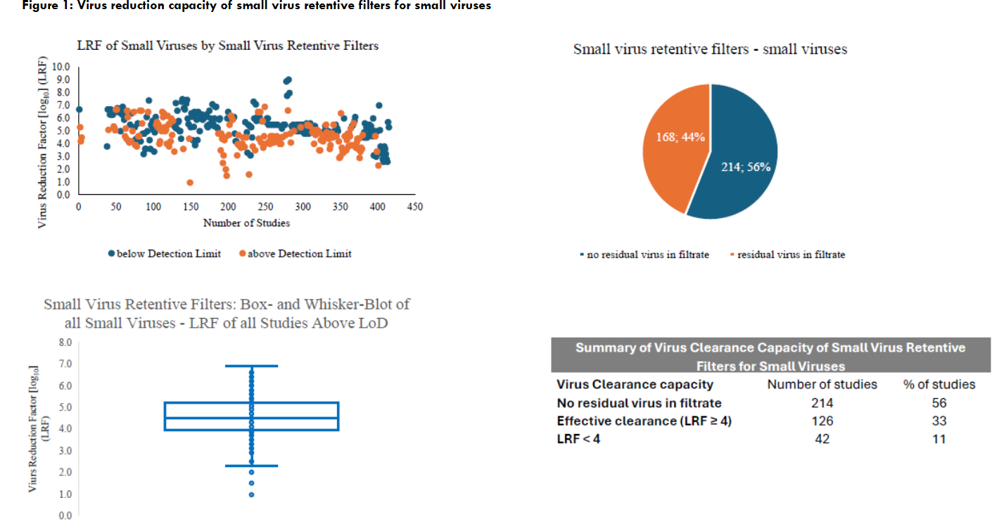
Small virus retentive filters (Planova 15N, 20N, BioEX, Viresolve NFP, Viresolve Pro, Ultipor DV20, Pegasus SV4, Virosart CPV, Virosart HC studied) are primarily applied to remove small viruses from the product intermediate; however, due to the capacity to remove small viruses, also large viruses are effectively retained. As stated in the ICH Q5A(R2) guideline, parvoviruses “may be used as single worst-case model virus for larger spherical/icosahedral viruses and enveloped viruses at validation of virus filters”.
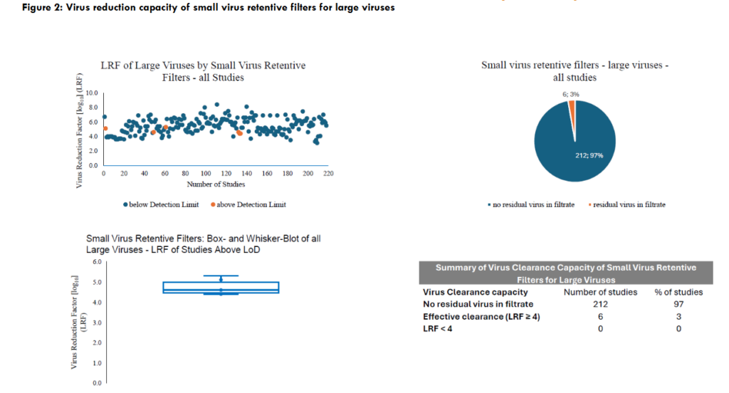
Small virus retentive filters remove small viruses effectively in the order of 90% of all studies and all large viruses, as expected, effectively as shown in the figures. The detection of large viruses in the filtrate of small virus retentive filters, despite the fact that the removal capacity was effective (LRF ≥ 4), cannot be explained; e.g. Ajayi et al. stated that no root cause for passing large viruses through small virus retentive filters could be identified. A cross-contamination of cell cultures used for quantification the virus load in the respective samples during handling the different filtrate fractions with virus or during the infectivity assays cannot be excluded. The studies showed that parvoviruses are within the group of small viruses the smallest viruses and, therefore, are a challenge for their removal even for small virus retentive filters. An assessment of the data published showed that no residual parvovirus vs. residual parvovirus in the filtrate could be detected in 47% vs 53% of all studies and for the other small viruses studied the relation was 92% vs. 8%.
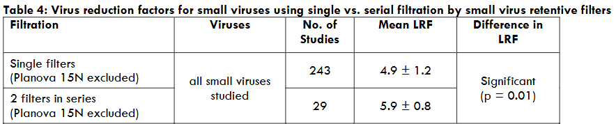
Serial filtration with 2 small virus retentive filters removed small viruses to a significant higher rate than a single filter.
3.2 LARGE VIRUS RETENTIVE FILTERS
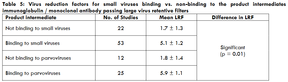
Due to the pore size of the large virus retentive filters, it is expected that small viruses are not retained to a high rate by these filters. The distribution of the virus reduction factors for small viruses is very high, a reason for this effect was assessed. As immunoglobulins and, partly, monoclonal antibodies (mAbs) bind to viruses enlarging their size considerably, this enlarging effect was assessed for small viruses and large virus retentive filters. There is no difference in the virus reduction capacity for small virus retentive filters in immunoglobulin intermediates compared to all studies covering small viruses; therefore, for small virus retentive filters enlarging the size of the virus by binding to immunoglobulins / mAbs is not relevant. However, the effect of enlarging a virus particle by the binding of antibodies is considerable when assessing large virus retentive filters. The effect of antibodies binding to the viruses studied [bovine parvovirus (BPV), parvovirus B19 (B19V), hepatitis A virus (HAV), hepatitis E virus (HEV) (stripped from the quasi-envelope), and poliovirus] versus not binding to the viruses studied [minute virus of mice (MVM), porcine parvovirus (PPV), HEV with quasi-envelope, and encephalomyocarditis virus (EMCV)] is shown in the table.
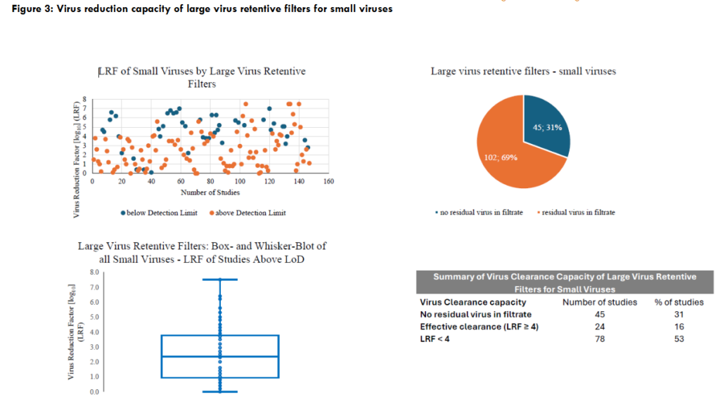
3.2 LARGE VIRUS RETENTIVE FILTERS
Due to the pore size of the large virus retentive filters, it is expected that small viruses are not retained to a high rate by these filters; Figure 3 shows the detail of study in 47% of all studies an effective virus reduction capacity for small viruses (LRF > 4) could be stated. As the distribution of the virus reduction factors for small viruses is very high, a reason for this effect was assessed. As immunoglobulins and, partly, monoclonal antibodies (mAbs) bind to viruses enlarging their size considerably, this enlarging effect was assessed for small viruses and large virus retentive filters. There is no difference in the virus reduction capacity for small virus retentive filters in immunoglobulin intermediates compared to all studies covering small viruses; therefore, for small virus retentive filters enlarging the size of the virus by binding to immunoglobulins / mAbs is not relevant. However, the effect of enlarging a virus particle by the binding of antibodies is considerable when assessing large virus retentive filters. The effect of antibodies binding to the viruses studied [bovine parvovirus (BPV), parvovirus B19 (B19V), hepatitis A virus (HAV), hepatitis E virus (HEV) (stripped from the quasi-envelope), and poliovirus] versus not binding to the viruses studied [minute virus of mice (MVM), porcine parvovirus (PPV), HEV with quasi-envelope, and encephalomyocarditis virus (EMCV)] is shown in Table 5; in addition, the binding / non-binding effect to different parvoviruses was also assessed.
All studies with residual infectivity in the filtrate of large virus retentive filters were mid-size viruses (BVDV, WNV, YFV, TBEV, HCV, SINV, ZIKV, CHIKV, MAYV, SV40, Reo3, and HBV – compare Table 2a) whereas the large viruses (HIV, MuLV, PI3, PRV, HSV, and VSV) were removed to below the limit of detection of the infectivity assay with the exception of 2 publications detecting HIV in the filtrate; the reported removal of BVDV, Reo3, SV40 and even BPV under the same conditions with no residual infectivity in the filtrate indicate a cross contamination of the cell culture with HIV during the experiment. In the second publication, in one of ten studies employing HIV this virus was detected in the filtrate.
Table 5: Virus reduction factors for small viruses binding vs. non-binding to the product intermediates
4. Discussion
In several publications data, partly not published, are compiled demonstrating for plasma-derived medicinal products as well as biotechnological products derived from cell cultures that virus retentive filtration removes viruses depending on the pore size of the respective filters highly effectively and robust. The volume per filter area, operating pressure and total protein concentration had had no significant impact on the efficacy of the virus removal capacity within the studied ranges. A multicompany collaboration with data compiled from CROs demonstrate that large viruses (MuLV, PRV, Reo3) were removed (primarily) by small virus retentive filters very effectively: No virus detected in the filtrate was reported for 97.3% of all runs (2311 runs in total), effective virus removal was reported for 99.1 % and only 0.9% of all runs resulted in a LRF < 4. Authors from CDER/FDA published data, extracted from the CMC section of IND and BLA applications, documenting that for the large virus MuLV the LRF was always above 2 and in only 1% of all studies the LRF was < 3 for small and large virus retentive filters. The removal of parvoviruses by small virus retention filters was stated to be filter type specific with one filter type (not disclosed) resulting in low LRFs. A further publication compiled studies from 10 biotechnology companies having employed small virus retentive filters of different manufacturers categorised into PES (polyethersulfone) and RC (regenerated cellulose). In all studies employing retro- and herpesviruses, no residual infectivity could be detected in the filtrate; the mid-size Reo3 was removed always effectively with only one experiment with residual infectivity in the filtrate and a LRF of 6.1. Parvoviruses were removed effectively by both, PES and RC filters (LRF of 5.9 (PES filters) vs. 5.0 (RC filters). The data analysis showed that the virus load per filter area has a considerable effect on the virus clearance capacity / virus infectivity in the filtrate for RC filters. Passage of the phage PP7 and ΦX-174 into the filtrate of small virus retentive filters (Viresolve NFP, Virosart CPV, Ultipor DV20 and Planova 20N) occurred in each filter type, particularly when overloaded with phage. The authors also reported brand-specific differences in flux decay due to phage overload and concluded that small virus retentive filters should not be viewed as absolute in their capacity to clear virus and they should not be viewed as interchangeable between brands.
The effect of overloading virus filters with viruses was studied intensively with the Planova 20N filter (regenerated cellulose) resulting in the fact that non-infectious MVM, i.e., also empty particles, can cause an overload of the filters resulting in a breakthrough of (infectious) viruses. It was concluded that a total particle number of more than approx. 12 log10/m² filter area should not be used in virus validation studies, just to achieve a very high LRF using an unrealistic high virus load. Compilation of data assessing also the so-called second generation of small virus retentive filters (Planova BioEX, Viresolve Pro) showed that these improved filters were able to remove e.g., parvoviruses to a higher degree as the so-called first generation of small virus retentive filters (Planova 15N, Planova 20N, Viresolve NFP, Virosart CPV, Ultipor DV20) and appeared to have less variability in the reported LRFs. In the meantime further small virus retentive filters were developed, e.g., Planova S20N, prepared, as Planova 20N, also from regenerated cellulose but with increased thickness of the membrane structure of the hollow fibre withstanding a higher membrane pressure and the Virosart HC, consisting of two asymmetric membranes oriented in opposite direction.
These review publications support the data accumulated from the published data reviewed here documenting a robust and effective clearance of viruses by virus retentive filters. It has to be considered, however, that certain parameters of the filtration procedure may impact the virus clearance capacity of such filters as pressure, flow decay and flow interruption, volumetric throughput of product intermediate and buffer flush. The impact of volume / filter area, protein load and operating pressure on the virus clearance capacity was negligible, documented for plasma-derived medicinal products. Flow decay due to blocking filters (e.g., fouling) should be considered a relevant parameter as under such conditions the virus clearance capacity may be reduced; therefore, based on virus clearance studies, a minimum flow rate (LMH – litre per m² and hour) for production conditions should be defined. A flow decay to zero LMH may occur when switching from product feed stream to buffer flush; this flow interruption is known to impact considerably the overall virus clearance capacity assessing the pooled filtrate of the product intermediate and the buffer flush to recover as much product as possible. This flow-interruption associated virus breakthrough is because viruses may migrate into deeper membrane layers, partly based on the membrane specific pore interconnectivity.
The ICH Q5A(R2) guideline defines potential critical parameters in virus filtration as volumetric throughput of product intermediate as well as of buffer flush and pressure including pressure/flow interruption due to prior knowledge / in-house experience. As summarised here, these parameters are important for distinct filter types and have to be controlled. Therefore, a change of filter brands, especially in a post-approval change has to be carefully assessed also regarding these parameters. The size-exclusion mechanism of (small) virus retentive filters is also able to remove prions, the causative agent of TSEs (transmissible spongiform encephalopathies) as (variant) Creutzfeldt-Jakob disease ((v)CJD) to a high degree. In the extremely unlikely situation that prions would be present in the product intermediate of plasma-derived medicinal products, the infectious prion material is a multimeric protein aggregate – not the monomeric protein – and this material can be removed. The challenge of prion removal data are the physicochemical properties of the prion spike material as the nature of the infectious agent in blood, if present, is currently not known. Different spike preparations were used in prion evaluation studies with a considerable removal capacity.
Continuous manufacturing is increasingly applied in the manufacturing process of biologicals in order to reduce costs and the footprint of the equipment used throughout the production facility. Challenges for virus filtration studies are discussed in general in the ICH guidelines Q5A(R2) and Q13. Virus filtration under constant flow, commonly at low pressure, extended volumetric throughputs and processing times, and, especially, product feed for the virus retention filter with fluctuations in protein and buffer concentrations have to be addressed properly. Furthermore, inline virus spiking has to be performed. In order to avoid significant variations in the feed stream, especially post chromatography steps, surge tanks for temporarily hold of the continuous stream can be considered; this approach will then mimic a classical batch process for virus filtration. Under these conditions, replacing filters during the process would be simpler; such filter replacement may be required during the long duration of the continuous process to avoid blocking of the filter and other, including unexpected, process events.
5. Conclusion
Published data on virus clearance by virus retentive filters demonstrate that virus removal is based on size exclusion, i.e., large viruses are very effectively removed by small virus retentive filters. Therefore, the LRF demonstrated for small viruses as parvoviruses can be applied to large viruses as retroviruses. Parameters considered potentially critical in virus filtration according to the ICH Q5A(R2) guideline are volumetric throughput of product intermediate and buffer flush as well as pressure including pressure/flow interruption. The presented data confirm, depending on the filter brand, this assessment which should include also flow decay. These brand specific differences show that brands cannot be changed (post-approval change) without appropriate validation. Each filter brand effectively removes viruses based on the pore size of the filter, i.e., large virus retentive filters remove large viruses and small virus retentive filters remove small and large viruses effectively having implemented the above-mentioned parameters for high virus removal capacity of the respective virus retentive filters, assessed in virus validation studies. The assessment of these parameters, preferably in a Design of Experiment approach covering virus clearance, are one of the bases of the specification of these critical process parameters (“Established Conditions”) resulting in a platform validation approach when the process step is predictable and robust in virus removal capacity based also on prior knowledge.
6 Conflict of Interest
No conflict of interest to be disclosed
7 Funding Statement
No funding to be reported
References
- CPMP/BWP/268/95. Note for guidance on virus validation studies: The design, contribution and interpretation of studies validating the inactivation and removal of viruses. 14 February 1996. Accessed December 2, 2024. Link
- ICH Harmonised Guideline. Viral Safety Evaluation of Biotechnology Products Derived from Cell Lines of Human or Animal Origin Q5A(R2), adopted on 1 November 2023. Accessed December 2, 2024. Link
- EMA/CHMP/BWP/706271/2010. Guideline on plasma-derived medicinal products. 21 July 2011. Accessed December 2, 2024. Link
- Johnson SA, Chen S, Bolton G, et al. Virus filtration: a review of current and future practices in bioprocessing. Biotechnol Bioeng. 2022;119(3):743-761. doi: 10.1002/bit.28017
- Ajayi OO, Johnson SA, Faison T, et al. An updated analysis of viral clearance unit operations for biotechnology manufacturing. Curr Res. Biotechnol. 2022;4:190-202. doi.org/10.1016/j.crbiot.2022.03.002
- Roth NJ, Dichtelmüller HO, Fabbrizzi F, et al. Nanofiltration as a robust method contributing to viral safety of plasma-derived therapeutics: 20 years’ experience of the plasma protein manufacturers. Transfusion. 2020;60(11):2661-2674. doi: 10.1111/trf.16022
- Virus Retentive Filtration. Technical report No. 41 (Revised 2022). Parenteral Drug Association. 2022.
- Wieser A, Modrof J, Kreil TR. Protection of biomanufacturing processes from virus contamination through upstream virus filtration of cell culture media. Biotechnol Bioeng 2023;120(10):2917-2924. doi: 10.1002/bit.28473
- Chen D, Bolton G. Proceedings of the 2017 Viral Clearance Symposium, Session 1.2: Upstream Mitigation, Part 2 – Virus Barrier Filter and HTST. PDA J Pharm Sci Technol 2018;72(5):462-469. doi:10.5731/pdajpst.2018.009092
- Burnouf-Radosevich M, Appourchaux P, Huart JJ, Burnoug T. Nanofiltration, a new specific virus elimination method applied to high-purity Factor IX and Factor XI concentrates. Vox Sang 1994;67(2):132-138. doi: 10.1111/j.1423-0410
- Römisch J, List W, Bernhardt D, et al. Nanofiltration bei der Herstellung des PPSB-Konzentrates Beriplex® P/N. Hämostaseologie 1995;15(3):171-178. DOI: 10.1055/s-0038-1655307
- Lute S, Riordan W, Pease LF 3rd, et al. A consensus rating method for small virus-retentive filters. I. Method development. PDA J Pharm Sci Technol 2008;62(5):318-333. PMID: 19055228.
- Brorson K, Lute S, Haque M, et al. A consensus rating method for small virus-retentive filters. II. Method evaluation. PDA J Pharm Sci Technol 2008;62(5):334-343. PMID: 19055229
- Brorson K, Sofer G, Aranha H. Nomenclature standardization for ‘large pore size’ virus-retentive filters. PDA J Pharm Sci Technol 2005;59(6):341-345. PMID: 16471421
- Bao R-M, Shibuya A, Uehira T, et al. Successful removal of porcine circovirus-1 from immunoglobulin G formulated in glycine solution using nanofiltration. Biologicals 2018;51:32-36. doi:10.1016/j.biologicals.2017.10.006
- Hilfenhaus J, Gröner A, Nowak T, Weimer T. Analysis of human plasma products: polymerase chain reaction does not discriminate between live and inactivated viruses. Transfusion 1997;37(9):935-940. doi: 10.1046/j.1537-2995.1997.37997454021.x.
- Brorson K, Krejci S, Lee K, Hamilton E, Steil K, Xu Y. Bracketed generic inactivation of rodent retroviruses by low pH treatment for monoclonal antibodies and recombinant proteins. Biotechnol Bioeng 2003;82(3):321-329. doi:10.1002/bit.10574
- Anwaruzzaman M, Wang W, Wang E, Erfe L, Lee J, Lui S. Evaluation of infectivity and reverse transcriptase real-time polymerase chain reaction assays for detection of xenotropic murine leukemia virus used in virus clearance validation. Biologicals 2015;43(4):256-265. doi:10.1016/j.biologicals.2015.04.001
- Tsujikawa M, Ohkubo Y, Masuda M, et al. Caution in evaluation of removal of virus by filtration: Misinterpretation due to detection of viral genome fragments by PCR. J Virol Methods 2011;178(1-2):39-43. doi:10.1016/j.jviromet.2011.08.009
- Zhao X, Bailey MR, Emery WR, Lambooy PK, Chen D. Evaluation of viral removal by nanofiltration using real-time quantitative polymerase chain reaction. Biotechnol Appl Biochem 2007;47(Pt 2):97-104. doi:10.1042/BA20060195
- Blümel J, Musso D, Teitz S, et al. Inactivation and removal of Zika virus during manufacturing of plasma-derived medicinal products. Transfusion 2017;57(3pt2):790-796. doi:10.1111/trf.13873
- Yue C, Teitz S, Miyabashi T, et al. Inactivation and removal of Chikungunya virus and Mayaro virus from plasma-derived medicinal products. Viruses. 2019;11(3):234. doi:10.3390/v11030234
- Preparation of virus spike used for virus clearance studies. Technical Report No. 47. Parenteral Drug Association. 2010
- Slocum A, Burnham M, Genest P, Venkiteshwaran A, Chen D, Hughes J. Impact of virus preparation quality on parvovirus filter performance. Biotechnol Bioeng. 2013;110(1):229-239. doi:10.1002/bit.
- Roush DJ, Myrold A, Burnham MS, And JV, Hughes JV. Limits in virus filtration capacity? Impact of virus quality and spike level on virus removal with xenotropic murine leukemia virus. Biotechnol Prog. 2015;31(1):135-144. doi:10.1002/btpr.2020
- Hongo-Hirasaki T, Yamaguchi K, Yanagida K, Hayashida H, Ide S. Effects of varying virus-spike conditions on a virus-removal filter Planova 20N in a virus validation study of antibody solutions. Biotechnol Prog. 2011;27(1):162-169. doi:10.1002/btpr.533
- Kozaili J, Shah A, Robbins D, et al. Serial filtration: a case study evaluating the product-dependent impact of control strategies on process efficiency. Biotechnol J. 2023;18(9):e2200599. doi:10.1002/biot.202200599
- Omar A, Kempf C. Removal of neutralized model parvoviruses and enteroviruses in human IgG solutions by nanofiltration. Transfusion. 2002;42(8):1005-1010. doi:10.1046/j.1537-2995.2002.00145.x
- Kreil TR, Wieser A, Berting A, et al. Removal of small nonenveloped viruses by antibody-enhanced nanofiltration during the manufacturing of plasma derivatives. Transfusion. 2006;46(7):1143-1151. doi:10.1111/j.1537-2995.2006.00864.x
- Kapsch A-M, Farcet MR, Wieser A, et al. Antibody-enhanced hepatitis E virus nanofiltration during the manufacture of human immunoglobulin. Transfusion. 2020;60(11):2500-2507. doi:10.1111/trf.16014
- Burnouf-Radosevich M, Appourchaux P, Huart JJ, Burnouf T. Nanofiltration, a new specific virus elimination method applied to high-purity factor IX and factor XI concentrates. Vox Sang. 1994;67(2):132-138. doi:10.1111/j.1423-0410.1994.tb01647.x
- Eibl J, Barrett N, Hämmerle T, Dorner F. Nanofiltration of immunoglobulin with 35-nm filters fails to remove substantial amounts of HCV. Biologicals. 1996;24(3):285-287. doi:10.1006/biol.1996.0036
- Stanley B, Holmes V, Manzari R, et al. Twenty plus years of data demonstrating virus filtration as an effective and robust step for large virus removal. PDA J Pharm Sci Technol. 2022;76(1):1-8. doi:10.5731/pdajpst.2020.012591
- Miesegaes G, Lute S, Brorson K. Analysis of virus clearance unit operations for monoclonal antibodies. Biotechnol Bioeng. 2010;106(2):238-246. doi:10.1002/bit.22662
- Mattila J, Clark M, Liu S, et al. Erratum for John Mattila, Mike Clark, Shengjiang Liu, et al.: “Retrospective Evaluation of Low-pH Viral Inactivation and Viral Filtration Data from a Multiple Company Collaboration”. PDA J Pharm Sci Technol. 2018;72(4):451. doi:10.5731/pdajpst.2017.007963
- Lute S, Bailey M, Combs J, Sukumar M, Brorson K. Phage passage after extended processing in small-virus-retentive filters. Biotechnol Appl Biochem. 2007;47(3):141-151. doi:10.1042/BA20060254
- Kayukawa T, Yanagibashi A, Hongo-Hirasaki T, Yanagida K. Particle-based analysis elucidates the real retention capacities of virus filters and enables optimal virus clearance study design with evaluation systems of diverse virological characteristics. Biotechnol Prog. 2022;38(2):e3237. doi:10.1002/btpr.3237
- Barnard JG, Kahn D, Cetlin D, Randolph TW, Carpenter JF. Investigations into the fouling mechanism of parvovirus filters during filtration of freeze-thawed mAb drug substance solutions. J Pharm Sci. 2014;103(3):890-899. doi:10.1002/jps.23881
- Isu S, Qian X, Zydney AL, Wickramasinghe SR. Process- and product-related foulants in virus filtration. Bioengineering (Basel). 2022;9(4):155. doi:10.3390/bioengineering9040155
- Bolton G, Cabatingan M, Rubino M, Lute S, Brorson K, Bailey M. Normal-flow virus filtration: detection and assessment of the endpoint in bio-processing. Biotechnol Appl Biochem. 2005;42(Pt 2):133-142. doi:10.1042/BA20050056
- Wieser A, Berting A, Medek C, Poelsler G, Kreil TR. Virus filtration and flow variation: an approach to evaluate any potential impact on virus retention. PDA J Pharm Sci Technol. 2016;70(4):325-331. doi:10.5731/pdajpst.2015.006346
- Nazem-Bokaee H, Chen D, O’Donnell SM, Zydney AL. New insights into the performance characteristics of the Planova-series hollow-fiber parvovirus filters using confocal and electron microscopy. Biotechnol Bioeng. 2019;116(8):2010-2017. doi:10.1002/bit.26991
- Leisi R, Widmer E, Gooch B, Roth NJ, Ros C. Mechanistic insight into flow-dependent virus retention in different nanofilter membranes. J Membr Sci 2021;636:119548. doi.org/10.1016/j.memsci.2021.119548
- Leisi R, Rostami I, Laughhunn A, et al. Visualizing protein fouling and its impact on parvovirus retention within distinct filter membrane morphologies. J Membr Sci 2022;659:120791. doi.org/10.1016/j.memsci.2022.120791
- LaCasse D, Genest P, Pizzelli K, Greenhalgh P, Mullin L, Slocum A. Impact of process interruption on virus retention of small-virus filters. BioProcess Int 2013;11(10):34-44
- Dishari SK, Venkiteshwaran A, Zydney AL. Probing effects of pressure release on virus capture during virus filtration using confocal microscopy. Biotechnol Bioeng. 2015;112(10):2115-2122. doi:10.1002/bit.25614
- Yamamoto A, Hongo-Hirasaki T, Uchi Y, Hayashida H, Nagoya F. Effect of hydrodynamic forces on virus removal capability of Planova filters. AIChE J 2014;60:2286-97. doi.org/10.1002/aic.14392
- Gustafsson O, Gustafsson S, Manukyan L, Mihranyan A. Significance of Brownian motion for nanoparticle and virus capture in nanocellulose-based filter paper. Membranes (Basel). 2018;8(4):90. doi:10.3390/membranes8040090
- Fallahianbijan F, Giglia S, Carbrello C, Zydney AL. Quantitative analysis of internal flow distribution and pore interconnectivity within asymmetric virus filtration membranes. J Membr Sci 2020;595:117578.
- Silveira JR, Raymond GJ, Hughson AG, et al. The most infectious prion protein particles. Nature. 2005;437(7056):257-261. doi:10.1038/nature03989
- CPMP/BWP/CPMP/5136/03. Guideline on the investigation of manufacturing processes for plasma-derived medicinal products with regard to vCJD risk. 21.October 2004. Accessed December 2, 2024. Link
- Truchot L, Arnaud T, Bloy C, Perret-Liaudet A. CJD PrPsc removal by nanofiltration process: Application to a therapeutic immunoglobulin solution (Lymphoglobuline®). Biologicals. 2006;34(3):227-231. doi:10.1016/j.biologicals.2005.11.007
- Cardone F, Simoneau S, Arzel A, et al. Comparison of nanofiltration efficacy in reducing infectivity of centrifuged versus ultracentrifuged 263K scrapie-infected brain homogenates in “spiked” albumin solutions. Transfusion. 2012;52(5):953-962. doi:10.1111/j.1537-2995.2011.03425.x
- Yunoki M, Tanaka H, Urayama T, et al. Prion removal by nanofiltration under different experimental conditions. Biologicals. 2008;36(1):27-36. doi:10.1016/j.biologicals.2007.04.005
- Stucki M, Boschetti N, Schäfer W, et al. Investigations of prion and virus safety of a new liquid IVIG product. Biologicals. 2008;36(4):239-247. doi:10.1016/j.biologicals.2008.01.004
- Yunoki M, Tanaka H, Urayama T, et al. Infectious prion protein in the filtrate even after 15 nm filtration. Biologicals. 2010;38(2):311-313. doi:10.1016/j.biologicals.2009.10.018
- Diez JM, Caballero S, Belda F, Otegui M, Gajardo R, Jorquera JI. Capacity of the manufacturing process of Flebogamma(®) DIF, a new human high purity intravenous immunoglobulin, to remove a TSE model agent. Biologicals. 2010;38(6):670-674. doi:10.1016/j.biologicals.2010.08.003
- Goussen C, Simoneau S, Bérend S, et al. Biological safety of a highly purified 10% liquid intravenous immunoglobulin preparation from human plasma. BioDrugs. 2017;31(3):251-261. doi:10.1007/s40259-017-0222-9
- Roberts PL, Dalton J, Evans D, et al. Removal of TSE agent from plasma products manufactured in the United Kingdom. Vox Sang. 2013;104(4):299-308. doi:10.1111/vox.12004
- Winge S, Yderland L, Kannicht C, et al. Development, upscaling and validation of the purification process for human-cl rhFVIII (Nuwiq®), a new generation recombinant factor VIII produced in a human cell-line. Protein Expr Purif. 2015;115:165-175. doi:10.1016/j.pep.2015.08.023
- Garger S, Severs J, Regan L, et al. BAY 81-8973, a full-length recombinant factor VIII: manufacturing processes and products characteristics. Haemophilia. 2017;23(2):e67-e78. doi:10.1111/hae.13148
- Schulz PM, Gehringer W, Nöhring S, et al. Biochemical characterization, stability, and pathogen safety of a new fibrinogen concentrate (fibryga®). Biologicals. 2018;52:72-77. doi:10.1016/j.biologicals.2017.12.003
- ICH Harmonised Guideline. Continuous manufacturing of drug substances and drug products Q13, Adopted on 16 November 2022. Accessed December 2, 2024. Link
- Lute S, Kozaili J, Johnson S, Kobayashi K, Strauss D. Development of small-scale models to understand the impact of continuous downstream bioprocessing of integrated virus filtration. Biotechnol Prog. 2020;36(3):e2962. doi:10.1002/btpr.2962
- Kozaili J, Rayfield W, Gospodarek A, Brower M, Strauss D. Adapting virus filtration to continuous processing: Effects of product and process variability on filtration performance. Biotechnol Prog. 2024;40(2):e3407. doi:10.1002/btpr.3407
- Hwang M, Wang J, Jung SY. Understanding the residence time distribution in a transient inline spiking system: Modeling, experiments, and simulations. Membranes (Basel). 2023;13(4):375. doi:10.3390/membranes13040375
- Kerr A, Nims R. Adventitious viruses detected in biopharmaceutical bulk harvest samples over a 10 year period. PDA J Pharm Sci Technol. 2010;64(5):481-485.
- Barone PW, Wiebe ME, Leung JC, et al. Viral contamination in biologic manufacture and implications for emerging therapies. Nat Biotechnol. 2020;38(5):563-572. doi:10.1038/s41587-020-0507-2
- Schleh M, Romanowski P, Bhebe P, et al. Susceptibility of mouse minute virus to inactivation by heat in two cell culture media types. Biotechnol Prog. 2009;25(3):854-860. doi:10.1002/btpr.181
- Cao X, Stimpfl G, Wen Z-Q, Frank G, Hunter G. Identification and root cause analysis of cell culture media precipitates in the viral deactivation treatment with high-temperature/short-time method. PDA J Pharm Sci Technol. 2013;67(1):63-73. doi:10.5731/pdajpst.2013.00894
- Yen S, Sokolenko S, Manocha B, Blondeel EJM, Aucoin MG. Treating cell culture media with UV irradiation against adventitious agents: Minimal impact on CHO performance. Biotechnol Prog. 2014;30(5):1190-1195. doi:10.1002/btpr.1942
- Gauvin G, Nims R. Gamma-irradiation of serum for the inactivation of adventitious contaminants. Biotechnol Prog. 2014;30(5):1190-1195. doi:10.1002/btpr.1942
- Mann K, Royce J, Carbrello C, et al. Protection of bioreactor culture from virus contamination by use of a virus barrier filter. BMC Proceedings 2015;9(Suppl 9):P22. doi:10.1186/1753-6561-9-S9-P22