

Thyroid Ultrasound Findings in Saudi University Students
Thyroid Ultrasonography Screening Findings: A Comparison between Male and Female University Students
Mahmoud S. Babiker (PhD) ¹, Awatif M. Omer (PhD) ¹, Meaad Elbashir (PhD) ², Bushra Hussain Sharahili (B.Sc.) ¹, Amel F.H Alzain (PhD) ¹, Fathelrehman A. Alamin (MD) ¹, and Mohamed Abdalla Eltahir (PhD) ³
Abstract
Objective: to compare the results of US thyroid screening findings between the male and female cohort of asymptomatic Saudi Arabian university students.
Methods: One hundred and fifty six university students were enrolled in a quantitative descriptive study from November 2024 to January 2025. They were selected through a simple sampling. An Esaote MyLab40 US machine with a 7 MHz probe was used. The thyroid was bilaterally evaluated with the US for length, echotexture, and abnormalities. The Independent T-test, Pearson’s correlations, cross-tabulation, and descriptive statistics were applied for the data analysis.
Results: the mean age of the participants was 21.3±1.3 years, and the mean BMI was 23.7 ±6.3. The right thyroid lobe’s mean length and anteroposterior (AP) diameter were 3.8 ± 0.48 cm and 1.2 ± 0.26 cm, respectively. The left lobe mean length and AP diameter were 3.5 ±0.49 cm and 1.1± 0.30 cm, respectively. 84.6% of the participants had normal findings; the positive findings were 15.4 % (n = 24): cystic nodules found in 10.3%, predominantly solid, thyroiditis, and dilated cervical lymph nodes were noted in a similar percentage (1.3%). The positive findings were more frequent in the female students (87.5% (out of 24)). The statistical analysis indicated a significant association between the US findings and the BMI p =0.04. The mean length of the Rt. and Lt. male thyroid lobes was greater than the females, t = 5.204 and 5.354, respectively, and p < 0.05.
Conclusion: The study concluded that the thyroid US incidental findings were significant, more frequent in females, and associated significantly with the BMI. The male’s mean thyroid length was greater than that of the females.
Keywords: Ultrasound, Thyroid Screening, Thyroid, Body Mass Index, Echotexture.
Introduction
Sonography is an important diagnostic tool for thyroid echotexture screening programs in asymptomatic individuals. Some authors indicated that ultrasound (US) is the modality of choice for thyroid evaluation. Based on neck palpation as a clinical test, (US) can help describe the nature of a palpable lesion or any changes in thyroid texture. Fine needle aspiration cytology (FNA) is useful in confirming the diagnosis when the US findings indicate a suspicious like hypoechogenicity, irregular margins, and microcalcifications.
Gnarini VL et al. suggested that incidental thyroid disorders were detected in 50.3% of the asymptomatic participants. A study of a male cohort of asymptomatic Saudi Arabian university students concluded that the thyroid US finding is very limited.
The published literature indicated that the average regular thyroid length is 4 to 4.8 cm in sagittal, 1 to 1.8 cm in transverse, and 0.8 to 1.6 cm in anteroposterior dimensions.
The description of thyroid nodules was performed according to the 2021 Korean Society of Thyroid Radiology consensus statement and recommendations for managing thyroid nodules. A nodule is solid when it has no obvious cystic component, predominantly solid when its cystic portion is ≤ 50%, predominantly when its cystic portion is > 50%, and cystic when it has no obvious solid component. According to published literature, the diagnostic US features of thyroiditis include hypo echogenicity, heterogeneity, and hypervascularity of the intrathyroidal gland.
The current study aimed to compare the thyroid US screening findings of the male and female cohorts of asymptomatic Saudi Arabian university students.
Methodology
A quantitative descriptive prospective study conducted at a Saudi Arabian university. The participants included a cohort of colleges of applied medical and nursery science students. The inclusion criteria included asymptomatic participants (male and female students), and a simple sampling was used to select the participants. The exclusion criteria included students unwilling to participate in the study.
All participants were subjected to thyroid US screening by an expert sonologist with more than ten years of sonographic scanning experience. The US images were obtained with the participant lying on an examination table supine with his/her neck elevated and tilted to the opposite side of the targeted one. If needed, both right (Rt.) and left (Lt.) thyroid lobes were scanned using grayscale and color Doppler. Thyroid size measurements and echotexture checks were performed. The length and anteroposterior size measurements were obtained for each thyroid lobe. The participants with positive findings through this study were directed to receive additional testing in the local governmental hospital.
A structured data sheet was used to record the study data and variables such as age, weight, height, thyroid length, and echo texture. The participant’s BMI was calculated using the formula (BMI = weight (kg)/height, m2)
Statistical Analysis
The data was analyzed using industry standard tools, Microsoft Excel (2021) and the Statistical Package for the Social Sciences (SPSS) program Version 26 (IBM, Armonk, New York, USA). Categorical and continuous variables were presented as percentages, frequencies, and descriptive statistics. The chi squared test, and Pearson’s correlations were used to explore the relationship between BMI and the length of the Rt. and Lt. thyroid lobes. A t-test was used to compare the male and female findings. The pvalue was set for significance at < .05.
Results
Table 1 summarizes the descriptive statistics of the study, which showed that the mean age of the participants was 21.3 ±1.3 years, and the mean BMI was 23.7 ±6.3. The right thyroid lobe’s mean length and anteroposterior (AP) diameter were 3.8 ± 0.48 cm and 1.2 ± 0.26 cm, respectively. The left lobe mean length and AP diameter were 3.5 ±0.49 cm and 1.1± 0.30 cm, respectively.
| Age | Rt. Lobe length/cm | Rt. lobe AP/cm | Lt. lobe length/cm | Lt. lobe AP/cm | Body max index | |
|---|---|---|---|---|---|---|
| Mean | 21.3269 | 3.7680 | 1.2475 | 3.5445 | 1.1187 | 23.7414 |
| Std. Deviation | 1.31082 | .48098 | .26058 | .48598 | .30229 | 6.37189 |
| Minimum | 18.00 | 2.47 | .20 | 2.20 | .30 | 14.69 |
| Maximum | 26.00 | 4.70 | 1.93 | 4.70 | 2.10 | 50.17 |
Table 2 demonstrates the US findings; the unremarkable diagnosis was 84.6% (out of 156), and the positive findings were 15.4 % (n = 24) as follows: cystic nodules found in 10.3%, predominantly solid, thyroiditis, and dilated cervical lymph nodes were noted in similar percentages (1.3%). The positive findings were more frequent in the female students (87.5% (out of 24)). The statistical analysis indicated a significant association between the US findings and the BMI p =0.04.
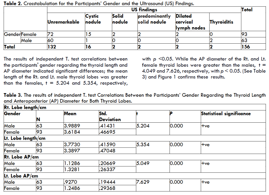
The results of independent T. test correlations between the participants’ gender regarding the thyroid length and AP diameter indicated significant differences; the mean length of the Rt. and Lt. male thyroid lobes was greater than the females, t = 5.204 and 5.354, respectively, with p <0.05. While the AP diameter of the Rt. and Lt. female thyroid lobes were greater than the males, t = 4.049 and 7.626, respectively, with p < 0.05.
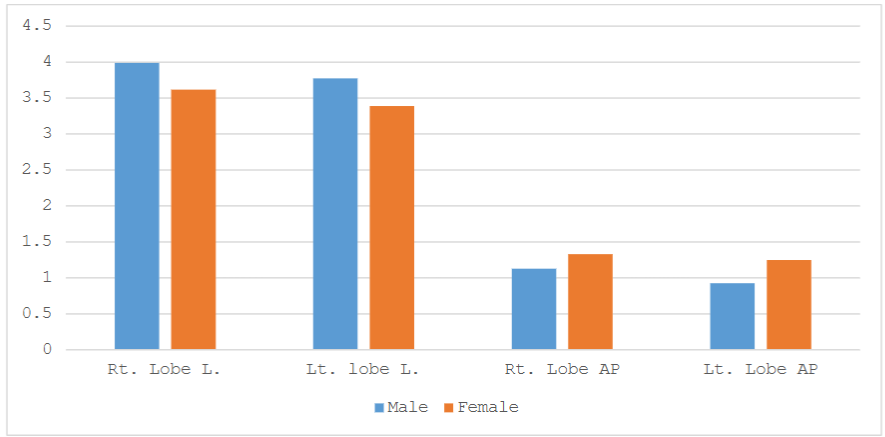
Table 4 summarizes the Pearson correlations between the right and left thyroid lobe lengths and the BMI, which showed no significant statistical association (Pearson coefficient = 0.155 and 0.133, respectively, p = .053 and .098, respectively).
Table 4: The Statistical Correlations Between the Thyroid Length and the Body Mass Index (BMI), for the 156 Participants.
| Rt. Lobe length/cm | Body max index | Lt. lobe length/cm | Body max index | |
|---|---|---|---|---|
| Pearson Correlation | 1 | .155 | 1 | .133 |
| Sig. (2-tailed) | .053 | .098 |
Table 5 shows the Pearson correlations between the US findings and the BMI. Statistical analysis revealed a significant statistical association between the US findings and the BMI (Pearson Coefficient = 0.163, p = .053 and .041).
| US Findings | Body max index | ||
|---|---|---|---|
| US Findings | Pearson Correlation | 1 | -.163 |
| Sig. (2-tailed) | .041 |
Discussion
Both Rt. and Lt. thyroid texture, length, and anteroposterior (AP) diameter measurements were obtained in the current study. The results revealed that the mean age of the participants was 21.3±1.3 years, and the mean BMI was 23.7 ±6.3. The right thyroid lobe’s mean length and anteroposterior (AP) diameter were 3.8 ± 0.48 cm and 1.2 ± 0.26 cm, respectively. The left lobe mean length and AP diameter were 3.5 ±0.49 cm and 1.1± 0.30 cm, respectively.
Regarding the US findings, the current study indicates a high percentage of unremarkable US findings. These results align with Saeed Bafaraj et al., which suggested that 75.3% of the participants had no findings through a thyroid US screening study among university students. In contrast, Gnarini, V. L. et al. reported that incidental thyroid disorders were detected in 50.3% of the asymptomatic participants.
Although, some institutions recommend against screening for thyroid cancer in asymptomatic adults the current study results indicated the usefulness of this screening, positive US findings were noted in 15.4 % (out of 156) they had thyroid lesions as follows; 83.3% (out of 24) were thyroid nodules, and they were more prevalent in females. Relatively, this percentage is low in comparison to other previous studies; the results of Kim, S. J. et al. in female thyroid screening indicated that 42% of the participants had thyroid abnormalities. Senashova O. et al. have reported that sonography can detect thyroid nodules in 19%-70% of the population, even in asymptomatic subjects, depending on their age and co-morbidities’ prevalence. They also showed that these nodules were more likely found in females, which is consistent with the current findings.
The current study showed that only 0.6% of the participants were diagnosed with thyroiditis. These results were considered relatively low prevalence in comparison to other authors’ findings. Mm, M., et al. reported that 30.1% of the participants had thyroiditis, indicating a higher prevalence.
All participants with positive findings directed to proceed additional investigation for confirmation and suitable treatments. Usually, some nodules should be evaluated by fine needle aspiration cytology (FNA). Strongly this suggestion supported by Ji Yang Kim et al., who proposed that US findings with suspicion should be considered indicators for FNA, regardless of their size. In contrast Remonti LR et al. have another opinion, they suggested that US criteria alone do not provide reliable information for a selection of nodules that should have an FNA performed. Bin Saeedan M et al. stated that the US is the modality of choice for thyroid evaluation. In the same context Lee MK et al. have reported that the US is essential in directing the most appropriate monitoring strategy for patients with nodular thyroid disease.
The study showed that the male mean thyroid length was larger than that of the females. These results agree with Basnet P et al. and Alsaqer, F. A. et al., who reported that the thyroid gland volume was found to be larger in males than in females. Despite the statistical correlations of the current results, the female mean AP diameter was slightly larger in females than in males.
Regarding the correlations between the thyroid lengths and the BMI, the current results revealed no significant statistical associations between the thyroid lengths and the BMI. In contrast, Alsaqer, F. A. et al. suggested that the estimated thyroid volume has negative significant statistical associations between the participants’ thyroid volume and BMI.
The current results indicated significant statistical correlations between the thyroid US findings and BMI. These findings are strongly consistent with Xu, W. et al. results, which concluded that the presence of thyroid nodules was positively associated with weight, height, and BMI.
Limitation:
A small sample size of the study should be considered a limitation.
Conclusion
The study concluded that the thyroid US screening revealed incidental findings in around 15% of the participants, and they were more frequent in the females. Moreover, these findings had a significant association with the participants’ BMI. The males’ thyroid mean length was greater than that of the females. The study’s outcome included that all positive cases were directed to have additional investigations in governmental hospitals to proceed with a final diagnosis and suitable treatment.
Further future studies were recommended, and more extensive and diverse sample sizes should be included through a random selection. The benefit of the association between incidentally discovered thyroid findings and BMI serves as a valuable avenue for understanding potential correlations between anthropometric measures and thyroid abnormalities. This association offers insights into whether body weight or height variations might predispose individuals to an increased likelihood of thyroid irregularities.
References
- Lee MK, Na DG, Joo L, et al. Standardized Imaging and Reporting for Thyroid Ultrasound: Korean Society of Thyroid Radiology Consensus Statement and Recommendation. Korean Journal of Radiology. 2023; 24 (1):22. doi:10.3348/kjr.2022.0894
- Durante C, Grani G, Lamartina L, Filetti S, Mandel SJ, Cooper DS. The diagnosis and management of thyroid nodules. JAMA. 2018; 319 (9):914. doi:10.1001/jama.2018.0898
- Gnarini VL, Brigante G, Della Valle E, et al. Very high prevalence of ultrasound thyroid scan abnormalities in healthy volunteers in Modena, Italy. PubMed. Published online October 1, 2013. doi:10.3275/8931
- Babiker MS, Alamin FA, Almteri M, Alradaddi A, Alharbi W. Thyroid ultrasonography screening of a cohort of male university students. Journal of Diagnostic Medical Sonography. Published online November 16, 2024. doi:10.1177/87564793241293444
- Can AS, Rehman A: Goiter. St. Petersburg, FL: StatPearls; 2020. Accessed October 23, 2024. https://www.ncbi.nlm.nih.gov/books/NBK562161/.
- Ha EJ, Chung SR, Na DG, et al. 2021 Korean Thyroid Imaging Reporting and Data System and Imaging-Based Management of Thyroid Nodules: Korean Society of Thyroid Radiology Consensus Statement and Recommendations. Korean Journal of Radiology. 2021; 22(12):2094. doi:10.3348/kjr.2021.0713
- opoulos N, Goulis DG, Chrisogonidis I, Giannoula E, Iakovou I. Ultrasound characteristics of Hashimoto’s thyroiditis in the subclinical stages of the disease. WFUMB Ultrasound Open. 2023; 1(2):100022. doi:10.1016/j.wfumbo.2023.100022
- Bafaraj S, Awad I, Jastaniah S, Abbas H, Musa A. Screening for thyroid diseases among students of applied medical sciences at King Abdulaziz University, Saudi Arabia. Saudi Medical Journal. 2018; 39(3):311-314. doi:10.15537/smj.2018.3.22137
- Bibbins-Domingo K, Grossman DC, Curry SJ, et al. Screening for thyroid cancer. JAMA. 2017; 317(18):1882. doi:10.1001/jama.2017.4011
- Kim SJ, Moon WK, Cho N. Sonographic criteria for fine-needle aspiration cytology in a korean female population undergoing thyroid ultrasound screening. Acta Radiologica. 2010; 51(5):475-481. doi:10.3109/02841851003641834
- Senashova O, Samuels M. Diagnosis and management of nodular thyroid disease. Techniques in Vascular and Interventional Radiology. 2022; 25(2):100816. doi:10.1016/j.tvir.2022.100816
- Mm M, Ma EA, Ta S. Thyroid ultrasound findings and its associated parameters in Saudi people. Annals of Thyroid Research. 2021; 7(2). doi:10.26420/annalsthyroidres.2021.1078
- Ji Yang Kim, Chang Hyun Lee, Soo Young Kim, et al. Radiologic and Pathologic Findings of Nonpalpable Thyroid Carcinomas Detected by Ultrasonography in a Medical Screening Center. Journal of ultrasound in medicine. 2008; 27(2):215-223. doi:https://doi.org/10.7863/jum.2008.27.2.215
- Remonti LR, Kramer CK, Leitao CB, Pinto LCF, Gross JL. Thyroid Ultrasound Features and Risk of Carcinoma: A Systematic Review and Meta-Analysis of Observational Studies. Thyroid. 2015; 25(5):538-550. doi:https://doi.org/10.1089/thy.2014.0353
- Bin Saeedan M, Aljohani IM, Khushaim AO, Bukhari SQ, Elnaas ST. Thyroid computed tomography imaging: pictorial review of variable pathologies. Insights into Imaging. 2016;7(4):601-617. doi:https://doi.org/10.1007/s13244-016-0506-5
- Lee MK, Na DG, Joo L, et al. Standardized Imaging and Reporting for Thyroid Ultrasound: Korean Society of Thyroid Radiology Consensus Statement and Recommendation. Korean Journal of Radiology. 2023;24(1):22. doi:https://doi.org/10.3348/kjr.2022.0894
- Basnet P, Singh AK, Uphadhayay HP, Chaulagain R. Ultrasound Evaluation of Normal Thyroid Size. JCMS Nepal. 2022; 18(4), 315. https://doi.org/10.3126/jcmsn.v18i4.50275
- Alsaqer FA, Kulaib WA, Alkhorayef M, Mahmoud MZ, Sulieman A. Effects of body weight, height, and body mass index on thyroid volume among healthy undergraduate Saudi males using ultrasound. Biomedical Research – India. 2018; 29(9). doi:10.4066/biomedicalresearch.29-18-419
- Xu W, Chen Z, Li N, et al. Relationship of anthropometric measurements to thyroid nodules in a Chinese population. BMJ Open. 2015; 5(12):e008452. doi:10.1136/bmjopen-2015-008452